|
Median Eminence
The median eminence, part of the inferior boundary of the hypothalamus in the brain, is attached to the infundibulum. The median eminence is a small swelling on the tuber cinereum, posterior to and atop the pituitary stalk; it lies in the area roughly bounded on its posterolateral region by the cerebral peduncles, and on its anterolateral region by the optic chiasm. As one of the seven areas of the brain devoid of a blood–brain barrier, the median eminence is a circumventricular organ having permeable capillaries. Its main function is as a gateway for release of hypothalamic hormones, although it does share contiguous perivascular spaces with the adjacent hypothalamic arcuate nucleus, indicating a potential sensory role. __TOC__ Physiology The median eminence is a part of the hypothalamus from which regulatory hormones are released. It is integral to the hypophyseal portal system, which connects the hypothalamus with the pituitary gland. The pars nervosa (part of the posterior p ... [...More Info...] [...Related Items...] OR: [Wikipedia] [Google] [Baidu] |
Hypothalamus
The hypothalamus () is a part of the brain that contains a number of small nuclei with a variety of functions. One of the most important functions is to link the nervous system to the endocrine system via the pituitary gland. The hypothalamus is located below the thalamus and is part of the limbic system. In the terminology of neuroanatomy, it forms the ventral part of the diencephalon. All vertebrate brains contain a hypothalamus. In humans, it is the size of an almond. The hypothalamus is responsible for regulating certain metabolic processes and other activities of the autonomic nervous system. It synthesizes and secretes certain neurohormones, called releasing hormones or hypothalamic hormones, and these in turn stimulate or inhibit the secretion of hormones from the pituitary gland. The hypothalamus controls body temperature, hunger, important aspects of parenting and maternal attachment behaviours, thirst, fatigue, sleep, and circadian rhythms. Structure T ... [...More Info...] [...Related Items...] OR: [Wikipedia] [Google] [Baidu] |
Arcuate Nucleus
The arcuate nucleus of the hypothalamus (also known as ARH, ARC, or infundibular nucleus) is an aggregation of neurons in the mediobasal hypothalamus, adjacent to the third ventricle and the median eminence. The arcuate nucleus includes several important and diverse populations of neurons that help mediate different neuroendocrine and physiological functions, including neuroendocrine neurons, centrally projecting neurons, and astrocytes. The populations of neurons found in the arcuate nucleus are based on the hormones they secrete or interact with and are responsible for hypothalamic function, such as regulating hormones released from the pituitary gland or secreting their own hormones. Neurons in this region are also responsible for integrating information and providing inputs to other nuclei in the hypothalamus or inputs to areas outside this region of the brain. These neurons, generated from the ventral part of the periventricular epithelium during embryonic development, locat ... [...More Info...] [...Related Items...] OR: [Wikipedia] [Google] [Baidu] |
Ventromedial Nucleus
The ventromedial nucleus of the hypothalamus (VMN, also sometimes referred to as the ventromedial hypothalamus, VMH) is a Nucleus (neuroanatomy), nucleus of the hypothalamus. "The ventromedial hypothalamus (VMH) is a distinct morphological nucleus involved in terminating hunger, fear, thermoregulation, and sexual activity." This nuclear region is involved in the recognition of the feeling of fullness. Structure It has four subdivisions: * Anterior (VMHa) * Dorsomedial (VMHdm) * Ventrolateral (VMHvl) * Central (VMHc) These subdivisions differ Anatomy, anatomically, neurochemically, and behaviorally. Function The ventromedial nucleus (VMN) is most commonly associated with satiety. Early studies showed that VMN lesions caused over-eating and obesity in rats. However, the interpretation of these experiments was summarily discredited when Gold's research demonstrated that precision lesioning of the VMN did not result in hyperphagia. Nevertheless, numerous studies have shown that ... [...More Info...] [...Related Items...] OR: [Wikipedia] [Google] [Baidu] |
Anterior Pituitary
A major organ of the endocrine system, the anterior pituitary (also called the adenohypophysis or pars anterior) is the glandular, anterior lobe that together with the posterior lobe (posterior pituitary, or the neurohypophysis) makes up the pituitary gland (hypophysis). The anterior pituitary regulates several physiological processes, including stress, growth, reproduction, and lactation. Proper functioning of the anterior pituitary and of the organs it regulates can often be ascertained via blood tests that measure hormone levels. Structure The pituitary gland sits in a protective bony enclosure called the sella turcica (''Turkish chair/saddle''). It is composed of three lobes: the anterior, intermediate, and posterior lobes. In many animals, these lobes are distinct. However, in humans, the intermediate lobe is but a few cell layers thick and indistinct; as a result, it is often considered part of the anterior pituitary. In all animals, the fleshy, glandular anterior pitui ... [...More Info...] [...Related Items...] OR: [Wikipedia] [Google] [Baidu] |
Dopamine
Dopamine (DA, a contraction of 3,4-dihydroxyphenethylamine) is a neuromodulatory molecule that plays several important roles in cells. It is an organic compound, organic chemical of the catecholamine and phenethylamine families. Dopamine constitutes about 80% of the catecholamine content in the brain. It is an amine synthesized by removing a carboxyl group from a molecule of its precursor (chemistry), precursor chemical, L-DOPA, which is biosynthesis, synthesized in the brain and kidneys. Dopamine is also synthesized in plants and most animals. In the brain, dopamine functions as a neurotransmitter—a chemical released by neurons (nerve cells) to send signals to other nerve cells. Neurotransmitters are synthesized in specific regions of the brain, but affect many regions systemically. The brain includes several distinct dopaminergic pathway, dopamine pathways, one of which plays a major role in the motivational component of reward system, reward-motivated behavior. The anticipa ... [...More Info...] [...Related Items...] OR: [Wikipedia] [Google] [Baidu] |
Growth Hormone-releasing Hormone
Growth may refer to: Biology * Auxology, the study of all aspects of human physical growth * Bacterial growth * Cell growth * Growth hormone, a peptide hormone that stimulates growth * Human development (biology) * Plant growth * Secondary growth, growth that thickens woody plants Economics * Economic growth, the increase in the inflation-adjusted market value of the goods and services * Growth investing, a style of investment strategy focused on capital appreciation Mathematics * Exponential growth, also called geometric growth * Hyperbolic growth * Linear growth, refers to two distinct but related notions * Logistic growth, characterized as an S curve Social science * Developmental psychology * Erikson's stages of psychosocial development * Human development (humanity) * Personal development * Population growth Other uses * ''Growth'' (film), a 2010 American horror film * Izaugsme (''Growth''), a Latvian political party * ''Grown'' (album), by 2PM See also * Grow (disambi ... [...More Info...] [...Related Items...] OR: [Wikipedia] [Google] [Baidu] |
Thyrotropin-releasing Hormone
Thyrotropin-releasing hormone (TRH) is a hypophysiotropic hormone produced by neurons in the hypothalamus that stimulates the release of thyroid-stimulating hormone (TSH) and prolactin from the anterior pituitary. TRH has been used clinically for the treatment of spinocerebellar degeneration and disturbance of consciousness in humans. Its pharmaceutical form is called protirelin (INN) (). Synthesis and release TRH is synthesized within parvocellular neurons of the paraventricular nucleus of the hypothalamus. It is translated as a 242-amino acid precursor polypeptide that contains 6 copies of the sequence -Gln-His-Pro-Gly-, flanked by Lys-Arg or Arg-Arg sequences. To produce the mature form, a series of enzymes are required. First, a protease cleaves to the C-terminal side of the flanking Lys-Arg or Arg-Arg. Second, a carboxypeptidase removes the Lys/Arg residues leaving Gly as the C-terminal residue. Then, this Gly is converted into an amide residue by a series of enzy ... [...More Info...] [...Related Items...] OR: [Wikipedia] [Google] [Baidu] |
Gonadotropin-releasing Hormone
Gonadotropin-releasing hormone (GnRH) is a releasing hormone responsible for the release of follicle-stimulating hormone (FSH) and luteinizing hormone (LH) from the anterior pituitary. GnRH is a tropic peptide hormone synthesized and released from GnRH neurons within the hypothalamus. The peptide belongs to gonadotropin-releasing hormone family. It constitutes the initial step in the hypothalamic–pituitary–gonadal axis. Structure The identity of GnRH was clarified by the 1977 Nobel Laureates Roger Guillemin and Andrew V. Schally: pyroGlu-His-Trp-Ser-Tyr-Gly-Leu-Arg-Pro-Gly-NH2 As is standard for peptide representation, the sequence is given from amino terminus to carboxyl terminus; also standard is omission of the designation of chirality, with assumption that all amino acids are in their L- form. The abbreviations are the standard abbreviations for the corresponding proteinogenic amino acids, except for ''pyroGlu'', which refers to pyroglutamic acid, a derivative of g ... [...More Info...] [...Related Items...] OR: [Wikipedia] [Google] [Baidu] |
Corticotropin-releasing Factor
Corticotropin-releasing factor family, CRF family is a family of related neuropeptides in vertebrates. This family includes corticotropin-releasing hormone (also known as CRF), urotensin-I, urocortin, and sauvagine. The family can be grouped into 2 separate paralogous lineages, with urotensin-I, urocortin and sauvagine in one group and CRH forming the other group. Urocortin and sauvagine appear to represent orthologues of fish urotensin-I in mammals and amphibians, respectively. The peptides have a variety of physiological effects on stress and anxiety, vasoregulation, thermoregulation, growth and metabolism, metamorphosis and reproduction in various species, and are all released as prohormones. Corticotropin-releasing hormone (CRH) is a releasing hormone found mainly in the paraventricular nucleus of the mammalian hypothalamus that regulates the release of corticotropin (ACTH) from the pituitary gland. The paraventricular nucleus transports CRH to the anterior pituitary, stimul ... [...More Info...] [...Related Items...] OR: [Wikipedia] [Google] [Baidu] |
Pars Nervosa
The posterior pituitary (or neurohypophysis) is the posterior lobe of the pituitary gland which is part of the endocrine system. The posterior pituitary is not glandular as is the anterior pituitary. Instead, it is largely a collection of axonal projections from the hypothalamus that terminate behind the anterior pituitary, and serve as a site for the secretion of neurohypophysial hormones ( oxytocin and vasopressin) directly into the blood. The hypothalamic–neurohypophyseal system is composed of the hypothalamus (the paraventricular nucleus and supraoptic nucleus), posterior pituitary, and these axonal projections. Structure The posterior pituitary consists mainly of neuronal projections (axons) of magnocellular neurosecretory cells extending from the supraoptic and paraventricular nuclei of the hypothalamus. These axons store and release neurohypophysial hormones oxytocin and vasopressin into the neurohypophyseal capillaries, from there they get into the systemic circula ... [...More Info...] [...Related Items...] OR: [Wikipedia] [Google] [Baidu] |
Hypophyseal Portal System
The hypophyseal portal system is a system of blood vessels in the microcirculation at the base of the brain, connecting the hypothalamus with the anterior pituitary. Its main function is to quickly transport and exchange hormones between the hypothalamus arcuate nucleus and anterior pituitary gland. The capillaries in the portal system are fenestrated (have many small channels with high vascular permeability) which allows a rapid exchange between the hypothalamus and the pituitary. The main hormones transported by the system include gonadotropin-releasing hormone, corticotropin-releasing hormone, growth hormone–releasing hormone, and thyrotropin-releasing hormone. Structure The blood supply and direction of flow in the hypophyseal portal system has been studied over many years on laboratory animals and human cadaver specimens with injection and vascular corrosion casting methods. Short portal vessels between the neural and anterior pituitary lobes provide an avenue for rapid ho ... [...More Info...] [...Related Items...] OR: [Wikipedia] [Google] [Baidu] |
Perivascular Space
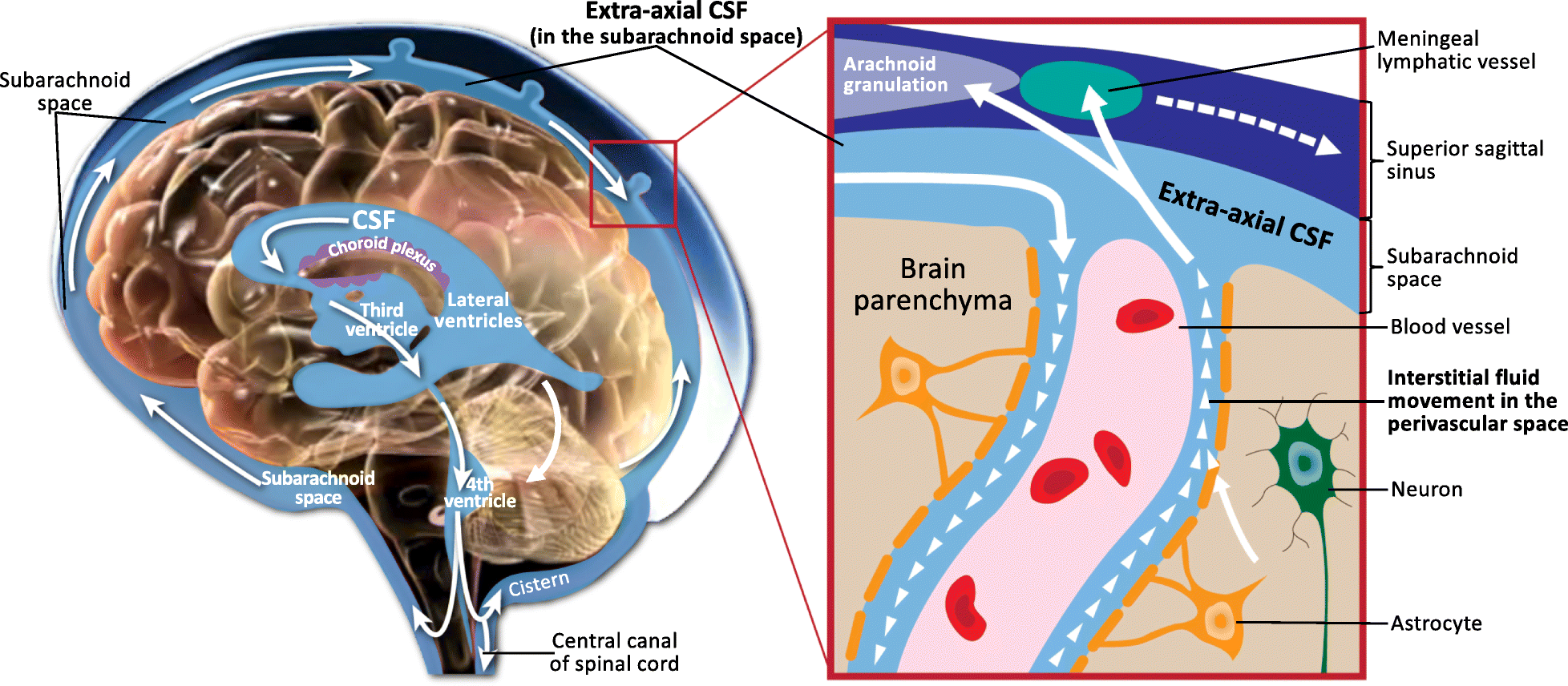
A perivascular space, also known as a Virchow–Robin space, is a fluid-filled space surrounding certain blood vessels in several organs, including the brain, potentially having an immunological function, but more broadly a dispersive role for neural and blood-derived messengers. The brain pia mater is reflected from the surface of the brain onto the surface of blood vessels in the subarachnoid space. In the brain, ''perivascular cuffs'' are regions of leukocyte aggregation in the perivascular spaces, usually found in patients with viral encephalitis. Perivascular spaces vary in dimension according to the type of blood vessel. In the brain where most capillaries have an imperceptible perivascular space, select structures of the brain, such as the circumventricular organs, are notable for having large perivascular spaces surrounding highly permeable capillaries, as observed by microscopy. The median eminence, a brain structure at the base of the hypothalamus, contains capillar ... [...More Info...] [...Related Items...] OR: [Wikipedia] [Google] [Baidu] |


