|
Superior Mesenteric Artery
In human anatomy, the superior mesenteric artery (SMA) is an artery which arises from the anterior surface of the abdominal aorta, just inferior to the origin of the celiac trunk, and supplies blood to the intestine from the lower part of the duodenum through two-thirds of the transverse colon, as well as the pancreas. Structure It arises anterior to lower border of vertebra L1 in an adult. It is usually 1 cm lower than the celiac trunk. It initially travels in an anterior/inferior direction, passing behind/under the neck of the pancreas and the splenic vein. Located under this portion of the superior mesenteric artery, between it and the aorta, are the following: * left renal vein - travels between the left kidney and the inferior vena cava (can be compressed between the SMA and the abdominal aorta at this location, leading to nutcracker syndrome). * the third part of the duodenum, a segment of the small intestines (can be compressed by the SMA at this location, lea ... [...More Info...] [...Related Items...] OR: [Wikipedia] [Google] [Baidu] |
Superior Mesenteric Vein
In human anatomy, the superior mesenteric vein (SMV) is a blood vessel that drains blood from the small intestine (jejunum and ileum). Behind the neck of the pancreas, the superior mesenteric vein combines with the splenic vein to form the hepatic portal vein. The superior mesenteric vein lies to the right of the similarly named artery, the superior mesenteric artery, which originates from the abdominal aorta. Structure Tributaries of the superior mesenteric vein drain the small intestine, large intestine, stomach, pancreas and appendix and include: * Right gastro-omental vein (also known as the right gastro-epiploic vein) * inferior pancreaticoduodenal veins * veins from jejunum * veins from ileum * middle colic vein – drains the transverse colon * right colic vein – drains the ascending colon * ileocolic vein The superior mesenteric vein combines with the splenic vein to form the portal vein. Clinical significance Thrombosis of the superior mesenteric vein is quite rare ... [...More Info...] [...Related Items...] OR: [Wikipedia] [Google] [Baidu] |
Splenic Vein
The spleen is an organ (biology), organ found in almost all vertebrates. Similar in structure to a large lymph node, it acts primarily as a blood filter. The word spleen comes .σπλήν Henry George Liddell, Robert Scott, ''A Greek-English Lexicon'', on Perseus Digital Library The spleen plays very important roles in regard to red blood cells (erythrocytes) and the immune system. It removes old red blood cells and holds a reserve of blood, which can be valuable in case of Shock (circulatory), hemorrhagic shock, and also Human iron metabolism, recycles iron. As a part of the mononuclear phagocyte system, it metabolizes hemoglobin removed from senescent red blood cells. The globin portion of hemoglobin is degraded to its constitutive amino acids, and the h ... [...More Info...] [...Related Items...] OR: [Wikipedia] [Google] [Baidu] |
Ileocecal
{{SIA, animals ...
In many Animalia, including humans, an ileocolic structure or problem is something that concerns the region of the gastrointestinal tract from the ileum to the colon. In Animalia that have ceca, the ileocecal region is a subset of the ileocolic region, and the entire range can also be described as ileocecocolic, whereas in some Animalia, the ileocolic region contains no cecum, as the ileum joins the colon directly. Things that are ileocolic, ileocecal, or both include the following: * Ileocecal fold * Ileocecal/ileocolic intussusception * Ileocecal valve * Ileocolic artery * Ileocolic lymph nodes * Ileocolic vein The ileocolic vein is a vein which drains the ileum, colon, and cecum The cecum or caecum is a pouch within the peritoneum that is considered to be the beginning of the large intestine. It is typically located on the right side of the body (t ... [...More Info...] [...Related Items...] OR: [Wikipedia] [Google] [Baidu] |
Vermiform Appendix
The appendix (or vermiform appendix; also cecal r caecalappendix; vermix; or vermiform process) is a finger-like, blind-ended tube connected to the cecum, from which it develops in the embryo. The cecum is a pouch-like structure of the large intestine, located at the junction of the small and the large intestines. The term "vermiform" comes from Latin and means "worm-shaped". The appendix was once considered a vestigial organ, but this view has changed since the early 2000s. Research suggests that the appendix may serve an important purpose. In particular, it may serve as a reservoir for beneficial gut bacteria. Structure The human appendix averages in length but can range from . The diameter of the appendix is , and more than is considered a thickened or inflamed appendix. The longest appendix ever removed was long. The appendix is usually located in the lower right quadrant of the abdomen, near the right hip bone. The base of the appendix is located beneath the ileoce ... [...More Info...] [...Related Items...] OR: [Wikipedia] [Google] [Baidu] |
Cecum
The cecum or caecum is a pouch within the peritoneum that is considered to be the beginning of the large intestine. It is typically located on the right side of the body (the same side of the body as the appendix (anatomy), appendix, to which it is joined). The word cecum (, plural ceca ) stems from the Latin ''wikt:caecus, caecus'' meaning blindness, blind. It receives chyme from the ileum, and connects to the ascending colon of the large intestine. It is separated from the ileum by the ileocecal valve (ICV) or Bauhin's valve. It is also separated from the Large intestine#Structure, colon by the cecocolic junction. While the cecum is usually intraperitoneal, the ascending colon is Retroperitoneal space, retroperitoneal. In herbivores, the cecum stores food material where bacteria are able to break down the cellulose. In humans, the cecum is involved in absorption of salts and electrolytes and lubricates the solid waste that passes into the large intestine. Structure Develo ... [...More Info...] [...Related Items...] OR: [Wikipedia] [Google] [Baidu] |
Jejunum
The jejunum is the second part of the small intestine in humans and most higher vertebrates, including mammals, reptiles, and birds. Its lining is specialised for the absorption by enterocytes of small nutrient molecules which have been previously digested by enzymes in the duodenum. The jejunum lies between the duodenum and the ileum and is considered to start at the suspensory muscle of the duodenum, a location called the duodenojejunal flexure. The division between the jejunum and ileum is not anatomically distinct. In adult humans, the small intestine is usually long (post mortem), about two-fifths of which (about ) is the jejunum. Structure The interior surface of the jejunum—which is exposed to ingested food—is covered in finger–like projections of mucosa, called villi, which increase the surface area of tissue available to absorb nutrients from ingested foodstuffs. The epithelial cells which line these villi have microvilli. The transport of nutrients across epi ... [...More Info...] [...Related Items...] OR: [Wikipedia] [Google] [Baidu] |
Ileum
The ileum () is the final section of the small intestine in most higher vertebrates, including mammals, reptiles, and birds. In fish, the divisions of the small intestine are not as clear and the terms posterior intestine or distal intestine may be used instead of ileum. Its main function is to absorb vitamin B12, bile salts, and whatever products of digestion that were not absorbed by the jejunum. The ileum follows the duodenum and jejunum and is separated from the cecum by the ileocecal valve (ICV). In humans, the ileum is about 2–4 m long, and the pH is usually between 7 and 8 (neutral or slightly basic). ''Ileum ''is derived from the Greek word ''eilein'', meaning "to twist up tightly". Structure The ileum is the third and final part of the small intestine. It follows the jejunum and ends at the ileocecal junction, where the terminal ileum communicates with the cecum of the large intestine through the ileocecal valve. The ileum, along with the jejunum, is suspended ... [...More Info...] [...Related Items...] OR: [Wikipedia] [Google] [Baidu] |
Intestinal Arteries
The intestinal arteries arise from the convex side of the superior mesenteric artery. They are usually from twelve to fifteen in number, and are distributed to the jejunum and ileum. Nomenclature The term "intestinal arteries" can be confusing, because these arteries only serve a small portion of the intestines. * They do not supply any of the large intestine. The large intestine is primarily supplied by the right colic artery, middle colic artery, and left colic artery. * They do not supply the duodenum of the small intestine. The duodenum is primarily supplied by the inferior pancreaticoduodenal artery and superior pancreaticoduodenal artery. For clarity, some sources prefer instead using the more specific terms ileal arteries and jejunal arteries. Path They run nearly parallel with one another between the layers of the mesentery, each vessel dividing into two branches, which unite with adjacent branches, forming a series of arches (arterial arcades), the convexities of which a ... [...More Info...] [...Related Items...] OR: [Wikipedia] [Google] [Baidu] |
Uncinate Process Of Pancreas
The uncinate process is a small part of the pancreas. The uncinate process is the formed prolongation of the angle of junction of the lower and left lateral borders in the head of the pancreas. The word "uncinate" comes from the Latin "uncinatus", meaning "hooked". Structure Development The pancreas arises as two separate bodies, the dorsal pancreas and the ventral pancreas. The dorsal pancreas appears first, at around day 26, opposite the developing hepatic duct, and grows into the dorsal mesentery. The ventral pancreas develops at the junction of the hepatic duct and the rest of the foregut. During development, differential growth of the wall of the stomach causes it to rotate to the left, and the liver and stomach undergo a lot of growth. This makes the two parts of the pancreas rotate around the duodenum. They then fuse; the dorsal pancreatic bud becomes the body, tail, and isthmus of the pancreas. The isthmus (also called the central pancreas) is the region of the gland that ... [...More Info...] [...Related Items...] OR: [Wikipedia] [Google] [Baidu] |
Superior Mesenteric Artery Syndrome
Superior mesenteric artery (SMA) syndrome is a gastro-vascular disorder in which the third and final portion of the duodenum is compressed between the abdominal aorta (AA) and the overlying superior mesenteric artery. This rare, potentially life-threatening syndrome is typically caused by an angle of 6°–25° between the AA and the SMA, in comparison to the normal range of 38°–56°, due to a lack of retroperitoneal and visceral fat (mesenteric fat). In addition, the aortomesenteric distance is 2–8 millimeters, as opposed to the typical 10–20. However, a narrow SMA angle alone is not enough to make a diagnosis, because patients with a low BMI, most notably children, have been known to have a narrow SMA angle with no symptoms of SMA syndrome. SMA syndrome is also known as Wilkie's syndrome, cast syndrome, mesenteric root syndrome, chronic duodenal ileus and intermittent arterio-mesenteric occlusion. It is distinct from nutcracker syndrome, which is the entrapment of the l ... [...More Info...] [...Related Items...] OR: [Wikipedia] [Google] [Baidu] |
Small Intestines
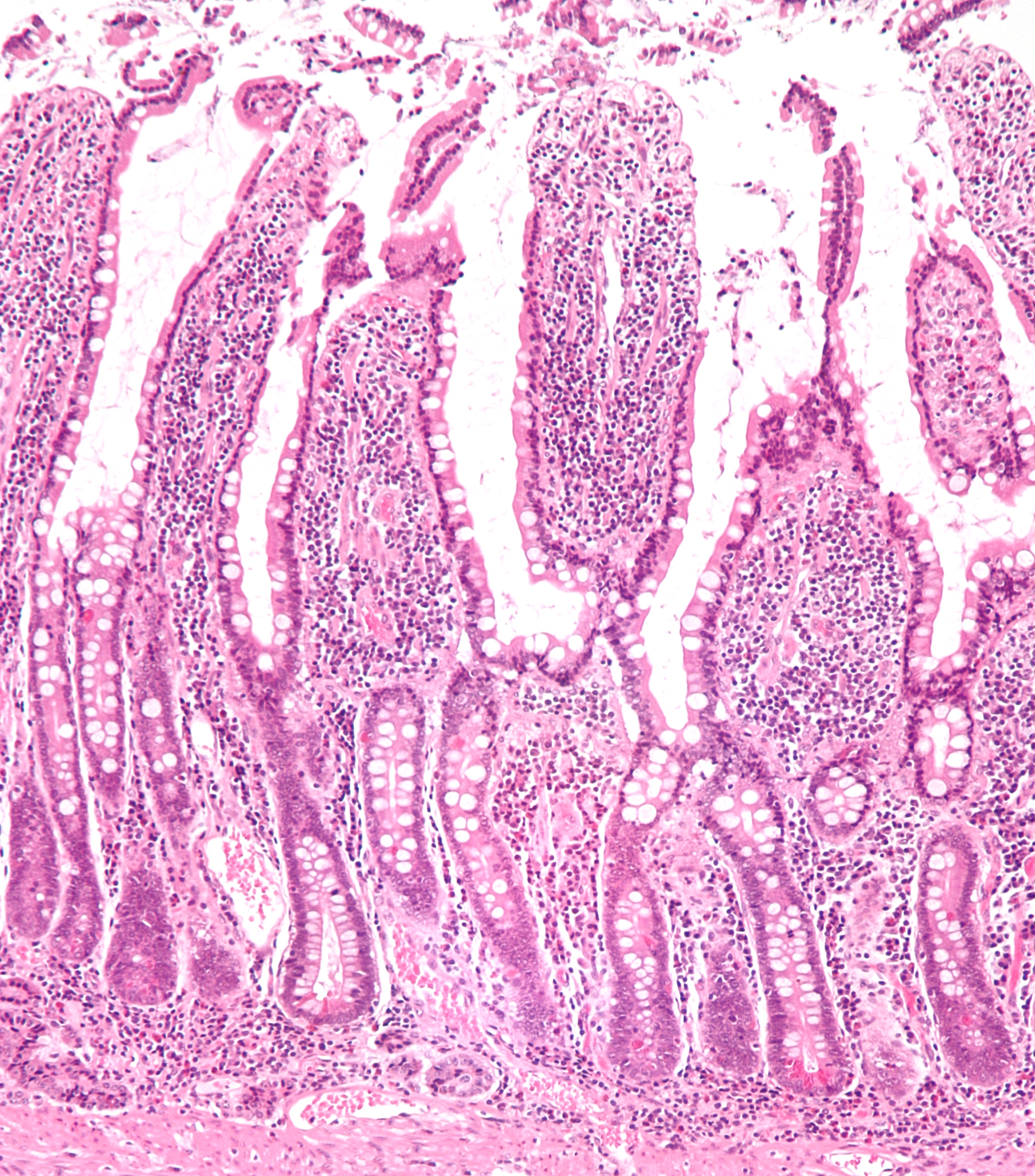
The small intestine or small bowel is an organ in the gastrointestinal tract where most of the absorption of nutrients from food takes place. It lies between the stomach and large intestine, and receives bile and pancreatic juice through the pancreatic duct to aid in digestion. The small intestine is about long and folds many times to fit in the abdomen. Although it is longer than the large intestine, it is called the small intestine because it is narrower in diameter. The small intestine has three distinct regions – the duodenum, jejunum, and ileum. The duodenum, the shortest, is where preparation for absorption through small finger-like protrusions called villi begins. The jejunum is specialized for the absorption through its lining by enterocytes: small nutrient particles which have been previously digested by enzymes in the duodenum. The main function of the ileum is to absorb vitamin B12, bile salts, and whatever products of digestion that were not absorbed by the jej ... [...More Info...] [...Related Items...] OR: [Wikipedia] [Google] [Baidu] |
Nutcracker Syndrome
The nutcracker syndrome (NCS) results most commonly from the compression of the left renal vein (LRV) between the abdominal aorta (AA) and superior mesenteric artery (SMA), although other variants exist. The name derives from the fact that, in the sagittal plane and/or transverse plane, the SMA and AA (with some imagination) appear to be a nutcracker crushing a nut (the renal vein). Furthermore, the venous return from the left gonadal vein returning to the left renal vein is blocked, thus causing testicular pain (colloquially referred to as "nut pain"). There is a wide spectrum of clinical presentations and diagnostic criteria are not well defined, which frequently results in delayed or incorrect diagnosis. The first clinical report of Nutcracker phenomenon appeared in 1950. This condition is not to be confused with superior mesenteric artery syndrome, which is the compression of the third portion of the duodenum by the SMA and the AA. Signs and symptoms The signs and symptoms of N ... [...More Info...] [...Related Items...] OR: [Wikipedia] [Google] [Baidu] |


