|
Somitogenesis
Somitogenesis is the process by which somites form. Somites are bilaterally paired blocks of paraxial mesoderm that form along the anterior-posterior axis of the developing embryo in segmented animals. In vertebrates, somites give rise to skeletal muscle, cartilage, tendons, endothelium, and dermis. Overview In somitogenesis, somites form from the paraxial mesoderm, a particular region of mesoderm in the neurulating embryo. This tissue undergoes convergent extension as the primitive streak regresses, or as the embryo gastrulates. The notochord extends from the base of the head to the tail; with it extend thick bands of paraxial mesoderm. As the primitive streak continues to regress, somites form from the paraxial mesoderm by "budding off" rostrally as somitomeres, or whorls of paraxial mesoderm cells, compact and separate into discrete bodies. The periodic nature of these splitting events has led many to say to that somitogenesis occurs via a clock-wavefront model, in which wav ... [...More Info...] [...Related Items...] OR: [Wikipedia] [Google] [Baidu] |
Clock And Wavefront Model
The clock and wavefront model is a model used to describe the process of somitogenesis in vertebrates. Somitogenesis is the process by which somites, blocks of mesoderm that give rise to a variety of connective tissues, are formed. The model describes the splitting off of somites from the paraxial mesoderm as the result of oscillating expression of particular proteins and their gradients. Overview Once the cells of the pre-somitic mesoderm are in place following by cell migration during gastrulation, oscillatory expression of many genes begins in these cells as if regulated by a developmental "clock." This has led many to conclude that somitogenesis is coordinated by a "clock and wave" mechanism. More technically, this means that somitogenesis occurs due to the largely cell-autonomous oscillations of a network of genes and gene products which causes cells to oscillate between a permissive and a non-permissive state in a consistently timed-fashion, like a clock. These genes includ ... [...More Info...] [...Related Items...] OR: [Wikipedia] [Google] [Baidu] |
Notch Signaling
The Notch signaling pathway is a highly Conserved sequence, conserved cell signaling system present in most animals. Mammals possess four different Notch proteins, notch receptors, referred to as NOTCH1, NOTCH2, Notch 3, NOTCH3, and NOTCH4. The notch receptor is a single-pass Cell surface receptor, transmembrane receptor protein. It is a hetero-oligomer composed of a large extracellular portion, which associates in a calcium-dependent, non-covalent interaction with a smaller piece of the notch protein composed of a short extracellular region, a single transmembrane-pass, and a small intracellular region. Notch signaling promotes proliferative signaling during neurogenesis, and its activity is inhibited by NUMB (gene), Numb to promote neural differentiation. It plays a major role in the regulation of embryonic development. Notch signaling is dysregulated in many cancers, and faulty notch signaling is implicated in many diseases, including T-cell acute lymphoblastic leukemia (Pre ... [...More Info...] [...Related Items...] OR: [Wikipedia] [Google] [Baidu] |
Sclerotome
The somites (outdated term: primitive segments) are a set of bilaterally paired blocks of paraxial mesoderm that form in the embryonic stage of somitogenesis, along the head-to-tail axis in segmented animals. In vertebrates, somites subdivide into the dermatomes, myotomes, sclerotomes and syndetomes that give rise to the vertebrae of the vertebral column, rib cage, part of the occipital bone, skeletal muscle, cartilage, tendons, and skin (of the back). The word ''somite'' is sometimes also used in place of the word '' metamere''. In this definition, the somite is a homologously-paired structure in an animal body plan, such as is visible in annelids and arthropods. Development The mesoderm forms at the same time as the other two germ layers, the ectoderm and endoderm. The mesoderm at either side of the neural tube is called paraxial mesoderm. It is distinct from the mesoderm underneath the neural tube which is called the chordamesoderm that becomes the notochord. The pa ... [...More Info...] [...Related Items...] OR: [Wikipedia] [Google] [Baidu] |
Somite
The somites (outdated term: primitive segments) are a set of bilaterally paired blocks of paraxial mesoderm that form in the embryonic stage of somitogenesis, along the head-to-tail axis in segmented animals. In vertebrates, somites subdivide into the dermatomes, myotomes, sclerotomes and syndetomes that give rise to the vertebrae of the vertebral column, rib cage, part of the occipital bone, skeletal muscle, cartilage, tendons, and skin (of the back). The word ''somite'' is sometimes also used in place of the word '' metamere''. In this definition, the somite is a homologously-paired structure in an animal body plan, such as is visible in annelids and arthropods. Development The mesoderm forms at the same time as the other two germ layers, the ectoderm and endoderm. The mesoderm at either side of the neural tube is called paraxial mesoderm. It is distinct from the mesoderm underneath the neural tube which is called the chordamesoderm that becomes the notochord. The pa ... [...More Info...] [...Related Items...] OR: [Wikipedia] [Google] [Baidu] |
Segmentation (biology)
Segmentation in biology is the division of some animal and plant body plans into a series of repetitive segments. This article focuses on the segmentation of animal body plans, specifically using the examples of the taxa Arthropoda, Chordata, and Annelida. These three groups form segments by using a "growth zone" to direct and define the segments. While all three have a generally segmented body plan and use a growth zone, they use different mechanisms for generating this patterning. Even within these groups, different organisms have different mechanisms for segmenting the body. Segmentation of the body plan is important for allowing free movement and development of certain body parts. It also allows for regeneration in specific individuals. Definition Segmentation is a difficult process to satisfactorily define. Many taxa (for example the molluscs) have some form of serial repetition in their units but are not conventionally thought of as segmented. Segmented animals are tho ... [...More Info...] [...Related Items...] OR: [Wikipedia] [Google] [Baidu] |
Paraxial Mesoderm
Paraxial mesoderm, also known as presomitic or somitic mesoderm is the area of mesoderm in the neurulating embryo that flanks and forms simultaneously with the neural tube. The cells of this region give rise to somites, blocks of tissue running along both sides of the neural tube, which form muscle and the tissues of the back, including connective tissue and the dermis. Formation and somitogenesis The paraxial and other regions of the mesoderm are thought to be specified by bone morphogenetic proteins, or BMPs, along an axis spanning from the center to the sides of the body. Members of the FGF family also play an important role, as does the WNT pathway. In particular, Noggin, a downstream target of the Wnt pathway, antagonizes BMP signaling, forming boundaries where antagonists meet and limiting this signaling to a particular region of the mesoderm. Together, these pathways provide the initial specification of the paraxial mesoderm and maintain this identity. This specific ... [...More Info...] [...Related Items...] OR: [Wikipedia] [Google] [Baidu] |
Sonic Hedgehog
Sonic hedgehog protein (SHH) is encoded for by the ''SHH'' gene. The protein is named after the character ''Sonic the Hedgehog''. This signaling molecule is key in regulating embryonic morphogenesis in all animals. SHH controls organogenesis and the organization of the central nervous system, limbs, digits and many other parts of the body. Sonic hedgehog is a morphogen that patterns the developing embryo using a concentration gradient characterized by the French flag model. This model has a non-uniform distribution of SHH molecules which governs different cell fates according to concentration. Mutations in this gene can cause holoprosencephaly, a failure of splitting in the cerebral hemispheres, as demonstrated in an experiment using SHH knock-out mice in which the forebrain midline failed to develop and instead only a single fused telencephalic vesicle resulted. Sonic hedgehog still plays a role in differentiation, proliferation, and maintenance of adult tissues. Abnormal activ ... [...More Info...] [...Related Items...] OR: [Wikipedia] [Google] [Baidu] |
Epithelial–mesenchymal Transition
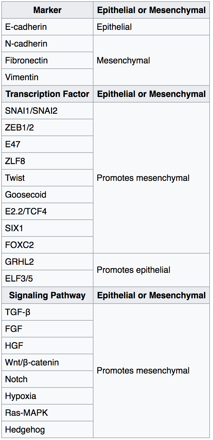
The epithelial–mesenchymal transition (EMT) is a process by which epithelial cells lose their cell polarity and cell–cell adhesion, and gain migratory and invasive properties to become mesenchymal stem cells; these are multipotent stromal cells that can differentiate into a variety of cell types. EMT is essential for numerous developmental processes including mesoderm formation and neural tube formation. EMT has also been shown to occur in wound healing, in organ fibrosis and in the initiation of metastasis in cancer progression. Introduction Epithelial–mesenchymal transition was first recognized as a feature of embryogenesis by Betty Hay in the 1980s. EMT, and its reverse process, MET ( mesenchymal-epithelial transition) are critical for development of many tissues and organs in the developing embryo, and numerous embryonic events such as gastrulation, neural crest formation, heart valve formation, secondary palate development, and myogenesis. Epithelial and mesenchyma ... [...More Info...] [...Related Items...] OR: [Wikipedia] [Google] [Baidu] |
Signal Transduction
Signal transduction is the process by which a chemical or physical signal is transmitted through a cell as a series of molecular events, most commonly protein phosphorylation catalyzed by protein kinases, which ultimately results in a cellular response. Proteins responsible for detecting stimuli are generally termed receptors, although in some cases the term sensor is used. The changes elicited by ligand binding (or signal sensing) in a receptor give rise to a biochemical cascade, which is a chain of biochemical events known as a signaling pathway. When signaling pathways interact with one another they form networks, which allow cellular responses to be coordinated, often by combinatorial signaling events. At the molecular level, such responses include changes in the transcription or translation of genes, and post-translational and conformational changes in proteins, as well as changes in their location. These molecular events are the basic mechanisms controlling cell growth, ... [...More Info...] [...Related Items...] OR: [Wikipedia] [Google] [Baidu] |
Negative Feedback
Negative feedback (or balancing feedback) occurs when some function (Mathematics), function of the output of a system, process, or mechanism is feedback, fed back in a manner that tends to reduce the fluctuations in the output, whether caused by changes in the input or by other disturbances. Whereas positive feedback tends to lead to instability via exponential growth, oscillation or chaos theory, chaotic behavior, negative feedback generally promotes stability. Negative feedback tends to promote a settling to List of types of equilibrium, equilibrium, and reduces the effects of perturbations. Negative feedback loops in which just the right amount of correction is applied with optimum timing can be very stable, accurate, and responsive. Negative feedback is widely used in mechanical and electronic engineering, and also within living organisms, and can be seen in many other fields from chemistry and economics to physical systems such as the climate. General negative feedback ... [...More Info...] [...Related Items...] OR: [Wikipedia] [Google] [Baidu] |
Fibronectin
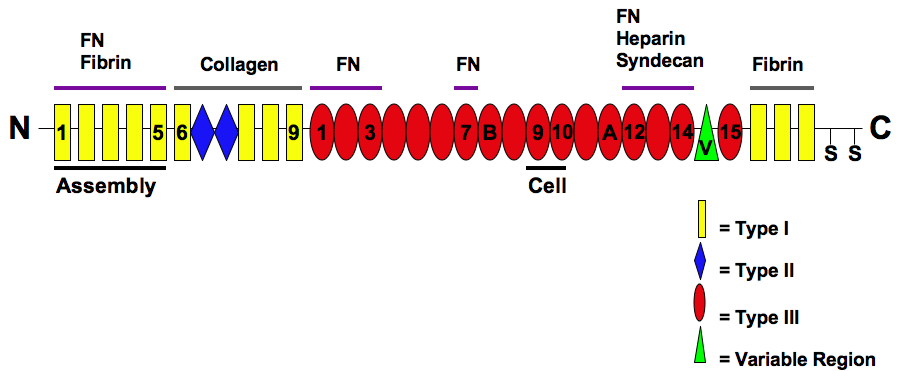
Fibronectin is a high- molecular weight (~500-~600 kDa) glycoprotein of the extracellular matrix that binds to membrane-spanning receptor proteins called integrins. Fibronectin also binds to other extracellular matrix proteins such as collagen, fibrin, and heparan sulfate proteoglycans (e.g. syndecans). Fibronectin exists as a protein dimer, consisting of two nearly identical monomers linked by a pair of disulfide bonds. The fibronectin protein is produced from a single gene, but alternative splicing of its pre-mRNA leads to the creation of several isoforms. Two types of fibronectin are present in vertebrates: * soluble plasma fibronectin (formerly called "cold-insoluble globulin", or CIg) is a major protein component of blood plasma (300 μg/ml) and is produced in the liver by hepatocytes. * insoluble cellular fibronectin is a major component of the extracellular matrix. It is secreted by various cells, primarily fibroblasts, as a soluble protein dimer and is then ass ... [...More Info...] [...Related Items...] OR: [Wikipedia] [Google] [Baidu] |
Eph Receptor
Eph receptors (Ephs, after erythropoietin-producing human hepatocellular receptors) are a group of receptors that are activated in response to binding with Eph receptor-interacting proteins (Ephrins). Ephs form the largest known subfamily of receptor tyrosine kinases (RTKs). Both Eph receptors and their corresponding ephrin ligands are membrane-bound proteins that require direct cell-cell interactions for Eph receptor activation. Eph/ephrin signaling has been implicated in the regulation of a host of processes critical to embryonic development including axon guidance, formation of tissue boundaries, cell migration, and segmentation. Additionally, Eph/ephrin signaling has been identified to play a critical role in the maintenance of several processes during adulthood including long-term potentiation, angiogenesis, and stem cell differentiation and cancer. Subclasses Ephs can be divided into two subclasses, EphAs and EphBs (encoded by the genetic loci designated ''EPHA' ... [...More Info...] [...Related Items...] OR: [Wikipedia] [Google] [Baidu] |


