|
Sex Determination And Differentiation (human)
Sexual differentiation in humans is the process of development of sex differences in humans. It is defined as the development of phenotypic structures consequent to the action of hormones produced following gonadal determination. Sexual differentiation includes development of different genitalia and the internal genital tracts and body hair plays a role in sex identification. The development of sexual differences begins with the XY sex-determination system that is present in humans, and complex mechanisms are responsible for the development of the phenotypic differences between male and female humans from an undifferentiated zygote. Females typically have two X chromosomes, and males typically have a Y chromosome and an X chromosome. At an early stage in embryonic development, both sexes possess equivalent internal structures. These are the mesonephric ducts and paramesonephric ducts. The presence of the SRY gene on the Y chromosome causes the development of the testes in males, ... [...More Info...] [...Related Items...] OR: [Wikipedia] [Google] [Baidu] |
Fertilization
Fertilisation or fertilization (see spelling differences), also known as generative fertilisation, syngamy and impregnation, is the fusion of gametes to give rise to a new individual organism or offspring and initiate its development. Processes such as insemination or pollination which happen before the fusion of gametes are also sometimes informally called fertilisation. The cycle of fertilisation and development of new individuals is called sexual reproduction. During double fertilisation in angiosperms the haploid male gamete combines with two haploid polar nuclei to form a triploid primary endosperm nucleus by the process of vegetative fertilisation. History In Antiquity, Aristotle conceived the formation of new individuals through fusion of male and female fluids, with form and function emerging gradually, in a mode called by him as epigenetic. In 1784, Spallanzani established the need of interaction between the female's ovum and male's sperm to form a zygote in frog ... [...More Info...] [...Related Items...] OR: [Wikipedia] [Google] [Baidu] |
Ovaries
The ovary is an organ in the female reproductive system that produces an ovum. When released, this travels down the fallopian tube into the uterus, where it may become fertilized by a sperm. There is an ovary () found on each side of the body. The ovaries also secrete hormones that play a role in the menstrual cycle and fertility. The ovary progresses through many stages beginning in the prenatal period through menopause. It is also an endocrine gland because of the various hormones that it secretes. Structure The ovaries are considered the female gonads. Each ovary is whitish in color and located alongside the lateral wall of the uterus in a region called the ovarian fossa. The ovarian fossa is the region that is bounded by the external iliac artery and in front of the ureter and the internal iliac artery. This area is about 4 cm x 3 cm x 2 cm in size.Daftary, Shirish; Chakravarti, Sudip (2011). Manual of Obstetrics, 3rd Edition. Elsevier. pp. 1-16. . The ovarie ... [...More Info...] [...Related Items...] OR: [Wikipedia] [Google] [Baidu] |
Testes
A testicle or testis (plural testes) is the male reproductive gland or gonad in all bilaterians, including humans. It is homologous to the female ovary. The functions of the testes are to produce both sperm and androgens, primarily testosterone. Testosterone release is controlled by the anterior pituitary luteinizing hormone, whereas sperm production is controlled both by the anterior pituitary follicle-stimulating hormone and gonadal testosterone. Structure Appearance Males have two testicles of similar size contained within the scrotum, which is an extension of the abdominal wall. Scrotal asymmetry, in which one testicle extends farther down into the scrotum than the other, is common. This is because of the differences in the vasculature's anatomy. For 85% of men, the right testis hangs lower than the left one. Measurement and volume The volume of the testicle can be estimated by palpating it and comparing it to ellipsoids of known sizes. Another method is to use cali ... [...More Info...] [...Related Items...] OR: [Wikipedia] [Google] [Baidu] |
Gonads
A gonad, sex gland, or reproductive gland is a mixed gland that produces the gametes and sex hormones of an organism. Female reproductive cells are egg cells, and male reproductive cells are sperm. The male gonad, the testicle, produces sperm in the form of spermatozoa. The female gonad, the ovary, produces egg cells. Both of these gametes are haploid cells. Some hermaphroditic animals have a type of gonad called an ovotestis. Evolution It is hard to find a common origin for gonads, but gonads most likely evolved independently several times. Regulation The gonads are controlled by luteinizing hormone and follicle-stimulating hormone, produced and secreted by gonadotropes or gonadotrophins in the anterior pituitary gland. This secretion is regulated by gonadotropin-releasing hormone produced in the hypothalamus. Development Gonads start developing as a common primordium (an organ in the earliest stage of development), in the form of genital ridges, which are only late ... [...More Info...] [...Related Items...] OR: [Wikipedia] [Google] [Baidu] |
Labia (genitalia)
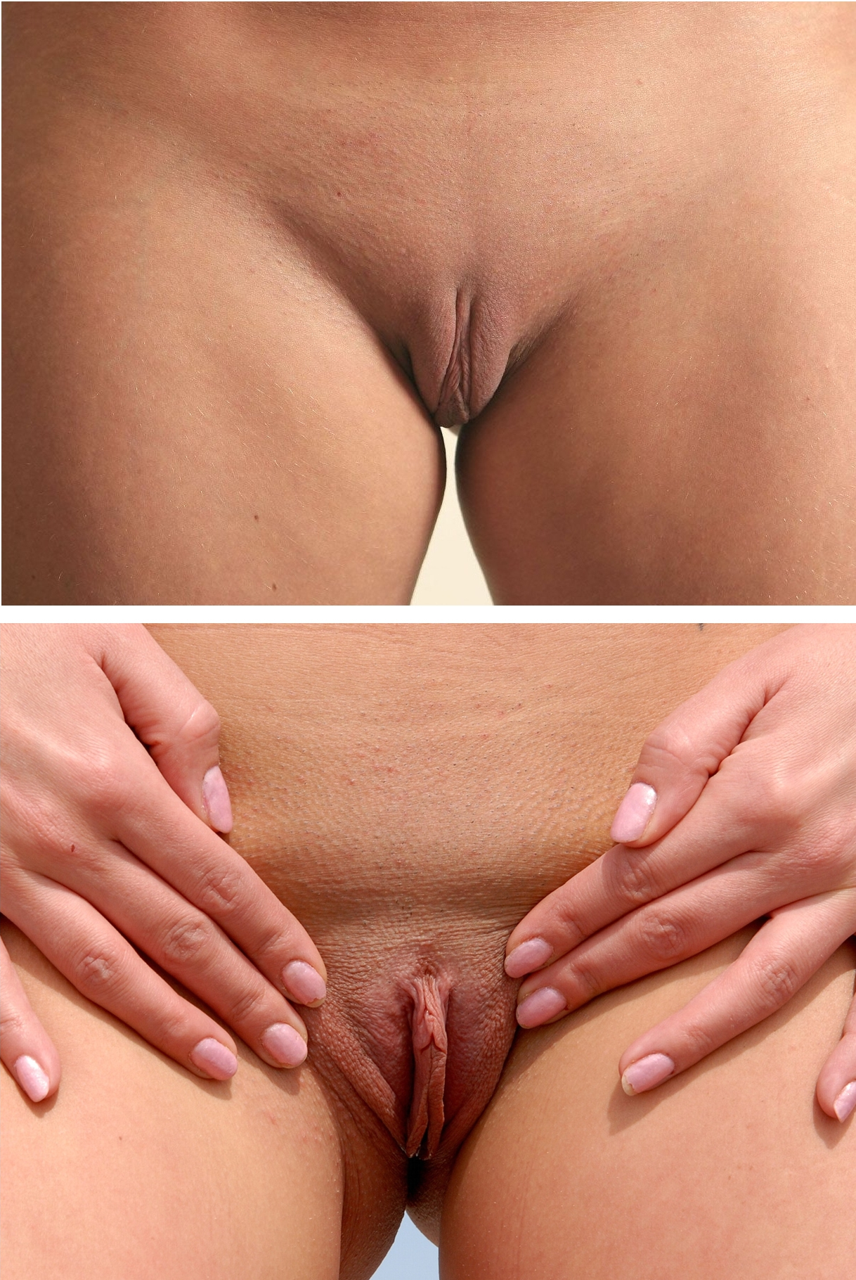
The labia are part of the female genitalia; they are the major externally visible portions of the vulva. In humans, there are two pairs of labia: the ''labia majora'' (or the outer labia) are larger and thicker, while the ''labia minora'' are folds of skin between the outer labia. The labia surround and protect the clitoris and the openings of the vagina and the urethra. Etymology ''Labium'' (plural ''labia'') is a Latin-derived term meaning "lip". ''Labium'' and its derivatives (including labial, labrum) are used to describe any lip-like structure, but in the English language, ''labium'' often specifically refers to parts of the vulva. Anatomy The labia majora, also commonly called outer labia or outer lips, are lip-like structures consisting mostly of skin and adipose (fatty) tissue, which extend on either side of the vulva to form the pudendal cleft through the middle. The labia majora often have a plump appearance, and are thicker towards the anterior. The anterior junction ... [...More Info...] [...Related Items...] OR: [Wikipedia] [Google] [Baidu] |
Vagina
In mammals, the vagina is the elastic, muscular part of the female genital tract. In humans, it extends from the vestibule to the cervix. The outer vaginal opening is normally partly covered by a thin layer of mucosal tissue called the hymen. At the deep end, the cervix (neck of the uterus) bulges into the vagina. The vagina allows for sexual intercourse and birth. It also channels menstrual flow, which occurs in humans and closely related primates as part of the menstrual cycle. Although research on the vagina is especially lacking for different animals, its location, structure and size are documented as varying among species. Female mammals usually have two external openings in the vulva; these are the urethral opening for the urinary tract and the vaginal opening for the genital tract. This is different from male mammals, who usually have a single urethral opening for both urination and reproduction. The vaginal opening is much larger than the nearby urethral opening, an ... [...More Info...] [...Related Items...] OR: [Wikipedia] [Google] [Baidu] |
Urethra
The urethra (from Greek οὐρήθρα – ''ourḗthrā'') is a tube that connects the urinary bladder to the urinary meatus for the removal of urine from the body of both females and males. In human females and other primates, the urethra connects to the urinary meatus above the vagina, whereas in marsupials, the female's urethra empties into the urogenital sinus. Females use their urethra only for urinating, but males use their urethra for both urination and ejaculation. The external urethral sphincter is a striated muscle that allows voluntary control over urination. The internal sphincter, formed by the involuntary smooth muscles lining the bladder neck and urethra, receives its nerve supply by the sympathetic division of the autonomic nervous system. The internal sphincter is present both in males and females. Structure The urethra is a fibrous and muscular tube which connects the urinary bladder to the external urethral meatus. Its length differs between the sexes, ... [...More Info...] [...Related Items...] OR: [Wikipedia] [Google] [Baidu] |
Clitoris
The clitoris ( or ) is a female sex organ present in mammals, ostriches and a limited number of other animals. In humans, the visible portion – the glans – is at the front junction of the labia minora (inner lips), above the opening of the urethra. Unlike the penis, the male homologue (equivalent) to the clitoris, it usually does not contain the distal portion (or opening) of the urethra and is therefore not used for urination. In most species, the clitoris lacks any reproductive function. While few animals urinate through the clitoris or use it reproductively, the spotted hyena, which has an especially large clitoris, urinates, mates, and gives birth via the organ. Some other mammals, such as lemurs and spider monkeys, also have a large clitoris. The clitoris is the human female's most sensitive erogenous zone and generally the primary anatomical source of human female sexual pleasure. In humans and other mammals, it develops from an outgrowth in the embry ... [...More Info...] [...Related Items...] OR: [Wikipedia] [Google] [Baidu] |
Labioscrotal Folds
The labioscrotal swellings (genital swellings or labioscrotal folds) are paired structures in the human embryo that represent the final stage of development of the caudal end of the external genitals before sexual differentiation. In both males and females, the two swellings merge: * In the ''female'', they become the posterior labial commissure. The sides of the genital tubercle grow backward as the genital swellings, which ultimately form the labia majora; the tubercle itself becomes the mons pubis. In contrast, the labia minora are formed by the urogenital folds. * In the ''male'', they become the scrotum The scrotum or scrotal sac is an anatomical male reproductive structure located at the base of the penis that consists of a suspended dual-chambered sac of skin and smooth muscle. It is present in most terrestrial male mammals. The scrotum co .... References External links "Development of Male External Genitalia", at mcgill.caDiagram at mhhe.com* * {{Authority ... [...More Info...] [...Related Items...] OR: [Wikipedia] [Google] [Baidu] |
Genital Tubercle
A genital tubercle or phallic tubercle is a body of tissue present in the development of the reproductive system. It forms in the ventral, caudal region of mammalian embryos of both sexes, and eventually develops into a primordial phallus. In the human fetus, the genital tubercle develops around week 4 of gestation, and by week 9 becomes recognizably either a clitoris or penis. This should not be confused with the sinus tubercle which is a proliferation of endoderm induced by paramesonephric ducts. Even after the phallus is developed, the term genital tubercle remains, but only as the terminal end of it, which develops into either the glans penis or the glans clitoridis. The genital tubercle is sensitive to dihydrotestosterone and rich in 5-alpha-reductase, so that the amount of fetal testosterone present after the second month is a major determinant of phallus size at birth. See also *Sexual differentiation Sexual differentiation is the process of development of the sex differ ... [...More Info...] [...Related Items...] OR: [Wikipedia] [Google] [Baidu] |


.png)