|
Superior Parietal Lobule
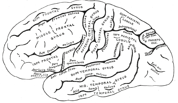
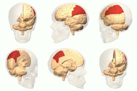
The superior parietal lobule is bounded in front by the upper part of the postcentral sulcus, but is usually connected with the postcentral gyrus above the end of the sulcus. The superior parietal lobule contains Brodmann's areas brodmann area 5, 5 and brodmann area 7, 7. Behind it is the lateral part of the parietooccipital fissure, around the end of which it is joined to the occipital lobe by a curved gyrus, the arcus parietooccipitalis. Below, it is separated from the inferior parietal lobule by the horizontal portion of the intraparietal sulcus. The superior parietal lobule is involved with spatial orientation, and receives a great deal of visual input as well as sensory input from one's hand. It is also involved with other functions of the parietal lobe in general. There are major white matter pathway connections with the superior parietal lobule such as the Cingulum (brain), Cingulum, SLF I, superior parietal lobule connections of the Medial longitudinal fasciculus and othe ... [...More Info...] [...Related Items...] OR: [Wikipedia] [Google] [Baidu] |
Postcentral Sulcus
The postcentral sulcus of the parietal lobe lies parallel to, and behind, the central sulcus in the human brain. (A ''sulcus'' is one of the prominent grooves on the surface of the brain.) The postcentral sulcus divides the postcentral gyrus from the remainder of the parietal lobe. Additional images File:Gray725 postcentral sulcus.png, Lateral surface of left cerebral hemisphere, viewed from above. File:ParietCapts lateral.png, Gyri and sulci of right cerebral hemisphere. Postcentral sulcus labeled in red at top center. File:Cerebral Hemisphere Demonstration - Sanjoy Sanyal - Neuroscience Lab Fall 2013 (Cropped from 27m17s to 27m54s) Postcentral sulcus.webm, Human brain dissection video (36 sec). Demonstrating position of the postcentral sulcus of the left cerebral hemisphere. External links * - "Cerebral Hemisphere, Superior View" Cerebrum Sulci (neuroanatomy) Articles containing video clips {{neuroanatomy-stub ... [...More Info...] [...Related Items...] OR: [Wikipedia] [Google] [Baidu] |
Postcentral Gyrus
In neuroanatomy, the postcentral gyrus is a prominent gyrus in the lateral parietal lobe of the human brain. It is the location of the primary somatosensory cortex, the main sensory receptive area for the somatosensory system, sense of touch. Like other sensory areas, there is a map of sensory space in this location, called the ''sensory homunculus''. The primary somatosensory cortex was initially defined from surface stimulation studies of Wilder Penfield, and parallel surface potential studies of Bard, Woolsey, and Marshall. Although initially defined to be roughly the same as Brodmann areas Brodmann area 3, 3, Brodmann area 1, 1 and Brodmann area 2, 2, more recent work by Jon Kaas, Kaas has suggested that for homogeny with other sensory fields only area 3 should be referred to as "primary somatosensory cortex", as it receives the bulk of the Thalamocortical radiations, thalamocortical projections from the sensory input fields. Structure The lateral postcentral gyrus is bounded ... [...More Info...] [...Related Items...] OR: [Wikipedia] [Google] [Baidu] |
Brodmann Area 5
Brodmann area 5 is one of Brodmann's cytoarchitectural defined regions of the brain. It is involved in somatosensory processing, movement and association, and is part of the posterior parietal cortex. Human Brodmann area 5 is a subdivision of the parietal cortex, part of the cortex in the human brain. BA5 is part of the superior parietal lobule and part of the postcentral gyrus. It is situated immediately posterior to the primary somatosensory cortex. It is bounded cytoarchitecturally by Brodmann area 2, Brodmann area 7, Brodmann area 4, and Brodmann area 31. Monkey In guenon Brodmann area 5 is a subdivision of the parietal lobe defined on the basis of cytoarchitecture. It occupies primarily the superior parietal lobule. Brodmann-1909 considered it topologically and cytoarchitecturally homologous to the preparietal area 5 of the human. Distinctive features (Brodmann-1905): compared to area 4 of Brodmann-1909 area 5 has a thick self-contained internal granular layer (IV); l ... [...More Info...] [...Related Items...] OR: [Wikipedia] [Google] [Baidu] |
Brodmann Area 7
Brodmann is a German surname. Notable people with the surname include: *Ines Brodmann (birth date unknown), Swiss orienteer *Korbinian Brodmann (1868–1918), German neurologist *Mario Brodmann (born 1966), Swiss former ice hockey forward *René Brodmann (born 1933), Swiss football defender See also *Brodmann area A Brodmann area is a region of the cerebral cortex, in the human or other primate brain, defined by its cytoarchitecture, or histological structure and organization of cells. History Brodmann areas were originally defined and numbered by the ..., a region in the brain cortex * Michael L. Brodman, American gynecologist and obstetrician {{surname, Brodmann German-language surnames ... [...More Info...] [...Related Items...] OR: [Wikipedia] [Google] [Baidu] |
Occipital Lobe
The occipital lobe is one of the four major lobes of the cerebral cortex in the brain of mammals. The name derives from its position at the back of the head, from the Latin ''ob'', "behind", and ''caput'', "head". The occipital lobe is the visual processing center of the mammalian brain containing most of the anatomical region of the visual cortex. The primary visual cortex is Brodmann area 17, commonly called V1 (visual one). Human V1 is located on the medial side of the occipital lobe within the calcarine sulcus; the full extent of V1 often continues onto the occipital pole. V1 is often also called striate cortex because it can be identified by a large stripe of myelin, the Stria of Gennari. Visually driven regions outside V1 are called extrastriate cortex. There are many extrastriate regions, and these are specialized for different visual tasks, such as visuospatial processing, color differentiation, and motion perception. Bilateral lesions of the occipital lobe can lead ... [...More Info...] [...Related Items...] OR: [Wikipedia] [Google] [Baidu] |
Gyrus
In neuroanatomy, a gyrus (pl. gyri) is a ridge on the cerebral cortex. It is generally surrounded by one or more sulci (depressions or furrows; sg. ''sulcus''). Gyri and sulci create the folded appearance of the brain in humans and other mammals. Structure The gyri are part of a system of folds and ridges that create a larger surface area for the human brain and other mammalian brains. Because the brain is confined to the skull, brain size is limited. Ridges and depressions create folds allowing a larger cortical surface area, and greater cognitive function, to exist in the confines of a smaller cranium. Development The human brain undergoes gyrification during fetal and neonatal development. In embryonic development, all mammalian brains begin as smooth structures derived from the neural tube. A cerebral cortex without surface convolutions is lissencephalic, meaning 'smooth-brained'. As development continues, gyri and sulci begin to take shape on the fetal brain, with ... [...More Info...] [...Related Items...] OR: [Wikipedia] [Google] [Baidu] |
Arcus Parietooccipitalis
Arcus may refer to: Businesses and organizations *ARCUS, the Arctic Research Consortium of the United States, supporting Arctic policy in the U.S. *Arcus AS, a Norwegian producer of liquor * Arcus Co., a Bulgarian firearm manufacturer *Arcus Foundation, supporting great apes and LGBT rights * Arcus-Air, a German airline Gliders *Schempp-Hirth Arcus The Schempp-Hirth Arcus is a flapped Two Seater Class glider in production by Schempp-Hirth. It first flew 7 April 2009. It is offered in addition to the Duo Discus which is an unflapped 20 metre two-seater, whose fuselage it shares. The w ... a two-seat glider * Pegas Arcus, a Czech paraglider design * Swing Arcus, German paraglider design Human anatomy * Arcus anterior atlantis * Arcus aortae * Arcus corneae * Arcus costalis * Arcus dentalis * Arcus dentalis mandibularis * Arcus dentalis maxillaris * Arcus ductus thoracici * Arcus iliopectineus * Arcus inguinalis * Arcus lumbocostalis lateralis * Arcus lumbocos ... [...More Info...] [...Related Items...] OR: [Wikipedia] [Google] [Baidu] |
Inferior Parietal Lobule
The inferior parietal lobule (subparietal district) lies below the horizontal portion of the intraparietal sulcus, and behind the lower part of the postcentral sulcus. Also known as Geschwind's territory after Norman Geschwind, an American neurologist, who in the early 1960s recognised its importance. It is a part of the parietal lobe. Structure It is divided from rostral to caudal into two gyri: * One, the supramarginal gyrus, arches over the upturned end of the lateral fissure; it is continuous in front with the postcentral gyrus, and behind with the superior temporal gyrus. * The second, the angular gyrus, arches over the posterior end of the superior temporal sulcus, behind which it is continuous with the middle temporal gyrus. In macaque neuroanatomy, this region is often divided into caudal and rostral portions, cIPL and rIPL, respectively. The cIPL is further divided into areas Opt and PG whereas rIPL is divided into PFG and PF areas. Function Inferior parietal lobule has ... [...More Info...] [...Related Items...] OR: [Wikipedia] [Google] [Baidu] |
Intraparietal Sulcus
The intraparietal sulcus (IPS) is located on the lateral surface of the parietal lobe, and consists of an oblique and a horizontal portion. The IPS contains a series of functionally distinct subregions that have been intensively investigated using both single cell neurophysiology in primates and human functional neuroimaging. Its principal functions are related to perceptual-motor coordination (e.g., directing eye movements and reaching) and visual attention, which allows for visually-guided pointing, grasping, and object manipulation that can produce a desired effect. The IPS is also thought to play a role in other functions, including processing symbolic numerical information, visuospatial working memory and interpreting the intent of others. Function Five regions of the intraparietal sulcus (IPS): anterior, lateral, ventral, caudal, and medial * LIP & VIP: involved in visual attention and saccadic eye movements * VIP & MIP: visual control of reaching and pointing * AIP: visu ... [...More Info...] [...Related Items...] OR: [Wikipedia] [Google] [Baidu] |
Parietal Lobe
The parietal lobe is one of the four major lobes of the cerebral cortex in the brain of mammals. The parietal lobe is positioned above the temporal lobe and behind the frontal lobe and central sulcus. The parietal lobe integrates sensory information among various modalities, including spatial sense and navigation (proprioception), the main sensory receptive area for the sense of touch in the somatosensory cortex which is just posterior to the central sulcus in the postcentral gyrus, and the dorsal stream of the visual system. The major sensory inputs from the skin (touch, temperature, and pain receptors), relay through the thalamus to the parietal lobe. Several areas of the parietal lobe are important in language processing. The somatosensory cortex can be illustrated as a distorted figure – the cortical homunculus (Latin: "little man") in which the body parts are rendered according to how much of the somatosensory cortex is devoted to them. The superior parietal lobule and in ... [...More Info...] [...Related Items...] OR: [Wikipedia] [Google] [Baidu] |
Cingulum (brain)
In neuroanatomy, the cingulum is a nerve tract – a collection of axons – projecting from the cingulate gyrus to the entorhinal cortex in the brain, allowing for communication between components of the limbic system. It forms the white matter core of the cingulate gyrus, following it from the subcallosal gyrus of the frontal lobe beneath the rostrum of corpus callosum to the parahippocampal gyrus and uncus of the temporal lobe. Neurons of the cingulum receive afferent fibers from the parts of the thalamus that are associated with the spinothalamic tract. This, in addition to the fact that the cingulum is a central structure in learning to correct mistakes, indicates that the cingulum is involved in appraisal of pain and reinforcement of behavior that reduces it. Cingulotomy, the surgical severing of the anterior cingulum, is a form of psychosurgery used to treat depression and OCD. The cingulum was one of the earliest identified brain structures. Anatomy and function The ... [...More Info...] [...Related Items...] OR: [Wikipedia] [Google] [Baidu] |
SLF I , a bus
{{disambig ...
SLF may refer to: *Seattle Liberation Front, anti-Vietnam War organization *Shuttle Landing Facility, for the Space Shuttle *Social Liberal Forum, UK *Spotted lanternfly, an insect native to parts of China, India, and Vietnam, and recently invading parts of the eastern USA *Stiff Little Fingers, Northern Irish punk band * Subscriber Location Function in IP Multimedia Subsystem *Sun Life Financial, Canada *Super low frequency electromagnetic waves *Superior longitudinal fasciculus, an association fiber tract in the brain *UD SLF The UD SLF is a rear-engined single-decker bus, and also a semi-low-floor city bus made by the UD Trucks. The other city buses of UD is the UD BRT. The BRT is an articulated bus unlike the SLF, which is a rigid bus. About The SLF is named from ... [...More Info...] [...Related Items...] OR: [Wikipedia] [Google] [Baidu] |


