|
Superior Longitudinal Fasciculus
The superior longitudinal fasciculus (SLF) is an association tract in the brain that is composed of three separate components. It is present in both hemispheres and can be found lateral to the centrum semiovale and connects the frontal, occipital, parietal, and temporal lobes. This bundle of tracts (fasciculus) passes from the frontal lobe through the operculum to the posterior end of the lateral sulcus where they either radiate to and synapse on neurons in the occipital lobe, or turn downward and forward around the putamen and then radiate to and synapse on neurons in anterior portions of the temporal lobe. The SLF is composed of three distinct components SLF I, SLF II, and SLF III. SLF I SLF I is the dorsal component and originates in the superior and medial parietal cortex, passes around the cingulate sulcus and in the superior parietal and frontal white matter, and terminates in the dorsal and medial cortex of the frontal lobe ( Brodmann 6, 8, and 9) and in the supplem ... [...More Info...] [...Related Items...] OR: [Wikipedia] [Google] [Baidu] |
Cerebral Hemisphere
The vertebrate cerebrum (brain) is formed by two cerebral hemispheres that are separated by a groove, the longitudinal fissure. The brain can thus be described as being divided into left and right cerebral hemispheres. Each of these hemispheres has an outer layer of grey matter, the cerebral cortex, that is supported by an inner layer of white matter. In eutherian (placental) mammals, the hemispheres are linked by the corpus callosum, a very large bundle of nerve fibers. Smaller commissures, including the anterior commissure, the posterior commissure and the fornix, also join the hemispheres and these are also present in other vertebrates. These commissures transfer information between the two hemispheres to coordinate localized functions. There are three known poles of the cerebral hemispheres: the '' occipital pole'', the '' frontal pole'', and the '' temporal pole''. The central sulcus is a prominent fissure which separates the parietal lobe from the frontal lobe and ... [...More Info...] [...Related Items...] OR: [Wikipedia] [Google] [Baidu] |
Cingulate Sulcus
The cingulate sulcus is a sulcus (brain fold) on the cingulate cortex in the medial wall of the cerebral cortex. The frontal and parietal lobes are separated from the cingulate gyrus by the cingulate sulcus. It terminates as the marginal sulcus of the cingulate sulcus. It sends a ramus to separate the paracentral lobule from the frontal gyri, the paracentral sulcus. Additional images File:Cingulate sulcus animation small.gif, Position of cingulate sulcus (shown in red). File:LobesCaptsMedial1.png, Medial surface of right cerebral hemisphere. Cingulate sulcus (labeled as sulcus cinguli) and brain lobes. File:Slide2ZEN.JPG, Medial surface of cerebral hemisphere.Medial view.Deep dissection. File:Slide3ZEN.JPG, Medial surface of cerebral hemisphere.Medial view.Deep dissection. File:Slide4ZE.JPG, Medial surface of cerebral hemisphere.Medial view.Deep dissection. External links * NIF Search - Cingulate Sulcusvia the Neuroscience Information Framework The Neuroscience Informat ... [...More Info...] [...Related Items...] OR: [Wikipedia] [Google] [Baidu] |
Brodmann Area 44
Brodmann area 44, or BA44, is part of the frontal cortex in the human brain. Situated just anterior to premotor cortex ( BA6) and on the lateral surface, inferior to BA9. This area is also known as pars opercularis (of the inferior frontal gyrus), and it refers to a subdivision of the cytoarchitecturally defined frontal region of cerebral cortex. In the human it corresponds approximately to the opercular part of the inferior frontal gyrus. Thus, it is bounded caudally by the inferior precentral sulcus (H) and rostrally by the anterior ascending limb of lateral sulcus (H). It surrounds the diagonal sulcus (H). In the depth of the lateral sulcus it borders on the insula. Cytoarchitectonically it is bounded caudally and dorsally by the agranular frontal area 6, dorsally by the granular frontal area 9 and rostrally by the triangular part of inferior frontal gyrus ( Brodmann area 45 BA 45). Functions Together with left-hemisphere BA45, the left hemisphere BA44 comprises Broca ... [...More Info...] [...Related Items...] OR: [Wikipedia] [Google] [Baidu] |
Prefrontal Cortex
In mammalian brain anatomy, the prefrontal cortex (PFC) covers the front part of the frontal lobe of the cerebral cortex. The PFC contains the Brodmann areas BA8, BA9, BA10, BA11, BA12, BA13, BA14, BA24, BA25, BA32, BA44, BA45, BA46, and BA47. The basic activity of this brain region is considered to be orchestration of thoughts and actions in accordance with internal goals. Many authors have indicated an integral link between a person's will to live, personality, and the functions of the prefrontal cortex. This brain region has been implicated in executive functions, such as planning, decision making, short-term memory, personality expression, moderating social behavior and controlling certain aspects of speech and language. Executive function relates to abilities to differentiate among conflicting thoughts, determine good and bad, better and best, same and different, future consequences of current activities, working toward a defined goal, prediction of outc ... [...More Info...] [...Related Items...] OR: [Wikipedia] [Google] [Baidu] |
Supramarginal Gyrus
The supramarginal gyrus is a portion of the parietal lobe. This area of the brain is also known as Brodmann area 40 based on the brain map created by Korbinian Brodmann to define the structures in the cerebral cortex. It is probably involved with language perception and processing, and lesions in it may cause receptive aphasia. Important functions The supramarginal gyrus is part of the somatosensory association cortex, which interprets tactile sensory data and is involved in perception of space and limbs location. It is also involved in identifying postures and gestures of other people and is thus a part of the mirror neuron system. The right-hemisphere supramarginal gyrus appears to play a central role in controlling empathy towards other people. When this structure is not working properly or when individuals have to make very quick judgements, empathy becomes severely limited. Research has shown that disrupting the neurons in the right supramarginal gyrus causes humans to p ... [...More Info...] [...Related Items...] OR: [Wikipedia] [Google] [Baidu] |
Working Memory
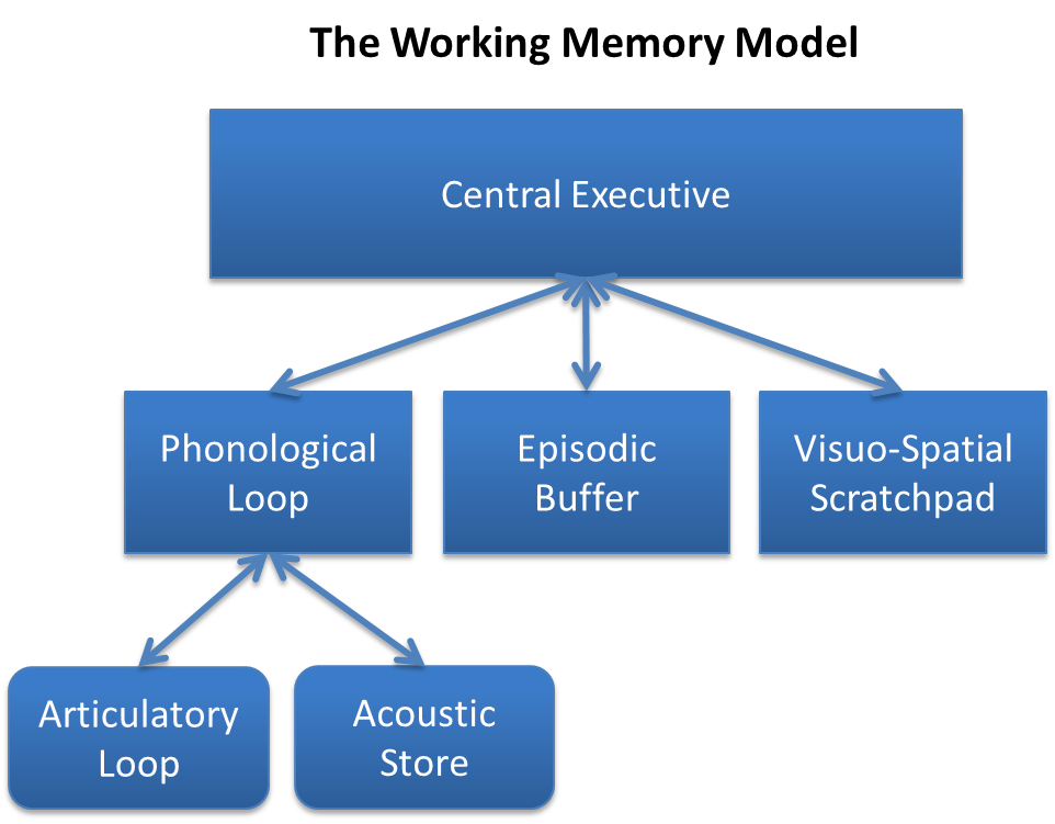
Working memory is a cognitive system with a limited capacity that can hold information temporarily. It is important for reasoning and the guidance of decision-making and behavior. Working memory is often used synonymously with short-term memory, but some theorists consider the two forms of memory distinct, assuming that working memory allows for the manipulation of stored information, whereas short-term memory only refers to the short-term storage of information. Working memory is a theoretical concept central to cognitive psychology, neuropsychology, and neuroscience. History The term "working memory" was coined by Miller, Galanter, and Pribram, and was used in the 1960s in the context of theories that likened the mind to a computer. In 1968, Atkinson and Shiffrin used the term to describe their "short-term store". What we now call working memory was formerly referred to variously as a "short-term store" or short-term memory, primary memory, immediate memory, operant me ... [...More Info...] [...Related Items...] OR: [Wikipedia] [Google] [Baidu] |
Visual Space
Visual space is the experience of space by an aware observer. It is the subjective counterpart of the space of physical objects. There is a long history in philosophy, and later psychology of writings describing visual space, and its relationship to the space of physical objects. A partial list would include René Descartes, Immanuel Kant, Hermann von Helmholtz, William James, to name just a few. Object Space and Visual Space. Space of physical objects The location and shape of physical objects can be accurately described with the tools of geometry. For practical purposes the space we occupy is Euclidean. It is three-dimensional and measurable using tools such as rulers. It can be quantified using co-ordinate systems like the Cartesian x,y,z, or polar coordinates with angles of elevation, azimuth and distance from an arbitrary origin. Space of visual percepts Percepts, the counterparts in the aware observer's conscious experience of objects in physical space, constitu ... [...More Info...] [...Related Items...] OR: [Wikipedia] [Google] [Baidu] |
Oculomotor
The oculomotor nerve, also known as the third cranial nerve, cranial nerve III, or simply CN III, is a cranial nerve that enters the orbit through the superior orbital fissure and innervates extraocular muscles that enable most movements of the eye and that raise the eyelid. The nerve also contains fibers that innervate the intrinsic eye muscles that enable pupillary constriction and accommodation (ability to focus on near objects as in reading). The oculomotor nerve is derived from the basal plate of the embryonic midbrain. Cranial nerves IV and VI also participate in control of eye movement. Structure The oculomotor nerve originates from the third nerve nucleus at the level of the superior colliculus in the midbrain. The third nerve nucleus is located ventral to the cerebral aqueduct, on the pre-aqueductal grey matter. The fibers from the two third nerve nuclei located laterally on either side of the cerebral aqueduct then pass through the red nucleus. From the red nucl ... [...More Info...] [...Related Items...] OR: [Wikipedia] [Google] [Baidu] |
Brodmann Area 46
Brodmann area 46, or BA46, is part of the frontal cortex in the human brain. It is between BA10 and BA45. BA46 is known as middle frontal area 46. In the human brain it occupies approximately the middle third of the middle frontal gyrus and the most rostral portion of the inferior frontal gyrus. Brodmann area 46 roughly corresponds with the dorsolateral prefrontal cortex (DLPFC), although the borders of area 46 are based on cytoarchitecture rather than function. The DLPFC also encompasses part of granular frontal area 9, directly adjacent on the dorsal surface of the cortex. Cytoarchitecturally, BA46 is bounded dorsally by the granular frontal area 9, rostroventrally by the frontopolar area 10 and caudally by the triangular area 45 (Brodmann-1909). There is some discrepancy between the extent of BA8 (Brodmann-1905) and the same area as described by Walker (1940). Function The dorsolateral prefrontal cortex plays a central role in sustaining attention and managing workin ... [...More Info...] [...Related Items...] OR: [Wikipedia] [Google] [Baidu] |
Dorsolateral Prefrontal Cortex
The dorsolateral prefrontal cortex (DLPFC or DL-PFC) is an area in the prefrontal cortex of the primate brain. It is one of the most recently derived parts of the human brain. It undergoes a prolonged period of maturation which lasts until adulthood. The DLPFC is not an anatomical structure, but rather a functional one. It lies in the middle frontal gyrus of humans (i.e., lateral part of Brodmann's area (BA) 9 and 46). In macaque monkeys, it is around the principal sulcus (i.e., in Brodmann's area 46). Other sources consider that DLPFC is attributed anatomically to BA 9 and 46 and BA 8, 9 and 10. The DLPFC has connections with the orbitofrontal cortex, as well as the thalamus, parts of the basal ganglia (specifically, the dorsal caudate nucleus), the hippocampus, and primary and secondary association areas of neocortex (including posterior temporal, parietal, and occipital areas). The DLPFC is also the end point for the dorsal pathway (stream), which is concerned with how t ... [...More Info...] [...Related Items...] OR: [Wikipedia] [Google] [Baidu] |
Premotor Cortex
The premotor cortex is an area of the motor cortex lying within the frontal lobe of the brain just anterior to the primary motor cortex. It occupies part of Brodmann's area 6. It has been studied mainly in primates, including monkeys and humans. The functions of the premotor cortex are diverse and not fully understood. It projects directly to the spinal cord and therefore may play a role in the direct control of behavior, with a relative emphasis on the trunk muscles of the body. It may also play a role in planning movement, in the spatial guidance of movement, in the sensory guidance of movement, in understanding the actions of others, and in using abstract rules to perform specific tasks. Different subregions of the premotor cortex have different properties and presumably emphasize different functions. Nerve signals generated in the premotor cortex cause much more complex patterns of movement than the discrete patterns generated in the primary motor cortex. Structure The pr ... [...More Info...] [...Related Items...] OR: [Wikipedia] [Google] [Baidu] |
Supplementary Motor Cortex
The supplementary motor area (SMA) is a part of the motor cortex of primates that contributes to the control of movement. It is located on the midline surface of the hemisphere just in front of (anterior to) the primary motor cortex leg representation. In monkeys the SMA contains a rough map of the body. In humans the body map is not apparent. Neurons in the SMA project directly to the spinal cord and may play a role in the direct control of movement. Possible functions attributed to the SMA include the postural stabilization of the body, the coordination of both sides of the body such as during bimanual action, the control of movements that are internally generated rather than triggered by sensory events, and the control of sequences of movements. All of these proposed functions remain hypotheses. The precise role or roles of the SMA is not yet known. For the discovery of the SMA and its relationship to other motor cortical areas, see the main article on the motor cortex. Subregi ... [...More Info...] [...Related Items...] OR: [Wikipedia] [Google] [Baidu] |
_-_inferiror_view.png)


