|
Subcoracoid Bursa
The subcoracoid bursa or subcoracoid bursa of Collas is a synovial bursa located in the shoulder. It is located anterior to the subscapularis muscle and inferior to the coracoid process. Its function is to reduce friction between the coracobrachialis, subscapularis and short head of the biceps tendons, thus facilitating internal and external rotation of the shoulder. The subcoracoid bursa does not communicate with the glenohumeral joint under normal circumstances, but may communicate with the subacromial bursa. As such, contrast fluid injected into the glenohumeral joint during an arthrogram that extends into the subcoracoid bursa is abnormal, and indirectly implies a full thickness rotator cuff The rotator cuff is a group of muscles and their tendons that act to stabilize the human shoulder and allow for its extensive range of motion. Of the seven scapulohumeral muscles, four make up the rotator cuff. The four muscles are the supraspi ... tear. References {{Bursae and she ... [...More Info...] [...Related Items...] OR: [Wikipedia] [Google] [Baidu] |
Synovial Bursa
Synovial () may refer to: * Synovial fluid * Synovial joint * Synovial membrane The synovial membrane (also known as the synovial stratum, synovium or stratum synoviale) is a specialized connective tissue that lines the inner surface of capsules of synovial joints and tendon sheath A tendon sheath is a layer of synovial m ... * Synovial bursa {{disambiguation ... [...More Info...] [...Related Items...] OR: [Wikipedia] [Google] [Baidu] |
Internal Rotation
Motion, the process of movement, is described using specific anatomical terms. Motion includes movement of organs, joints, limbs, and specific sections of the body. The terminology used describes this motion according to its direction relative to the anatomical position of the body parts involved. Anatomists and others use a unified set of terms to describe most of the movements, although other, more specialized terms are necessary for describing unique movements such as those of the hands, feet, and eyes. In general, motion is classified according to the anatomical plane it occurs in. ''Flexion'' and ''extension'' are examples of ''angular'' motions, in which two axes of a joint are brought closer together or moved further apart. ''Rotational'' motion may occur at other joints, for example the shoulder, and are described as ''internal'' or ''external''. Other terms, such as ''elevation'' and ''depression'', describe movement above or below the horizontal plane. Many anatomic ... [...More Info...] [...Related Items...] OR: [Wikipedia] [Google] [Baidu] |
Synovial Bursae
Synovial () may refer to: * Synovial fluid * Synovial joint * Synovial membrane * Synovial bursa Synovial () may refer to: * Synovial fluid * Synovial joint * Synovial membrane The synovial membrane (also known as the synovial stratum, synovium or stratum synoviale) is a specialized connective tissue that lines the inner surface of capsule ... {{disambiguation ... [...More Info...] [...Related Items...] OR: [Wikipedia] [Google] [Baidu] |
Rotator Cuff
The rotator cuff is a group of muscles and their tendons that act to stabilize the human shoulder and allow for its extensive range of motion. Of the seven scapulohumeral muscles, four make up the rotator cuff. The four muscles are the supraspinatus muscle, the infraspinatus muscle, teres minor muscle, and the subscapularis muscle. Structure Muscles composing rotator cuff The supraspinatus muscle spreads out in a horizontal band to insert on the superior facet of the greater tubercle of the humerus. The greater tubercle projects as the most lateral structure of the humeral head. Medial to this, in turn, is the lesser tubercle of the humeral head. The subscapularis muscle origin is divided from the remainder of the rotator cuff origins as it is deep to the scapula. The four tendons of these muscles converge to form the rotator cuff tendon. These tendinous insertions along with the articular capsule, the coracohumeral ligament, and the glenohumeral ligament complex, b ... [...More Info...] [...Related Items...] OR: [Wikipedia] [Google] [Baidu] |
Arthrogram
An arthrogram is a series of images of a joint after injection of a contrast medium, usually done by fluoroscopy or MRI. The injection is normally done under a local anesthetic such as Novocain or lidocaine. The radiologist or radiographer performs the study using fluoroscopy or x-ray to guide the placement of the needle into the joint and then injects around 10 ml of contrast based on age. There is some burning pain from the anesthetic and a painful bubbling feeling in the joint after the contrast is injected. This only lasts 20 – 30 hours until the Contrast is absorbed. During this time, while it is allowed, it is painful to use the limb for around 10 hours. After that the radiologist can more clearly see what is going on under your skin and can get results out within 24 to 48 hours. Types Conventional arthrography It is used primarily in the evaluation of menisci, cruciate ligaments, articular cartilage, and loose body within a joint. Fluoroscopic allows general view of th ... [...More Info...] [...Related Items...] OR: [Wikipedia] [Google] [Baidu] |
Gadolinium
Gadolinium is a chemical element with the symbol Gd and atomic number 64. Gadolinium is a silvery-white metal when oxidation is removed. It is only slightly malleable and is a ductile rare-earth element. Gadolinium reacts with atmospheric oxygen or moisture slowly to form a black coating. Gadolinium below its Curie point of is ferromagnetic, with an attraction to a magnetic field higher than that of nickel. Above this temperature it is the most paramagnetic element. It is found in nature only in an oxidized form. When separated, it usually has impurities of the other rare-earths because of their similar chemical properties. Gadolinium was discovered in 1880 by Jean Charles de Marignac, who detected its oxide by using spectroscopy. It is named after the mineral gadolinite, one of the minerals in which gadolinium is found, itself named for the Finnish chemist Johan Gadolin. Pure gadolinium was first isolated by the chemist Paul-Émile Lecoq de Boisbaudran around 1886. Gadolini ... [...More Info...] [...Related Items...] OR: [Wikipedia] [Google] [Baidu] |
Subacromial Bursa
The subacromial bursa is the synovial cavity located just below the acromion, which communicates with the subdeltoid bursa in most individuals, forming the so-called subacromial-subdeltoid bursa (SSB). The SSB bursa is located deep to the deltoid muscle and the coracoacromial arch and extends laterally beyond the humeral attachment of the rotator cuff, anteriorly to overlie the intertubercular groove, medially to the acromioclavicular joint, and posteriorly over the rotator cuff. The SSB decreases friction, and allows free motion of the rotator cuff relative to the coracoacromial arch and the deltoid muscle. French anatomist and surgeon Jean-François Jarjavay is credited as the first to describe morbid processes of the SSB in 1867. Since then, histologic studies have documented that synovial membrane may undergo inflammatory and/or degenerative changes and many now believe that they correspond to different stages in the spectrum of disease, with long-lasting inflammation le ... [...More Info...] [...Related Items...] OR: [Wikipedia] [Google] [Baidu] |
Glenohumeral Joint
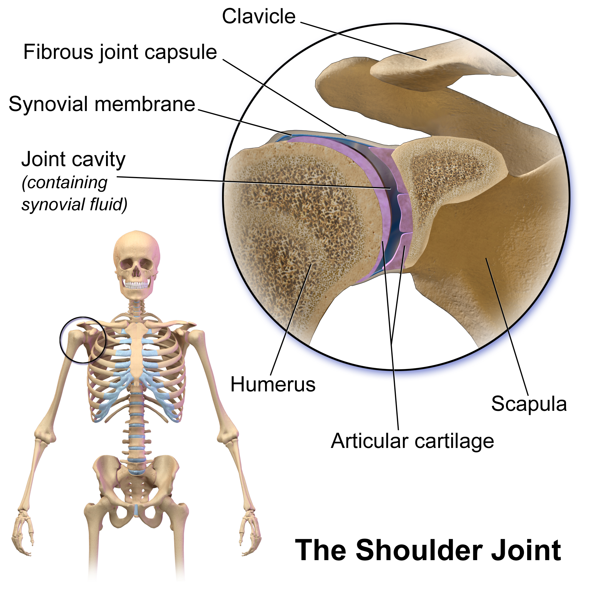
The shoulder joint (or glenohumeral joint from Greek ''glene'', eyeball, + -''oid'', 'form of', + Latin ''humerus'', shoulder) is structurally classified as a synovial ball-and-socket joint and functionally as a diarthrosis and multiaxial joint. It involves an articulation between the glenoid fossa of the scapula (shoulder blade) and the head of the humerus (upper arm bone). Due to the very loose joint capsule that gives a limited interface of the humerus and scapula, it is the most mobile joint of the human body. Structure The shoulder joint is a ball-and-socket joint between the scapula and the humerus. The socket of the glenoid fossa of the scapula is itself quite shallow, but it is made deeper by the addition of the glenoid labrum. The glenoid labrum is a ring of cartilaginous fibre attached to the circumference of the cavity. This ring is continuous with the tendon of the biceps brachii above. Spaces Significant joint spaces are: * The normal glenohumeral space is ... [...More Info...] [...Related Items...] OR: [Wikipedia] [Google] [Baidu] |
External Rotation
Motion, the process of movement, is described using specific anatomical terms. Motion includes movement of organs, joints, limbs, and specific sections of the body. The terminology used describes this motion according to its direction relative to the anatomical position of the body parts involved. Anatomists and others use a unified set of terms to describe most of the movements, although other, more specialized terms are necessary for describing unique movements such as those of the hands, feet, and eyes. In general, motion is classified according to the anatomical plane it occurs in. ''Flexion'' and ''extension'' are examples of ''angular'' motions, in which two axes of a joint are brought closer together or moved further apart. ''Rotational'' motion may occur at other joints, for example the shoulder, and are described as ''internal'' or ''external''. Other terms, such as ''elevation'' and ''depression'', describe movement above or below the horizontal plane. Many anatomic ... [...More Info...] [...Related Items...] OR: [Wikipedia] [Google] [Baidu] |
Tendon
A tendon or sinew is a tough, high-tensile-strength band of dense fibrous connective tissue that connects muscle to bone. It is able to transmit the mechanical forces of muscle contraction to the skeletal system without sacrificing its ability to withstand significant amounts of tension. Tendons are similar to ligaments; both are made of collagen. Ligaments connect one bone to another, while tendons connect muscle to bone. Structure Histologically, tendons consist of dense regular connective tissue. The main cellular component of tendons are specialized fibroblasts called tendon cells (tenocytes). Tenocytes synthesize the extracellular matrix of tendons, abundant in densely packed collagen fibers. The collagen fibers are parallel to each other and organized into tendon fascicles. Individual fascicles are bound by the endotendineum, which is a delicate loose connective tissue containing thin collagen fibrils and elastic fibres. Groups of fascicles are bounded by the epit ... [...More Info...] [...Related Items...] OR: [Wikipedia] [Google] [Baidu] |
Shoulder
The human shoulder is made up of three bones: the clavicle (collarbone), the scapula (shoulder blade), and the humerus (upper arm bone) as well as associated muscles, ligaments and tendons. The articulations between the bones of the shoulder make up the shoulder joints. The shoulder joint, also known as the glenohumeral joint, is the major joint of the shoulder, but can more broadly include the acromioclavicular joint. In human anatomy, the shoulder joint comprises the part of the body where the humerus attaches to the scapula, and the head sits in the glenoid cavity. The shoulder is the group of structures in the region of the joint. The shoulder joint is the main joint of the shoulder. It is a ball and socket joint that allows the arm to rotate in a circular fashion or to hinge out and up away from the body. The joint capsule is a soft tissue envelope that encircles the glenohumeral joint and attaches to the scapula, humerus, and head of the biceps. It is lined by a ... [...More Info...] [...Related Items...] OR: [Wikipedia] [Google] [Baidu] |
Biceps
The biceps or biceps brachii ( la, musculus biceps brachii, "two-headed muscle of the arm") is a large muscle that lies on the front of the upper arm between the shoulder and the elbow. Both heads of the muscle arise on the scapula and join to form a single muscle belly which is attached to the upper forearm. While the biceps crosses both the shoulder and elbow joints, its main function is at the elbow where it flexes the forearm and supinates the forearm. Both these movements are used when opening a bottle with a corkscrew: first biceps screws in the cork (supination), then it pulls the cork out ( flexion). Structure The biceps is one of three muscles in the anterior compartment of the upper arm, along with the brachialis muscle and the coracobrachialis muscle, with which the biceps shares a nerve supply. The biceps muscle has two heads, the short head and the long head, distinguished according to their origin at the coracoid process and supraglenoid tubercle of t ... [...More Info...] [...Related Items...] OR: [Wikipedia] [Google] [Baidu] |


