|
Subclavian Loop
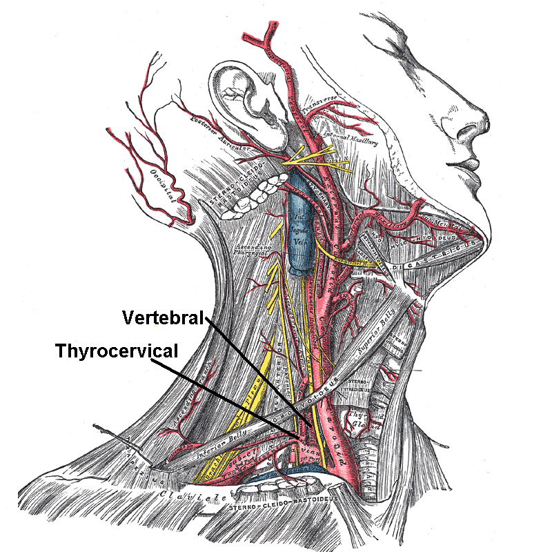
Subclavian loop (''ansa subclavia''), also known as Vieussens' ansa after French anatomist Raymond Vieussens (1635-1715), is a nerve cord that is a connection between the middle and inferior cervical ganglion which is commonly fused with the first thoracic ganglion and is then called the stellate ganglion. The subclavian ansa forms a loop around the subclavian artery; whence its name. This communicating branch downwards anteromedial to the vertebral artery makes a loop around the subclavian artery from anterior to posterior and then lies medially to the internal thoracic artery respectively. Sometimes there are two communicating branches encompassing the vertebral artery The vertebral arteries are major arteries An artery (plural arteries) () is a blood vessel in humans and most animals that takes blood away from the heart to one or more parts of the body (tissues, lungs, brain etc.). Most arteries carry o ..., one from anterior and the other from posterior. References D ... [...More Info...] [...Related Items...] OR: [Wikipedia] [Google] [Baidu] |
Anatomist
Anatomy () is the branch of biology concerned with the study of the structure of organisms and their parts. Anatomy is a branch of natural science that deals with the structural organization of living things. It is an old science, having its beginnings in prehistoric times. Anatomy is inherently tied to developmental biology, embryology, comparative anatomy, evolutionary biology, and phylogeny, as these are the processes by which anatomy is generated, both over immediate and long-term timescales. Anatomy and physiology, which study the structure and function of organisms and their parts respectively, make a natural pair of related disciplines, and are often studied together. Human anatomy is one of the essential basic sciences that are applied in medicine. The discipline of anatomy is divided into macroscopic and microscopic. Macroscopic anatomy, or gross anatomy, is the examination of an animal's body parts using unaided eyesight. Gross anatomy also includes the branch of ... [...More Info...] [...Related Items...] OR: [Wikipedia] [Google] [Baidu] |
Raymond Vieussens
Raymond Vieussens (ca. 1635 – 16 August 1715) was a French anatomist from Le Vigan. There is uncertainty regarding the exact year of Vieussens birth, with some sources placing it as late as 1641. He studied medicine at the University of Montpellier where he earned his degree in 1670. He later became head physician at Hôtel Dieu Saint-Eloi in Montpellier. Vieussens is remembered for his pioneer work in the field of cardiology, and his anatomical studies of the brain and spinal cord. He regarded English anatomist Thomas Willis (1621–1675) as a major influence towards his career. Vieussens is credited as being the first physician to give accurate descriptions of the left ventricle and several blood vessels of the heart. He was also the first to give a comprehensive description of mitral stenosis, as well as other types of heart disease and circulatory disorders. He also provided an early description of the brain's centrum semiovale, which is sometimes referred to as "Vieuss ... [...More Info...] [...Related Items...] OR: [Wikipedia] [Google] [Baidu] |
Nerve
A nerve is an enclosed, cable-like bundle of nerve fibers (called axons) in the peripheral nervous system. A nerve transmits electrical impulses. It is the basic unit of the peripheral nervous system. A nerve provides a common pathway for the electrochemical nerve impulses called action potentials that are transmitted along each of the axons to peripheral organs or, in the case of sensory nerves, from the periphery back to the central nervous system. Each axon, within the nerve, is an extension of an individual neuron, along with other supportive cells such as some Schwann cells that coat the axons in myelin. Within a nerve, each axon is surrounded by a layer of connective tissue called the endoneurium. The axons are bundled together into groups called fascicles, and each fascicle is wrapped in a layer of connective tissue called the perineurium. Finally, the entire nerve is wrapped in a layer of connective tissue called the epineurium. Nerve cells (often called neurons) are f ... [...More Info...] [...Related Items...] OR: [Wikipedia] [Google] [Baidu] |
Middle Cervical Ganglion
The middle cervical ganglion is the smallest of the three cervical ganglia, and is occasionally absent. It is placed opposite the sixth cervical vertebra, usually in front of, or close to, the inferior thyroid artery. It sends gray rami communicantes to the fifth and sixth cervical nerves, and gives off the middle cardiac nerve. It is probably formed by the coalescence of two ganglia corresponding to the fifth and sixth cervical nerves. Branches # Gray Rami Communicantes to the anterior rami of the fifth and sixth cervical nerves. # Thyroid Branches which pass along the inferior thyroid artery to the thyroid gland. # The middle cardiac branch, which descends in the neck and ends in the cardiac plexus in the thorax See also * Middle cardiac nerve The middle cardiac nerve (''great cardiac nerve''), the largest of the three cardiac nerves, arises from the middle cervical ganglion, or from the trunk between the middle and inferior ganglia. On the right side it descends behind the ... [...More Info...] [...Related Items...] OR: [Wikipedia] [Google] [Baidu] |
Inferior Cervical Ganglion
The inferior cervical ganglion is situated between the base of the transverse process of the last cervical vertebra and the neck of the first rib, on the medial side of the costocervical artery. Its form is irregular; it is larger in size than the middle cervical ganglion, and is frequently fused with the first thoracic ganglion, under which circumstances it is then called the "stellate ganglion." Structure It is connected to the middle cervical ganglion by two or more cords, one of which forms a loop around the subclavian artery and supplies offsets to it. This loop is named the ''ansa subclavia'' (Vieussenii). The ganglion sends gray rami communicantes to the seventh and eighth cervical nerves. Branches The inferior cervical ganglion gives off two branches: * The Inferior cardiac nerve * ''offsets to bloodvessels'' form plexuses on the subclavian artery and its branches. The plexus on the vertebral artery is continued on to the basilar, posterior cerebral, and cerebellar art ... [...More Info...] [...Related Items...] OR: [Wikipedia] [Google] [Baidu] |
Stellate Ganglion
The stellate ganglion (or cervicothoracic ganglion) is a sympathetic ganglion formed by the fusion of the inferior cervical ganglion and the first thoracic (superior thoracic sympathetic) ganglion, which exists in 80% of people. Sometimes, the second and the third thoracic ganglia are included in this fusion. The stellate ganglion is relatively big (10–12 x 8–20 mm) compared to much smaller thoracic, lumbar and sacral ganglia, and is polygonal in shape (). Stellate ganglion is located at the level of C7, anterior to the transverse process of C7 and the neck of the first rib, superior to the cervical pleura and just below the subclavian artery. It is superiorly covered by the prevertebral lamina of the cervical fascia and anteriorly in relation with common carotid artery, subclavian artery and the beginning of vertebral artery which sometimes leaves a groove at the apex of this ganglion (this groove can sometimes even separate the stellate ganglion into so called vertebral gangli ... [...More Info...] [...Related Items...] OR: [Wikipedia] [Google] [Baidu] |
Subclavian Artery
In human anatomy, the subclavian arteries are paired major arteries of the upper thorax, below the clavicle. They receive blood from the aortic arch. The left subclavian artery supplies blood to the left arm and the right subclavian artery supplies blood to the right arm, with some branches supplying the head and thorax. On the left side of the body, the subclavian comes directly off the aortic arch, while on the right side it arises from the relatively short brachiocephalic artery when it bifurcates into the subclavian and the right common carotid artery. The usual branches of the subclavian on both sides of the body are the vertebral artery, the internal thoracic artery, the thyrocervical trunk, the costocervical trunk and the dorsal scapular artery, which may branch off the transverse cervical artery, which is a branch of the thyrocervical trunk. The subclavian becomes the axillary artery at the lateral border of the first rib. Structure From its origin, the subclavian artery t ... [...More Info...] [...Related Items...] OR: [Wikipedia] [Google] [Baidu] |
Vertebral Artery
The vertebral arteries are major arteries An artery (plural arteries) () is a blood vessel in humans and most animals that takes blood away from the heart to one or more parts of the body (tissues, lungs, brain etc.). Most arteries carry oxygenated blood; the two exceptions are the pu ... of the neck. Typically, the vertebral arteries originate from the subclavian arteries. Each vessel courses superiorly along each side of the neck, merging within the skull to form the single, midline basilar artery. As the supplying component of the ''vertebrobasilar vascular system'', the vertebral arteries supply blood to the upper spinal cord, brainstem, cerebellum, and Cerebral circulation#Posterior cerebral circulation, posterior part of brain. Structure The vertebral arteries usually arise from the posterosuperior aspect of the central subclavian arteries on each side of the body, then enter deep to the transverse process at the level of the 6th cervical vertebrae (C6), or occasio ... [...More Info...] [...Related Items...] OR: [Wikipedia] [Google] [Baidu] |
Internal Thoracic Artery
In human anatomy, the internal thoracic artery (ITA), previously commonly known as the internal mammary artery (a name still common among surgeons), is an artery that supplies the anterior chest wall and the breasts. It is a paired artery, with one running along each side of the sternum, to continue after its bifurcation as the superior epigastric and musculophrenic arteries. Structure The internal thoracic artery arises from the anterior surface of the subclavian artery near its origin. It has a width of between 1-2 mm. It travels downward on the inside of the rib cage, approximately 1 cm from the sides of the sternum, and thus medial to the nipple. It is accompanied by the internal thoracic vein. It runs deep to the abdominal external oblique muscle, but superficial to the vagus nerve. In adults, internal thoracic artery lies closest to the sternum at the first intercoastal space. The gap between the artery and lateral border of the sternum increases when going downwards ... [...More Info...] [...Related Items...] OR: [Wikipedia] [Google] [Baidu] |


