|
Staging Of Cervical Cancer
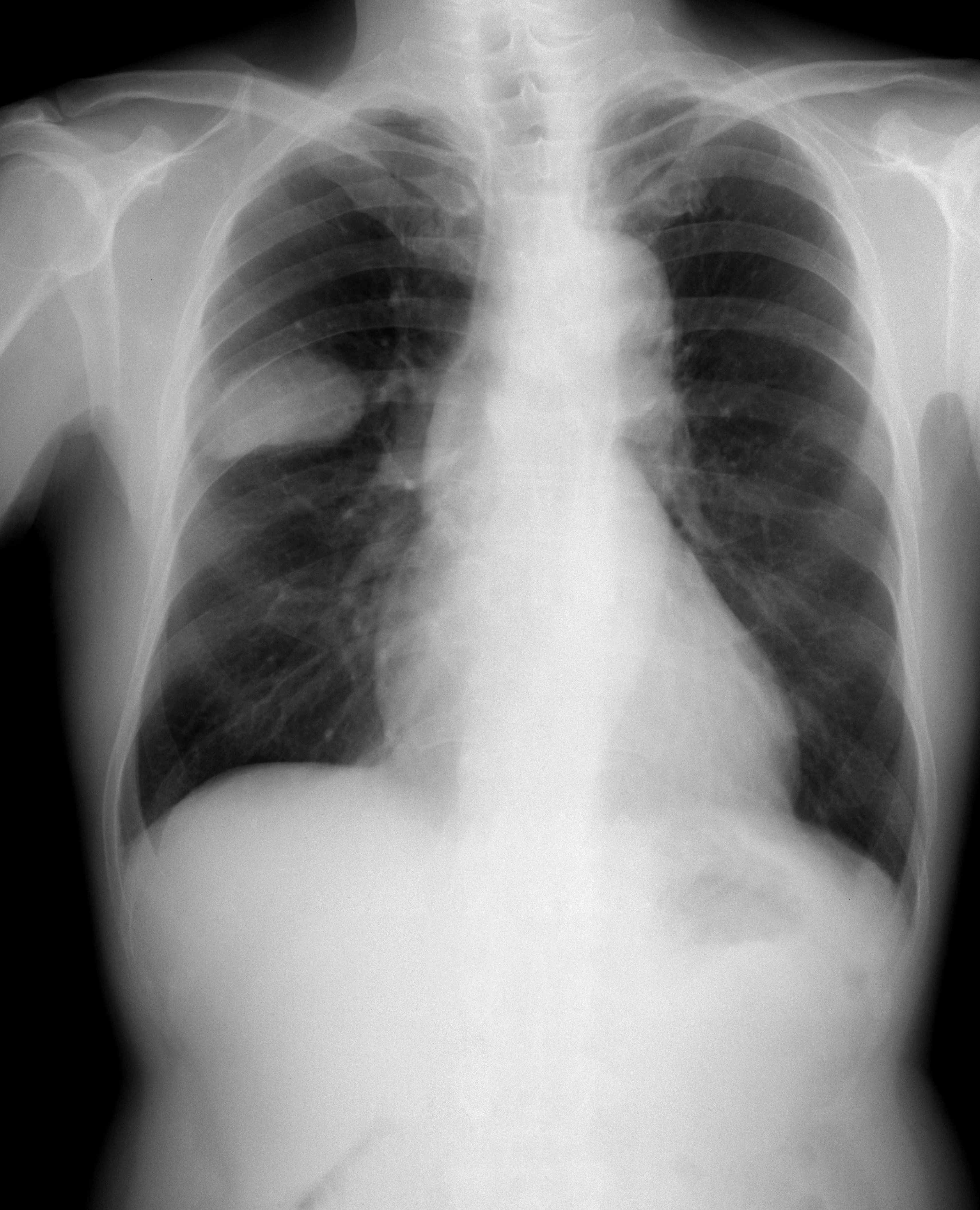
Cervical cancer staging is the assessment of cervical cancer to decide how far the disease has progressed. This is important for determining disease prognosis and treatment. Cancer staging generally runs from stage 0, which is pre-cancerous or non-invasive, to stage IV, in which the cancer has spread throughout a significant part of the body. Cervical cancer is staged by the International Federation of Gynecology and Obstetrics (FIGO) staging system. Prior to the 2018 update, FIGO staging of cervical cancer allowed only the following diagnostic tests to be used in determining the stage: palpation (feeling with the fingers), inspection, colposcopy, endocervical curettage, hysteroscopy, cystoscopy, proctoscopy, intravenous urography, and X-ray examination of the lungs and skeleton, and cervical conization. But with the 2018 update of FIGO staging of cervical cancer, imaging and pathology were added as allowable methods to assess disease stage. Overview of staging FIGO guidelines sug ... [...More Info...] [...Related Items...] OR: [Wikipedia] [Google] [Baidu] |
Cervical Cancer
Cervical cancer is a cancer arising from the cervix. It is due to the abnormal growth of cells that have the ability to invade or spread to other parts of the body. Early on, typically no symptoms are seen. Later symptoms may include abnormal vaginal bleeding, pelvic pain or pain during sexual intercourse. While bleeding after sex may not be serious, it may also indicate the presence of cervical cancer. Human papillomavirus infection (HPV) causes more than 90% of cases; most women who have had HPV infections, however, do not develop cervical cancer. HPV 16 and 18 strains are responsible for nearly 50% of high grade cervical pre-cancers. Other risk factors include smoking, a weak immune system, birth control pills, starting sex at a young age, and having many sexual partners, but these are less important. Genetic factors also contribute to cervical cancer risk. Cervical cancer typically develops from precancerous changes over 10 to 20 years. About 90% of cervical cancer cas ... [...More Info...] [...Related Items...] OR: [Wikipedia] [Google] [Baidu] |
Metastasis
Metastasis is a pathogenic agent's spread from an initial or primary site to a different or secondary site within the host's body; the term is typically used when referring to metastasis by a cancerous tumor. The newly pathological sites, then, are metastases (mets). It is generally distinguished from cancer invasion, which is the direct extension and penetration by cancer cells into neighboring tissues. Cancer occurs after cells are genetically altered to proliferate rapidly and indefinitely. This uncontrolled proliferation by mitosis produces a primary heterogeneic tumour. The cells which constitute the tumor eventually undergo metaplasia, followed by dysplasia then anaplasia, resulting in a malignant phenotype. This malignancy allows for invasion into the circulation, followed by invasion to a second site for tumorigenesis. Some cancer cells known as circulating tumor cells acquire the ability to penetrate the walls of lymphatic or blood vessels, after which they are abl ... [...More Info...] [...Related Items...] OR: [Wikipedia] [Google] [Baidu] |
Loop Electrical Excision Procedure
The loop electrosurgical excision procedure (LEEP) is one of the most commonly used approaches to treat high grade cervical dysplasia (CIN II/III, HGSIL) discovered on colposcopic examination. In the UK, it is known as large loop excision of the transformation zone (LLETZ). LEEP has many advantages including low cost and a high success rate. The procedure can be done in an office setting and usually only requires a local anesthetic, though sometimes IV sedation or a general anesthetic is used. Process When performing a LEEP, the physician uses a wire loop through which an electric current is passed at variable power settings. Various shapes and sizes of loop can be used depending on the size and orientation of the lesion. The cervical transformation zone and lesion are excised to an adequate depth, which in most cases is at least 8 mm, and extending 4 to 5 mm beyond the lesion. A second pass with a more narrow loop can also be done to obtain an endocervical specimen ... [...More Info...] [...Related Items...] OR: [Wikipedia] [Google] [Baidu] |
Small-cell Carcinoma
Small-cell carcinoma is a type of highly malignant cancer that most commonly arises within the lung, although it can occasionally arise in other body sites, such as the cervix, prostate, and gastrointestinal tract. Compared to non-small cell carcinoma, small cell carcinoma has a shorter doubling time, higher growth fraction, and earlier development of metastases. Extensive stage small cell lung cancer is classified as a rare disorder. Ten-year relative survival rate is 3.5%; however, women have a higher survival rate, 4.3%, and men lower, 2.8%. Survival can be higher or lower based on a combination of factors including stage, age, gender and race. Types of SCLC Small-cell lung carcinoma has long been divided into two clinicopathological stages, termed ''limited stage'' (LS) and ''extensive stage'' (ES). The stage is generally determined by the presence or absence of metastases, whether or not the tumor appears limited to the thorax, and whether or not the entire tumor burden wi ... [...More Info...] [...Related Items...] OR: [Wikipedia] [Google] [Baidu] |
Adenoid Cystic Carcinoma
Adenoid cystic carcinoma is a rare type of cancer that can exist in many different body sites. This tumor most often occurs in the salivary glands, but it can also be found in many anatomic sites, including the breast, lacrimal gland, lung, brain, bartholin gland, trachea, and the paranasal sinuses. It is the third-most common malignant salivary gland tumor overall (after mucoepidermoid carcinoma and polymorphous adenocarcinoma). It represents 28% of malignant submandibular gland tumors, making it the single most common malignant salivary gland tumor in this region. Patients may survive for years with metastases because this tumor is generally well-differentiated and slow growing. In a 1999 study of a cohort of 160 ACC patients, disease-specific survival was 89% at 5 years, but only 40% at 15 years, reflecting deaths from late-occurring metastatic disease. Cause Activation of the oncogenic transcription factor gene ''MYB'' is the key genomic event of ACC and seen in the vast m ... [...More Info...] [...Related Items...] OR: [Wikipedia] [Google] [Baidu] |
Glassy Cell Carcinoma Of The Cervix
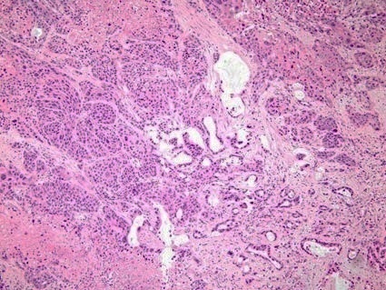
Glassy cell carcinoma of the cervix, also glassy cell carcinoma, is a rare aggressive malignant tumour of the uterine cervix. The tumour gets its name from its microscopic appearance; its cytoplasm has a glass-like appearance. Signs and symptoms The signs and symptoms are similar to other cervical cancers and may include post- coital bleeding and/or pain during intercourse ( dyspareunia). Early lesions may be completely asymptomatic. Cause Diagnosis The diagnosis is based on tissue examination, e.g. biopsy. Under the microscope, glassy cell carcinoma tumours are composed of cells with a glass-like cytoplasm, typically associated with an inflammatory infiltrate abundant in eosinophils and very mitotically active. PAS staining highlights the plasma membrane. Treatment The treatment is dependent on the stage. Advanced tumours are treated with surgery (radical hysterectomy and bilateral salpingo-opherectomy), radiation therapy and chemotherapy. See also * Cervix * Cervical ca ... [...More Info...] [...Related Items...] OR: [Wikipedia] [Google] [Baidu] |
Adenosquamous Carcinoma
Adenosquamous carcinoma is a type of cancer that contains two types of cells: squamous cells (thin, flat cells that line certain organs) and gland-like cells. It has been associated with more aggressive characteristics when compared to adenocarcinoma in certain cancers. It is responsible for 1% to 4% of exocrine forms of pancreas cancer. __TOC__ Diagnosis Light microscopy shows a combination of gland-like cells and squamous epithelial cells. On immunohistochemistry, it is typically positive for CK5/6, CK7 and p63, and negative for CK20, p16 and p53. On genetic testing, KRAS and p53 p53, also known as Tumor protein P53, cellular tumor antigen p53 (UniProt name), or transformation-related protein 53 (TRP53) is a regulatory protein that is often mutated in human cancers. The p53 proteins (originally thought to be, and often s ... are typically altered. References External links Adenosquamous carcinomaentry in the public domain NCI Dictionary of Cancer Terms Carcin ... [...More Info...] [...Related Items...] OR: [Wikipedia] [Google] [Baidu] |
Clear-cell Adenocarcinoma
Clear-cell adenocarcinoma is a type of adenocarcinoma that shows clear cells. Types include: * Clear-cell adenocarcinoma of the vagina * Clear-cell ovarian carcinoma * Uterine clear-cell carcinoma Uterine clear-cell carcinoma (CC) is a rare form of endometrial cancer with distinct morphological features on pathology; it is aggressive and has high recurrence rate. Like uterine papillary serous carcinoma CC does not develop from endometrial ... * Clear-cell adenocarcinoma of the lung (which is a type of Clear-cell carcinoma of the lung) See also * References External links Carcinoma {{oncology-stub ... [...More Info...] [...Related Items...] OR: [Wikipedia] [Google] [Baidu] |
Adenocarcinoma
Adenocarcinoma (; plural adenocarcinomas or adenocarcinomata ) (AC) is a type of cancerous tumor that can occur in several parts of the body. It is defined as neoplasia of epithelial tissue that has glandular origin, glandular characteristics, or both. Adenocarcinomas are part of the larger grouping of carcinomas, but are also sometimes called by more precise terms omitting the word, where these exist. Thus invasive ductal carcinoma, the most common form of breast cancer, is adenocarcinoma but does not use the term in its name—however, esophageal adenocarcinoma does to distinguish it from the other common type of esophageal cancer, esophageal squamous cell carcinoma. Several of the most common forms of cancer are adenocarcinomas, and the various sorts of adenocarcinoma vary greatly in all their aspects, so that few useful generalizations can be made about them. In the most specific usage (narrowest sense), the glandular origin or traits are exocrine; endocrine gland tumors, suc ... [...More Info...] [...Related Items...] OR: [Wikipedia] [Google] [Baidu] |
Squamous Cell Carcinoma
Squamous-cell carcinomas (SCCs), also known as epidermoid carcinomas, comprise a number of different types of cancer that begin in squamous cells. These cells form on the surface of the skin, on the lining of hollow organs in the body, and on the lining of the respiratory and digestive tracts. Common types include: * Squamous-cell skin cancer: A type of skin cancer * Squamous-cell carcinoma of the lung: A type of lung cancer * Squamous-cell thyroid carcinoma: A type of thyroid cancer * Esophageal squamous-cell carcinoma: A type of esophageal cancer * Squamous-cell carcinoma of the vagina: A type of vaginal cancer Despite sharing the name "squamous-cell carcinoma", the SCCs of different body sites can show differences in their presented symptoms, natural history, prognosis, and response to treatment. By body location Human papillomavirus infection has been associated with SCCs of the oropharynx, lung, fingers, and anogenital region. Head and neck cancer About 90% of cases ... [...More Info...] [...Related Items...] OR: [Wikipedia] [Google] [Baidu] |
Histopathology
Histopathology (compound of three Greek words: ''histos'' "tissue", πάθος ''pathos'' "suffering", and -λογία '' -logia'' "study of") refers to the microscopic examination of tissue in order to study the manifestations of disease. Specifically, in clinical medicine, histopathology refers to the examination of a biopsy or surgical specimen by a pathologist, after the specimen has been processed and histological sections have been placed onto glass slides. In contrast, cytopathology examines free cells or tissue micro-fragments (as "cell blocks"). Collection of tissues Histopathological examination of tissues starts with surgery, biopsy, or autopsy. The tissue is removed from the body or plant, and then, often following expert dissection in the fresh state, placed in a fixative which stabilizes the tissues to prevent decay. The most common fixative is 10% neutral buffered formalin (corresponding to 3.7% w/v formaldehyde in neutral buffered water, such as phosphate buf ... [...More Info...] [...Related Items...] OR: [Wikipedia] [Google] [Baidu] |
Fine-needle Aspiration
Fine-needle aspiration (FNA) is a diagnostic procedure used to investigate lumps or masses. In this technique, a thin (23–25 gauge (0.52 to 0.64 mm outer diameter)), hollow needle is inserted into the mass for sampling of cells that, after being stained, are examined under a microscope (biopsy). The sampling and biopsy considered together are called fine-needle aspiration biopsy (FNAB) or fine-needle aspiration cytology (FNAC) (the latter to emphasize that any aspiration biopsy involves cytopathology, not histopathology). Fine-needle aspiration biopsies are very safe minor surgical procedures. Often, a major surgical (excisional or open) biopsy can be avoided by performing a needle aspiration biopsy instead, eliminating the need for hospitalization. In 1981, the first fine-needle aspiration biopsy in the United States was done at Maimonides Medical Center. Today, this procedure is widely used in the diagnosis of cancer and inflammatory conditions. Aspiration is safer and ... [...More Info...] [...Related Items...] OR: [Wikipedia] [Google] [Baidu] |


