|
Spinocerebellar
The spinocerebellar tract is a nerve tract originating in the spinal cord and terminating in the same side (ipsilateral) of the cerebellum. Origins of proprioceptive information Proprioceptive information is obtained by Golgi tendon organs and muscle spindles. * Golgi tendon organs consist of a fibrous capsule enclosing tendon fascicles and bare nerve endings that respond to tension in the tendon by causing action potentials in type Ib afferents. These fibers are relatively large, myelinated, and quickly conducting. * Muscle spindles monitor the length within muscles and send information via faster Ia afferents. These axons are larger and faster than type Ib (from both nuclear bag fibers and nuclear chain fibers) and type II afferents (solely from nuclear chain fibers). All of these neurons are sensory (first order, or primary) and have their cell bodies in the dorsal root ganglia. They pass through Rexed laminae layers I-VI of the posterior grey column (dorsal horn) to form s ... [...More Info...] [...Related Items...] OR: [Wikipedia] [Google] [Baidu] |
Rostral Spinocerebellar Tract
The spinocerebellar tract is a nerve tract originating in the spinal cord and terminating in the same side (ipsilateral) of the cerebellum. Origins of proprioceptive information Proprioceptive information is obtained by Golgi tendon organs and muscle spindles. * Golgi tendon organs consist of a fibrous capsule enclosing tendon fascicles and bare nerve endings that respond to tension in the tendon by causing action potentials in type Ib afferents. These fibers are relatively large, myelinated, and quickly conducting. * Muscle spindles monitor the length within muscles and send information via faster Ia afferents. These axons are larger and faster than type Ib (from both nuclear bag fibers and nuclear chain fibers) and type II afferents (solely from nuclear chain fibers). All of these neurons are sensory (first order, or primary) and have their cell bodies in the dorsal root ganglia. They pass through Rexed laminae layers I-VI of the posterior grey column (dorsal horn) to form s ... [...More Info...] [...Related Items...] OR: [Wikipedia] [Google] [Baidu] |
Ventral Spinocerebellar Tract
The spinocerebellar tract is a nerve tract originating in the spinal cord and terminating in the same side (ipsilateral) of the cerebellum. Origins of proprioceptive information Proprioceptive information is obtained by Golgi tendon organs and muscle spindles. * Golgi tendon organs consist of a fibrous capsule enclosing tendon fascicles and bare nerve endings that respond to tension in the tendon by causing action potentials in type Ib afferents. These fibers are relatively large, myelinated, and quickly conducting. * Muscle spindles monitor the length within muscles and send information via faster Ia afferents. These axons are larger and faster than type Ib (from both nuclear bag fibers and nuclear chain fibers) and type II afferents (solely from nuclear chain fibers). All of these neurons are sensory (first order, or primary) and have their cell bodies in the dorsal root ganglia. They pass through Rexed laminae layers I-VI of the posterior grey column (dorsal horn) to form s ... [...More Info...] [...Related Items...] OR: [Wikipedia] [Google] [Baidu] |
Cuneocerebellar Tract
The spinocerebellar tract is a nerve tract originating in the spinal cord and terminating in the same side (ipsilateral) of the cerebellum. Origins of proprioceptive information Proprioceptive information is obtained by Golgi tendon organs and muscle spindles. * Golgi tendon organs consist of a fibrous capsule enclosing tendon fascicles and bare nerve endings that respond to tension in the tendon by causing action potentials in type Ib afferents. These fibers are relatively large, myelinated, and quickly conducting. * Muscle spindles monitor the length within muscles and send information via faster Ia afferents. These axons are larger and faster than type Ib (from both nuclear bag fibers and nuclear chain fibers) and type II afferents (solely from nuclear chain fibers). All of these neurons are sensory (first order, or primary) and have their cell bodies in the dorsal root ganglia. They pass through Rexed laminae layers I-VI of the posterior grey column (dorsal horn) to form ... [...More Info...] [...Related Items...] OR: [Wikipedia] [Google] [Baidu] |
Dorsal Spinocerebellar Tract
The spinocerebellar tract is a nerve tract originating in the spinal cord and terminating in the same side (ipsilateral) of the cerebellum. Origins of proprioceptive information Proprioceptive information is obtained by Golgi tendon organs and muscle spindles. * Golgi tendon organs consist of a fibrous capsule enclosing tendon fascicles and bare nerve endings that respond to tension in the tendon by causing action potentials in type Ib afferents. These fibers are relatively large, myelinated, and quickly conducting. * Muscle spindles monitor the length within muscles and send information via faster Ia afferents. These axons are larger and faster than type Ib (from both nuclear bag fibers and nuclear chain fibers) and type II afferents (solely from nuclear chain fibers). All of these neurons are sensory (first order, or primary) and have their cell bodies in the dorsal root ganglia. They pass through Rexed laminae layers I-VI of the posterior grey column (dorsal horn) to form ... [...More Info...] [...Related Items...] OR: [Wikipedia] [Google] [Baidu] |
Spinal Cord
The spinal cord is a long, thin, tubular structure made up of nervous tissue, which extends from the medulla oblongata in the brainstem to the lumbar region of the vertebral column (backbone). The backbone encloses the central canal of the spinal cord, which contains cerebrospinal fluid. The brain and spinal cord together make up the central nervous system (CNS). In humans, the spinal cord begins at the occipital bone, passing through the foramen magnum and then enters the spinal canal at the beginning of the cervical vertebrae. The spinal cord extends down to between the first and second lumbar vertebrae, where it ends. The enclosing bony vertebral column protects the relatively shorter spinal cord. It is around long in adult men and around long in adult women. The diameter of the spinal cord ranges from in the cervical and lumbar regions to in the thoracic area. The spinal cord functions primarily in the transmission of nerve signals from the motor cortex to the body, ... [...More Info...] [...Related Items...] OR: [Wikipedia] [Google] [Baidu] |
Proprioception
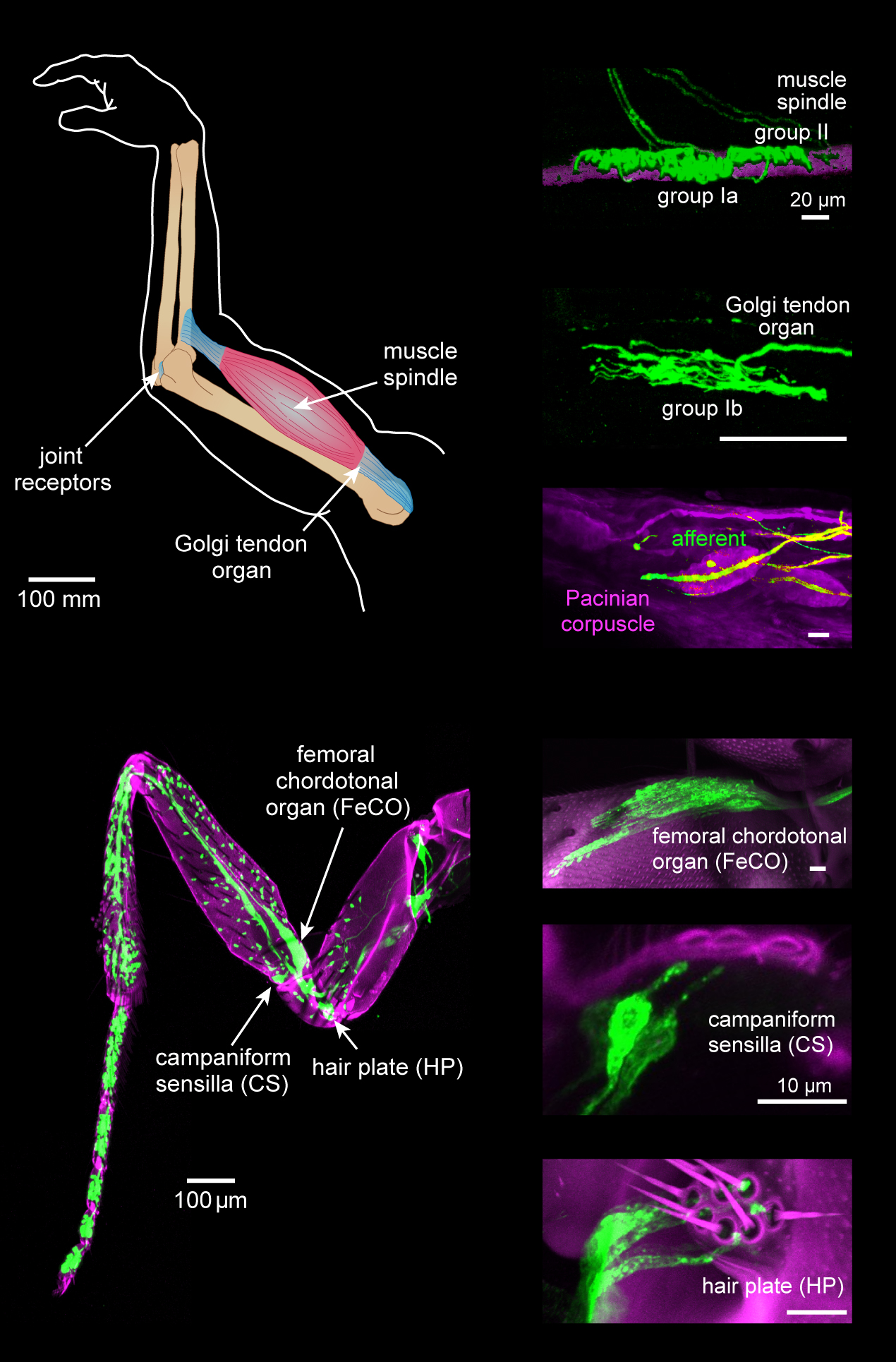
Proprioception ( ), also referred to as kinaesthesia (or kinesthesia), is the sense of self-movement, force, and body position. It is sometimes described as the "sixth sense". Proprioception is mediated by proprioceptors, mechanosensory neurons located within muscles, tendons, and joints. Most animals possess multiple subtypes of proprioceptors, which detect distinct kinematic parameters, such as joint position, movement, and load. Although all mobile animals possess proprioceptors, the structure of the sensory organs can vary across species. Proprioceptive signals are transmitted to the central nervous system, where they are integrated with information from other sensory systems, such as the visual system and the vestibular system, to create an overall representation of body position, movement, and acceleration. In many animals, sensory feedback from proprioceptors is essential for stabilizing body posture and coordinating body movement. System overview In vertebrates, limb ve ... [...More Info...] [...Related Items...] OR: [Wikipedia] [Google] [Baidu] |
Golgi Tendon Organs
The Golgi tendon organ (GTO) (also called Golgi organ, tendon organ, neurotendinous organ or neurotendinous spindle) is a proprioceptor – a type of sensory receptor that senses changes in muscle tension. It lies at the interface between a muscle and its tendon known as the musculotendinous junction also known as the myotendinous junction. It provides the sensory component of the Golgi tendon reflex. The Golgi tendon organ is one of several eponymous terms named after the Italian physician Camillo Golgi. Structure The body of the Golgi tendon organ is made up of braided strands of collagen (intrafusal fasciculi) that are less compact than elsewhere in the tendon and are encapsulated. The capsule is connected in series (along a single path) with a group of muscle fibers () at one end, and merge into the tendon proper at the other. Each capsule is about long, has a diameter of about , and is perforated by one or more afferent type Ib sensory nerve fibers ( Aɑ fiber), which are ... [...More Info...] [...Related Items...] OR: [Wikipedia] [Google] [Baidu] |
Golgi Organ
The Golgi tendon organ (GTO) (also called Golgi organ, tendon organ, neurotendinous organ or neurotendinous spindle) is a proprioceptor – a type of sensory receptor that senses changes in muscle tension. It lies at the interface between a muscle and its tendon known as the musculotendinous junction also known as the myotendinous junction. It provides the sensory component of the Golgi tendon reflex. The Golgi tendon organ is one of several eponymous terms named after the Italian physician Camillo Golgi. Structure The body of the Golgi tendon organ is made up of braided strands of collagen (intrafusal fasciculi) that are less compact than elsewhere in the tendon and are encapsulated. The capsule is connected in series (along a single path) with a group of muscle fibers () at one end, and merge into the tendon proper at the other. Each capsule is about long, has a diameter of about , and is perforated by one or more afferent type Ib sensory nerve fibers ( Aɑ fiber), which are ... [...More Info...] [...Related Items...] OR: [Wikipedia] [Google] [Baidu] |
Afferent Nerve Fiber
Afferent nerve fibers are the axons (nerve fibers) carried by a sensory nerve that relay sensory information from sensory receptors to regions of the brain. Afferent projections ''arrive'' at a particular brain region. Efferent nerve fibers are carried by efferent nerves and ''exit'' a region to act on muscles and glands. In the peripheral nervous system afferent and efferent nerve fibers are part of the somatic nervous system and arise from outside of the spinal cord. Sensory nerves carry the afferent fibers to enter into the spinal cord, and motor nerves carry the efferent fibers out of the spinal cord to act on skeletal muscles. In the central nervous system non-motor efferents are carried in efferent nerves to act on glands. Structure Afferent neurons are pseudounipolar neurons that have a single process leaving the cell body dividing into two branches: the long one towards the sensory organ, and the short one toward the central nervous system (e.g. spinal cord). The ... [...More Info...] [...Related Items...] OR: [Wikipedia] [Google] [Baidu] |
Golgi Tendon Organ
The Golgi tendon organ (GTO) (also called Golgi organ, tendon organ, neurotendinous organ or neurotendinous spindle) is a proprioceptor – a type of sensory receptor that senses changes in muscle tension. It lies at the interface between a muscle and its tendon known as the musculotendinous junction also known as the myotendinous junction. It provides the sensory component of the Golgi tendon reflex. The Golgi tendon organ is one of several eponymous terms named after the Italian physician Camillo Golgi. Structure The body of the Golgi tendon organ is made up of braided strands of collagen (intrafusal fasciculi) that are less compact than elsewhere in the tendon and are encapsulated. The capsule is connected in series (along a single path) with a group of muscle fibers () at one end, and merge into the tendon proper at the other. Each capsule is about long, has a diameter of about , and is perforated by one or more afferent type Ib sensory nerve fibers ( Aɑ fiber), which ... [...More Info...] [...Related Items...] OR: [Wikipedia] [Google] [Baidu] |

