|
Sphenopalatine Foramen
The sphenopalatine foramen is a foramen in the skull that connects the nasal cavity with the pterygopalatine fossa. Structure The processes of the superior border of the palatine bone are separated by the ''sphenopalatine notch'', which is converted into the sphenopalatine foramen by the under surface of the body of the sphenoid. In the articulated skull this foramen leads from the pterygopalatine fossa into the posterior part of the superior meatus of the nose, and transmits the sphenopalatine artery and vein and the posterior superior lateral nasal nerve and nasopalatine nerve The nasopalatine nerve (long sphenopalatine nerve) is a nerve of the head. It is a branch of the pterygopalatine ganglion, a continuation from the maxillary nerve (V2). It supplies parts of the palate and nasal septum. Structure The nasopalati ...s. Additional images File:Gray167.png, Articulation of left palatine bone with maxilla. File:Gray168.png, Left palatine bone. Nasal aspect. Enlarged. ... [...More Info...] [...Related Items...] OR: [Wikipedia] [Google] [Baidu] |
Palatine Bone
In anatomy, the palatine bones () are two irregular bones of the facial skeleton in many animal species, located above the uvula in the throat. Together with the maxillae, they comprise the hard palate. (''Palate'' is derived from the Latin ''palatum''.) Structure The palatine bones are situated at the back of the nasal cavity between the maxilla and the pterygoid process of the sphenoid bone. They contribute to the walls of three cavities: the floor and lateral walls of the nasal cavity, the roof of the mouth, and the floor of the orbits. They help to form the pterygopalatine and pterygoid fossae, and the inferior orbital fissures. Each palatine bone somewhat resembles the letter L, and consists of a horizontal plate, a perpendicular plate, and three projecting processes—the pyramidal process, which is directed backward and lateral from the junction of the two parts, and the orbital and sphenoidal processes, which surmount the vertical part, and are separated by a dee ... [...More Info...] [...Related Items...] OR: [Wikipedia] [Google] [Baidu] |
Foramina Of The Skull
This article lists foramina that occur in the human body. __TOC__ Skull The human skull has numerous openings (foramina), through which cranial nerves, arteries, veins, and other structures pass. These foramina vary in size and number, with age. Gray193.png , Base of the skull, upper surface Gray187.png , Base of the skull, inferior surface, attachment of muscles marked in red Spine Within the vertebral column (spine) of vertebrates, including the human spine, each bone has an opening at both its top and bottom to allow nerves, arteries, veins, etc. to pass through. Other * Apical foramen, the opening at the tip of the root of a tooth * Foramen ovale (heart), an opening between the venous and arterial sides of the fetal heart * Foramen transversarium, one of a pair of openings in each cervical vertebra, in which the vertebral artery travels * Greater sciatic foramen, a major foramen of the pelvis * Interventricular foramen, channels connecting ventricles in ... [...More Info...] [...Related Items...] OR: [Wikipedia] [Google] [Baidu] |
Nasal Cavity
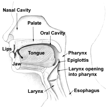
The nasal cavity is a large, air-filled space above and behind the nose in the middle of the face. The nasal septum divides the cavity into two cavities, also known as fossae. Each cavity is the continuation of one of the two nostrils. The nasal cavity is the uppermost part of the respiratory system and provides the nasal passage for inhaled air from the nostrils to the nasopharynx and rest of the respiratory tract. The paranasal sinuses surround and drain into the nasal cavity. Structure The term "nasal cavity" can refer to each of the two cavities of the nose, or to the two sides combined. The lateral wall of each nasal cavity mainly consists of the maxilla. However, there is a deficiency that is compensated for by the perpendicular plate of the palatine bone, the medial pterygoid plate, the labyrinth of ethmoid and the inferior concha. The paranasal sinuses are connected to the nasal cavity through small orifices called ostia. Most of these ostia communicate with the n ... [...More Info...] [...Related Items...] OR: [Wikipedia] [Google] [Baidu] |
Pterygopalatine Fossa
In human anatomy, the pterygopalatine fossa (sphenopalatine fossa) is a fossa in the skull. A human skull contains two pterygopalatine fossae—one on the left side, and another on the right side. Each fossa is a cone-shaped paired depression deep to the infratemporal fossa and posterior to the maxilla on each side of the skull, located between the pterygoid process and the maxillary tuberosity close to the apex of the orbit. It is the indented area medial to the pterygomaxillary fissure leading into the sphenopalatine foramen. It communicates with the nasal and oral cavities, infratemporal fossa, orbit, pharynx, and middle cranial fossa through eight foramina. Structure Boundaries It has the following boundaries: * ''anterior'': superomedial part of the infratemporal surface of maxilla * ''posterior'': root of the pterygoid process and adjoining anterior surface of the greater wing of sphenoid bone * ''medial'': perpendicular plate of the palatine bone and its orbital and sph ... [...More Info...] [...Related Items...] OR: [Wikipedia] [Google] [Baidu] |
Sphenoid Bone
The sphenoid bone is an unpaired bone of the neurocranium. It is situated in the middle of the skull towards the front, in front of the basilar part of occipital bone, basilar part of the occipital bone. The sphenoid bone is one of the seven bones that articulate to form the orbit (anatomy), orbit. Its shape somewhat resembles that of a butterfly or bat with its wings extended. Structure It is divided into the following parts: * a median portion, known as the body of sphenoid bone, containing the sella turcica, which houses the pituitary gland as well as the paired paranasal sinuses, the sphenoidal sinuses * two Greater wing of sphenoid bone, greater wings on the lateral side of the body and two Lesser wing of sphenoid bone, lesser wings from the anterior side. * Pterygoid processes of the sphenoides, directed downwards from the junction of the body and the greater wings. Two sphenoidal conchae are situated at the anterior and inferior part of the body. Intrinsic ligaments of ... [...More Info...] [...Related Items...] OR: [Wikipedia] [Google] [Baidu] |
Superior Meatus
In anatomy, the term nasal meatus can refer to any of the three meatuses (passages) through the skulls nasal cavity: the superior meatus (''meatus nasi superior''), middle meatus (''meatus nasi medius''), and inferior meatus (''meatus nasi inferior''). The nasal meatuses are located beneath each of the corresponding nasal conchae. In the case where a fourth, supreme nasal concha is present, there is a fourth supreme nasal meatus. Structure The superior meatus is the smallest of the three. It is a narrow cavity located obliquely below the superior concha. This meatus is short, lies above and extends from the middle part of the middle concha below. From behind, the sphenopalatine foramen opens into the cavity of the superior meatus and the meatus communicates with the posterior ethmoidal cells. Above and at the back of the superior concha is the sphenoethmoidal recess which the sphenoidal sinus opens into. The superior meatus occupies the middle third of the nasal cavity’s l ... [...More Info...] [...Related Items...] OR: [Wikipedia] [Google] [Baidu] |
Sphenopalatine Artery
The sphenopalatine artery (nasopalatine artery) is an artery of the head, commonly known as the artery of epistaxis. Course The sphenopalatine artery is a branch of the maxillary artery which passes through the sphenopalatine foramen into the cavity of the nose, at the back part of the superior meatus. Here it gives off its posterior lateral nasal branches. Crossing the under surface of the sphenoid, the sphenopalatine artery ends on the nasal septum as the posterior septal branches. Here it will anastomose with the branches of the greater palatine artery. Clinical significance The sphenopalatine artery is the artery responsible for the most serious, posterior nosebleeds (also known as epistaxis). It can be ligated surgically or blocked under image guidance with minimally invasive techniques by interventional radiologist using tiny microparticles to control such nosebleeds. See also *Kiesselbach's plexus Kiesselbach's plexus is an anastomotic arterial network (plexus) of four ... [...More Info...] [...Related Items...] OR: [Wikipedia] [Google] [Baidu] |
Posterior Superior Lateral Nasal Nerve , a relative future tense
{{disambiguation ...
Posterior may refer to: * Posterior (anatomy), the end of an organism opposite to its head ** Buttocks, as a euphemism * Posterior horn (other) * Posterior probability, the conditional probability that is assigned when the relevant evidence is taken into account * Posterior tense Relative tense and absolute tense are distinct possible uses of the grammatical category of Grammatical tense, tense. Absolute tense means the grammatical expression of time reference (usually past tense, past, present tense, present or future tense ... [...More Info...] [...Related Items...] OR: [Wikipedia] [Google] [Baidu] |
Nasopalatine Nerve
The nasopalatine nerve (long sphenopalatine nerve) is a nerve of the head. It is a branch of the pterygopalatine ganglion, a continuation from the maxillary nerve (V2). It supplies parts of the palate and nasal septum. Structure The nasopalatine nerve communicates with the corresponding nerve of the opposite side and with the greater palatine nerve. The medial superior posterior nasal branches of the maxillary nerve usually branch from the nasopalatine nerve. Origin The nasopalatine nerve is a branch of the pterygopalatine ganglion, a continuation from the maxillary nerve (V2), itself a branch of the trigeminal nerve. It enters the nasal cavity through the sphenopalatine foramen. Course It passes across the roof of the nasal cavity below the orifice of the sphenoidal sinus to reach the nasal septum. It then runs obliquely downward and forward between the periosteum and mucous membrane of the lower part of the nasal septum. It descends to the roof of the mouth through the ... [...More Info...] [...Related Items...] OR: [Wikipedia] [Google] [Baidu] |


