|
Simple Cells
A simple cell in the primary visual cortex is a cell that responds primarily to oriented edges and gratings (bars of particular orientations). These cells were discovered by Torsten Wiesel and David Hubel in the late 1950s. Such cells are tuned to different frequencies and orientations, even with different phase relationships, possibly for extracting disparity (depth) information and to attribute depth to detected lines and edges. This may result in a 3D 'wire-frame' representation as used in computer graphics. The fact that input from the left and right eyes is very close in the so-called cortical hypercolumns is an indication that depth processing occurs at a very early stage, aiding recognition of 3D objects. Later, many other cells with specific functions have been discovered: (a) end-stopped cells which are thought to detect singularities like line and edge crossings, vertices and line endings; (b) bar and grating cells. The latter are not linear operators because a bar cel ... [...More Info...] [...Related Items...] OR: [Wikipedia] [Google] [Baidu] |
Gabor Filter
In image processing, a Gabor filter, named after Dennis Gabor, is a linear filter used for texture analysis, which essentially means that it analyzes whether there is any specific frequency content in the image in specific directions in a localized region around the point or region of analysis. Frequency and orientation representations of Gabor filters are claimed by many contemporary vision scientists to be similar to those of the human visual system. They have been found to be particularly appropriate for texture representation and discrimination. In the spatial domain, a 2-D Gabor filter is a Gaussian kernel function modulated by a sinusoidal plane wave (see Gabor transform). Some authors claim that simple cells in the visual cortex of mammalian brains can be modeled by Gabor functions. Thus, image analysis with Gabor filters is thought by some to be similar to perception in the human visual system. Definition Its impulse response is defined by a sinusoidal wave (a plane ... [...More Info...] [...Related Items...] OR: [Wikipedia] [Google] [Baidu] |
Visual System
The visual system comprises the sensory organ (the eye) and parts of the central nervous system (the retina containing photoreceptor cells, the optic nerve, the optic tract and the visual cortex) which gives organisms the sense of sight (the ability to perception, detect and process visible light) as well as enabling the formation of several non-image photo response functions. It detects and interprets information from the optical spectrum perceptible to that species to "build a representation" of the surrounding environment. The visual system carries out a number of complex tasks, including the reception of light and the formation of monocular neural representations, colour vision, the neural mechanisms underlying stereopsis and assessment of distances to and between objects, the identification of a particular object of interest, motion perception, the analysis and integration of visual information, pattern recognition, accurate motor coordination under visual guidance, and mor ... [...More Info...] [...Related Items...] OR: [Wikipedia] [Google] [Baidu] |
Visual Cortex
The visual cortex of the brain is the area of the cerebral cortex that processes visual information. It is located in the occipital lobe. Sensory input originating from the eyes travels through the lateral geniculate nucleus in the thalamus and then reaches the visual cortex. The area of the visual cortex that receives the sensory input from the lateral geniculate nucleus is the primary visual cortex, also known as visual area 1 ( V1), Brodmann area 17, or the striate cortex. The extrastriate areas consist of visual areas 2, 3, 4, and 5 (also known as V2, V3, V4, and V5, or Brodmann area 18 and all Brodmann area 19). Both hemispheres of the brain include a visual cortex; the visual cortex in the left hemisphere receives signals from the right visual field, and the visual cortex in the right hemisphere receives signals from the left visual field. Introduction The primary visual cortex (V1) is located in and around the calcarine fissure in the occipital lobe. Each hemisphere's V1 ... [...More Info...] [...Related Items...] OR: [Wikipedia] [Google] [Baidu] |
Torsten Wiesel
Torsten Nils Wiesel (born 3 June 1924) is a Swedish neurophysiologist. With David H. Hubel, he received the 1981 Nobel Prize in Physiology or Medicine, for their discoveries concerning information processing in the visual system; the prize was shared with Roger W. Sperry for his independent research on the cerebral hemispheres. Career Wiesel was born in Uppsala, Sweden in 1924, the youngest of five children. In 1947, he began his scientific career in Carl Gustaf Bernhard's laboratory at the Karolinska Institute, where he received his medical degree in 1954. He went on to teach in the Institute's department of physiology and worked in the child psychiatry unit of the Karolinska Hospital. In 1955 he moved to the United States to work at Johns Hopkins School of Medicine under Stephen Kuffler. Wiesel began a fellowship in ophthalmology, and in 1958 he became an assistant professor. That same year, he met David Hubel, beginning a collaboration that would last over twenty years. In ... [...More Info...] [...Related Items...] OR: [Wikipedia] [Google] [Baidu] |
David Hubel
David Hunter Hubel (February 27, 1926 – September 22, 2013) was a Canadian American neurophysiologist noted for his studies of the structure and function of the visual cortex. He was co-recipient with Torsten Wiesel of the 1981 Nobel Prize in Physiology or Medicine (shared with Roger W. Sperry), for their discoveries concerning information processing in the visual system. For much of his career, Hubel worked as the Professor of Neurobiology at Johns Hopkins University and Harvard Medical School. In 1978, Hubel and Wiesel were awarded the Louisa Gross Horwitz Prize from Columbia University. In 1983, Hubel received the Golden Plate Award of the American Academy of Achievement. Early life and education David H. Hubel was born in Windsor, Ontario, Canada, to American parents in 1926. His grandfather emigrated as a child to the United States from the Bavarian town of Nördlingen. In 1929, his family moved to Montreal, where he spent his formative years. His father was a che ... [...More Info...] [...Related Items...] OR: [Wikipedia] [Google] [Baidu] |
Complex Cell
Complex cells can be found in the primary visual cortex (V1), the secondary visual cortex (V2), and Brodmann area 19 ( V3). Like a simple cell, a complex cell will respond primarily to oriented edges and gratings, however it has a degree of spatial invariance. This means that its receptive field cannot be mapped into fixed excitatory and inhibitory zones. Rather, it will respond to patterns of light in a certain orientation within a large receptive field, regardless of the exact location. Some complex cells respond optimally only to movement in a certain direction. These cells were discovered by Torsten Wiesel and David Hubel in the early 1960s. They refrained from reporting on the complex cells in (Hubel 1959) because they did not feel that they understood them well enough at the time. In Hubel and Wiesel (1962), they reported that complex cells were intermixed with simple cells and when excitatory and inhibitory regions could be established, the summation and mutual antagonis ... [...More Info...] [...Related Items...] OR: [Wikipedia] [Google] [Baidu] |
Lateral Geniculate Nucleus
In neuroanatomy, the lateral geniculate nucleus (LGN; also called the lateral geniculate body or lateral geniculate complex) is a structure in the thalamus and a key component of the mammalian visual pathway. It is a small, ovoid, ventral projection of the thalamus where the thalamus connects with the optic nerve. There are two LGNs, one on the left and another on the right side of the thalamus. In humans, both LGNs have six layers of neurons (grey matter) alternating with optic fibers (white matter). The LGN receives information directly from the ascending retinal ganglion cells via the optic tract and from the reticular activating system. Neurons of the LGN send their axons through the optic radiation, a direct pathway to the primary visual cortex. In addition, the LGN receives many strong feedback connections from the primary visual cortex. In humans as well as other mammals, the two strongest pathways linking the eye to the brain are those projecting to the dorsal part of th ... [...More Info...] [...Related Items...] OR: [Wikipedia] [Google] [Baidu] |
Retina
The retina (from la, rete "net") is the innermost, light-sensitive layer of tissue of the eye of most vertebrates and some molluscs. The optics of the eye create a focused two-dimensional image of the visual world on the retina, which then processes that image within the retina and sends nerve impulses along the optic nerve to the visual cortex to create visual perception. The retina serves a function which is in many ways analogous to that of the film or image sensor in a camera. The neural retina consists of several layers of neurons interconnected by synapses and is supported by an outer layer of pigmented epithelial cells. The primary light-sensing cells in the retina are the photoreceptor cells, which are of two types: rods and cones. Rods function mainly in dim light and provide monochromatic vision. Cones function in well-lit conditions and are responsible for the perception of colour through the use of a range of opsins, as well as high-acuity vision used for task ... [...More Info...] [...Related Items...] OR: [Wikipedia] [Google] [Baidu] |
Receptive Field
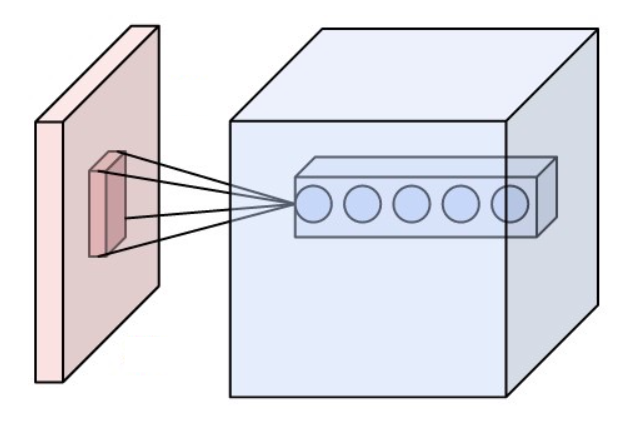
The receptive field, or sensory space, is a delimited medium where some physiological stimuli can evoke a sensory neuronal response in specific organisms. Complexity of the receptive field ranges from the unidimensional chemical structure of odorants to the multidimensional spacetime of human visual field, through the bidimensional skin surface, being a receptive field for touch perception. Receptive fields can positively or negatively alter the membrane potential with or without affecting the rate of action potentials. A sensory space can be dependent of an animal's location. For a particular sound wave traveling in an appropriate transmission medium, by means of sound localization, an auditory space would amount to a reference system that continuously shifts as the animal moves (taking into consideration the space inside the ears as well). Conversely, receptive fields can be largely independent of the animal's location, as in the case of place cells. A sensory space can also m ... [...More Info...] [...Related Items...] OR: [Wikipedia] [Google] [Baidu] |
Surround Suppression
Surround suppression is where the relative firing rate of a neuron may under certain conditions decrease when a particular stimulus is enlarged. It has been observed in electrophysiology studies of the brain and has been noted in many sensory neurons, most notably in the early visual system. Surround suppression is defined as a reduction in the activity of a neuron in response to a stimulus outside its classical receptive field. The necessary functional connections with other neurons influenced by stimulation outside a particular area and by dynamic processes in general, and the absence of a theoretical description of a system state to be treated as a baseline, deprive the term "classical receptive field" of functional meaning. The descriptor "surround suppression" suffers from a similar problem, as the activities of neurons in the "surround" of the "classical receptive field are similarly determined by connectivities and processes involving neurons beyond it.) This nonlinear effe ... [...More Info...] [...Related Items...] OR: [Wikipedia] [Google] [Baidu] |
Spike-triggered Average
The spike-triggered averaging (STA) is a tool for characterizing the response properties of a neuron using the spikes emitted in response to a time-varying stimulus. The STA provides an estimate of a neuron's linear receptive field. It is a useful technique for the analysis of electrophysiological data. Mathematically, the STA is the average stimulus preceding a spike.de Boer and Kuyper (1968) Triggered Correlation. ''IEEE Transact. Biomed. Eng.'', 15:169-179Marmarelis, P. Z. and Naka, K. (1972). White-noise analysis of a neuron chain: an application of the Wiener theory. ''Science'', 175:1276-1278Chichilnisky, E. J. (2001). A simple white noise analysis of neuronal light responses. ''Network: Computation in Neural Systems'', 12:199-213Simoncelli, E. P., Paninski, L., Pillow, J. & Swartz, O. (2004)."Characterization of neural responses with stochastic stimuli" In M. Gazzaniga (Ed.) ''The Cognitive Neurosciences, III'' (pp. 327-338). MIT press. To compute the STA, the stimulus ... [...More Info...] [...Related Items...] OR: [Wikipedia] [Google] [Baidu] |
Receptive Field
The receptive field, or sensory space, is a delimited medium where some physiological stimuli can evoke a sensory neuronal response in specific organisms. Complexity of the receptive field ranges from the unidimensional chemical structure of odorants to the multidimensional spacetime of human visual field, through the bidimensional skin surface, being a receptive field for touch perception. Receptive fields can positively or negatively alter the membrane potential with or without affecting the rate of action potentials. A sensory space can be dependent of an animal's location. For a particular sound wave traveling in an appropriate transmission medium, by means of sound localization, an auditory space would amount to a reference system that continuously shifts as the animal moves (taking into consideration the space inside the ears as well). Conversely, receptive fields can be largely independent of the animal's location, as in the case of place cells. A sensory space can also m ... [...More Info...] [...Related Items...] OR: [Wikipedia] [Google] [Baidu] |


