|
SUCLG2
Succinyl-CoA ligase DP-formingsubunit beta, mitochondrial is an enzyme that in humans is encoded by the ''SUCLG2'' gene on chromosome 3. This gene encodes a GTP-specific beta subunit of succinyl-CoA synthetase. Succinyl-CoA synthetase catalyzes the reversible reaction involving the formation of succinyl-CoA and succinate. Alternate splicing results in multiple transcript variants. Pseudogenes of this gene are found on chromosomes 5 and 12. rovided by RefSeq, Apr 2010ref name="entrez"/> Structure SCS, also known as succinyl CoA ligase (SUCL), is a heterodimer composed of a catalytic α subunit encoded by the '' SUCLG1'' gene and a β subunit encoded by either the '' SUCLA2'' gene or the ''SUCLG2'' gene, which determines the enzyme specificity for either ADP or GDP. SUCLG2 is the SCS variant containing the ''SUCLG2''-encoded β subunit. Amino acid sequence alignment of the two β subunit types reveals a homology of ~50% identity, with specific regions conserved throughout the ... [...More Info...] [...Related Items...] OR: [Wikipedia] [Google] [Baidu] |
SUCLA2
Succinyl-CoA ligase DP-formingsubunit beta, mitochondrial (SUCLA2), also known as ADP-forming succinyl-CoA synthetase (SCS-A), is an enzyme that in humans is encoded by the ''SUCLA2'' gene on chromosome 13. Succinyl-CoA synthetase (SCS) is a mitochondrial matrix enzyme that acts as a heterodimer, composed of an invariant alpha subunit and a substrate-specific beta subunit. The protein encoded by this gene is an ATP-specific SCS beta subunit that dimerizes with the SCS alpha subunit to form SCS-A, an essential component of the tricarboxylic acid cycle. SCS-A hydrolyzes ATP to convert succinyl-CoA to succinate. Defects in this gene are a cause of myopathic mitochondrial DNA depletion syndrome. A pseudogene of this gene has been found on chromosome 6. rovided by RefSeq, Jul 2008ref name="entrez"/> Structure SCS, also known as succinyl CoA ligase (SUCL), is a heterodimer composed of a catalytic α subunit encoded by the '' SUCLG1'' gene and a β subunit encoded by either the ... [...More Info...] [...Related Items...] OR: [Wikipedia] [Google] [Baidu] |
Substrate-level Phosphorylation
Substrate-level phosphorylation is a metabolism reaction that results in the production of ATP or GTP by the transfer of a phosphate group from a substrate directly to ADP or GDP. Transferring from a higher energy (whether phosphate group attached or not) into a lower energy product. This process uses some of the released chemical energy, the Gibbs free energy, to transfer a phosphoryl (PO3) group to ADP or GDP from another phosphorylated compound. Occurs in glycolysis and in the citric acid cycle. Unlike oxidative phosphorylation, oxidation and phosphorylation are not coupled in the process of substrate-level phosphorylation, and reactive intermediates are most often gained in the course of oxidation processes in catabolism. Most ATP is generated by oxidative phosphorylation in aerobic or anaerobic respiration while substrate-level phosphorylation provides a quicker, less efficient source of ATP, independent of external electron acceptors. This is the case in human erythro ... [...More Info...] [...Related Items...] OR: [Wikipedia] [Google] [Baidu] |
Enzyme
Enzymes () are proteins that act as biological catalysts by accelerating chemical reactions. The molecules upon which enzymes may act are called substrates, and the enzyme converts the substrates into different molecules known as products. Almost all metabolic processes in the cell need enzyme catalysis in order to occur at rates fast enough to sustain life. Metabolic pathways depend upon enzymes to catalyze individual steps. The study of enzymes is called ''enzymology'' and the field of pseudoenzyme analysis recognizes that during evolution, some enzymes have lost the ability to carry out biological catalysis, which is often reflected in their amino acid sequences and unusual 'pseudocatalytic' properties. Enzymes are known to catalyze more than 5,000 biochemical reaction types. Other biocatalysts are catalytic RNA molecules, called ribozymes. Enzymes' specificity comes from their unique three-dimensional structures. Like all catalysts, enzymes increase the reaction ra ... [...More Info...] [...Related Items...] OR: [Wikipedia] [Google] [Baidu] |
Ketone Body
Ketone bodies are water-soluble molecules that contain the ketone groups produced from fatty acids by the liver (ketogenesis). Ketone bodies are readily transported into tissues outside the liver, where they are converted into acetyl-CoA (acetyl-Coenzyme A)—which then enters the citric acid cycle (Krebs cycle) and is oxidized for energy. These liver-derived ketone groups include acetoacetic acid (acetoacetate), beta-hydroxybutyrate, and acetone, a spontaneous breakdown product of acetoacetate (see graphic). Ketone bodies are produced by the liver during periods of caloric restriction of various scenarios: low food intake (fasting), carbohydrate restrictive diets, starvation, prolonged intense exercise, alcoholism, or during untreated (or inadequately treated) type 1 diabetes mellitus. Ketone bodies are produced in liver cells by the breakdown of fatty acids. They are released into the blood ''after'' glycogen stores in the liver have been depleted. (Glycogen stores typica ... [...More Info...] [...Related Items...] OR: [Wikipedia] [Google] [Baidu] |
Oxidative Phosphorylation
Oxidative phosphorylation (UK , US ) or electron transport-linked phosphorylation or terminal oxidation is the metabolic pathway in which cells use enzymes to oxidize nutrients, thereby releasing chemical energy in order to produce adenosine triphosphate (ATP). In eukaryotes, this takes place inside mitochondria. Almost all aerobic organisms carry out oxidative phosphorylation. This pathway is so pervasive because it releases more energy than alternative fermentation processes such as anaerobic glycolysis. The energy stored in the chemical bonds of glucose is released by the cell in the citric acid cycle producing carbon dioxide, and the energetic electron donors NADH and FADH. Oxidative phosphorylation uses these molecules and O2 to produce ATP, which is used throughout the cell whenever energy is needed. During oxidative phosphorylation, electrons are transferred from the electron donors to a series of electron acceptors in a series of redox reactions ending in oxygen, who ... [...More Info...] [...Related Items...] OR: [Wikipedia] [Google] [Baidu] |
Mitochondrial DNA Depletion Syndrome
Mitochondrial DNA depletion syndrome (MDS or MDDS), or Alper's disease, is any of a group of autosomal recessive disorders that cause a significant drop in mitochondrial DNA in affected tissues. Symptoms can be any combination of myopathic, hepatopathic, or encephalomyopathic. These syndromes affect tissue in the muscle, liver, or both the muscle and brain, respectively. The condition is typically fatal in infancy and early childhood, though some have survived to their teenage years with the myopathic variant and some have survived into adulthood with the SUCLA2 encephalomyopathic variant. There is currently no curative treatment for any form of MDDS, though some preliminary treatments have shown a reduction in symptoms. Signs and symptoms All forms of MDDS are very rare. MDDS causes a wide range of symptoms, which can appear in newborns, infants, children, or adults, depending on the class of MDDS; within each class symptoms are also diverse. In MDDS associated with mutations ... [...More Info...] [...Related Items...] OR: [Wikipedia] [Google] [Baidu] |
Brain
A brain is an organ that serves as the center of the nervous system in all vertebrate and most invertebrate animals. It is located in the head, usually close to the sensory organs for senses such as vision. It is the most complex organ in a vertebrate's body. In a human, the cerebral cortex contains approximately 14–16 billion neurons, and the estimated number of neurons in the cerebellum is 55–70 billion. Each neuron is connected by synapses to several thousand other neurons. These neurons typically communicate with one another by means of long fibers called axons, which carry trains of signal pulses called action potentials to distant parts of the brain or body targeting specific recipient cells. Physiologically, brains exert centralized control over a body's other organs. They act on the rest of the body both by generating patterns of muscle activity and by driving the secretion of chemicals called hormones. This centralized control allows rapid and coordinated respon ... [...More Info...] [...Related Items...] OR: [Wikipedia] [Google] [Baidu] |
Oligodendrocyte
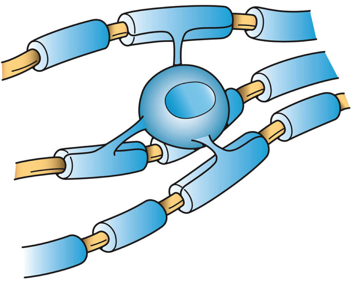
Oligodendrocytes (), or oligodendroglia, are a type of neuroglia whose main functions are to provide support and insulation to axons in the central nervous system of jawed vertebrates, equivalent to the function performed by Schwann cells in the peripheral nervous system. Oligodendrocytes do this by creating the myelin sheath. A single oligodendrocyte can extend its processes to 50 axons, wrapping approximately 1 μm of myelin sheath around each axon; Schwann cells, on the other hand, can wrap around only one axon. Each oligodendrocyte forms one segment of myelin for several adjacent axons. Oligodendrocytes are found only in the central nervous system, which comprises the brain and spinal cord. These cells were originally thought to have been produced in the ventral neural tube; however, research now shows oligodendrocytes originate from the ventral ventricular zone of the embryonic spinal cord and possibly have some concentrations in the forebrain. They are the last cell ... [...More Info...] [...Related Items...] OR: [Wikipedia] [Google] [Baidu] |
Microglia
Microglia are a type of neuroglia (glial cell) located throughout the brain and spinal cord. Microglia account for about 7% of cells found within the brain. As the resident macrophage cells, they act as the first and main form of active immune defense in the central nervous system (CNS). Microglia (and other neuroglia including astrocytes) are distributed in large non-overlapping regions throughout the CNS. Microglia are key cells in overall brain maintenance—they are constantly scavenging the CNS for plaques, damaged or unnecessary neurons and synapses, and infectious agents. Since these processes must be efficient to prevent potentially fatal damage, microglia are extremely sensitive to even small pathological changes in the CNS. This sensitivity is achieved in part by the presence of unique potassium channels that respond to even small changes in extracellular potassium. Recent evidence shows that microglia are also key players in the sustainment of normal brain functions und ... [...More Info...] [...Related Items...] OR: [Wikipedia] [Google] [Baidu] |
Astrocyte
Astrocytes (from Ancient Greek , , "star" + , , "cavity", "cell"), also known collectively as astroglia, are characteristic star-shaped glial cells in the brain and spinal cord. They perform many functions, including biochemical control of endothelial cells that form the blood–brain barrier, provision of nutrients to the nervous tissue, maintenance of extracellular ion balance, regulation of cerebral blood flow, and a role in the repair and scarring process of the brain and spinal cord following infection and traumatic injuries. The proportion of astrocytes in the brain is not well defined; depending on the counting technique used, studies have found that the astrocyte proportion varies by region and ranges from 20% to 40% of all glia. Another study reports that astrocytes are the most numerous cell type in the brain. Astrocytes are the major source of cholesterol in the central nervous system. Apolipoprotein E transports cholesterol from astrocytes to neurons and other glial ... [...More Info...] [...Related Items...] OR: [Wikipedia] [Google] [Baidu] |


