|
STED Microscopy
Stimulated emission depletion (STED) microscopy is one of the techniques that make up super-resolution microscopy. It creates super-resolution images by the selective deactivation of fluorophores, minimizing the area of illumination at the focal point, and thus enhancing the achievable resolution for a given system. It was developed by Stefan W. Hell and Jan Wichmann in 1994, and was first experimentally demonstrated by Hell and Thomas Klar in 1999. Hell was awarded the Nobel Prize in Chemistry in 2014 for its development. In 1986, V.A. Okhonin (Institute of Biophysics, USSR Academy of Sciences, Siberian Branch, Krasnoyarsk) had patented the STED idea. This patent was unknown to Hell and Wichmann in 1994. STED microscopy is one of several types of super resolution microscopy techniques that have recently been developed to bypass the diffraction limit of light microscopy to increase resolution. STED is a deterministic functional technique that exploits the non-linear response of ... [...More Info...] [...Related Items...] OR: [Wikipedia] [Google] [Baidu] |
STED Confocal Comparison 50nm HWFM
STED may refer to: * STED microscopy (stimulated emission depletion) * STED sewer (septic tank effluent drainage) * Septic tank effluent rainage system, a type of Sanitary sewer A sanitary sewer is an underground pipe or tunnel system for transporting sewage from houses and commercial buildings (but not stormwater) to a sewage treatment plant or disposal. Sanitary sewers are a type of gravity sewer and are part of ... See also * Stead (other) {{Disambiguation ... [...More Info...] [...Related Items...] OR: [Wikipedia] [Google] [Baidu] |
Tom20
Mitochondrial import receptor subunit TOM20 homolog is a protein that in humans is encoded by the ''TOMM20'' gene. TOM20 is one of the receptor systems of the TOM complex (translocase of the outer membrane) in the outer mitochondrial membrane (OMM). Function In mitochondrial protein import, TOM20 is closely associated with the pore-forming TOM40 complex and acts by recognizing and binding the N-terminal MTSs (matrix-targeting sequences), which form an amphipathic alpha helix and aid passage of the target proteins into the mitochondrial matrix. See also * Mitochondria Outer Membrane Translocase * TOMM22 * TOMM40 * TOMM70A Mitochondrial import receptor subunit TOM70 is a protein that in humans is encoded by the ''TOMM70A'' gene. The translocase of outer mitochondrial membrane (TOM) complex is a multisubunit complex involved in the recognition, unfolding, and translo ... References Further reading * * * * * * * * * * * * * * * {{gene-1-stub ... [...More Info...] [...Related Items...] OR: [Wikipedia] [Google] [Baidu] |
SNAP25
Synaptosomal-Associated Protein, 25kDa (SNAP-25) is a Target Soluble NSF (''N''-ethylmaleimide-sensitive factor) Attachment Protein Receptor (t-SNARE) protein encoded by the ''SNAP25'' gene found on chromosome 20p12.2 in humans. SNAP-25 is a component of the ''trans''-SNARE complex, which accounts for membrane fusion specificity and directly executes fusion by forming a tight complex that brings the synaptic vesicle and plasma membranes together. Structure and function SNAP-25, a Q-SNARE protein, is anchored to the cytosolic face of membranes via palmitoyl side chains covalently bound to cysteine amino acid residues in the central linker domain of the molecule. This means that SNAP-25 does not contain a trans-membrane domain. SNAP-25 has been identified to contribute two α-helices to the SNARE complex, a four-α-helix domain complex. The SNARE complex participates in vesicle fusion, which involves the docking, priming and merging of a vesicle with the cell membrane to in ... [...More Info...] [...Related Items...] OR: [Wikipedia] [Google] [Baidu] |
Tubulin
Tubulin in molecular biology can refer either to the tubulin protein superfamily of globular proteins, or one of the member proteins of that superfamily. α- and β-tubulins polymerize into microtubules, a major component of the eukaryotic cytoskeleton. Microtubules function in many essential cellular processes, including mitosis. Tubulin-binding drugs kill cancerous cells by inhibiting microtubule dynamics, which are required for DNA segregation and therefore cell division. In eukaryotes, there are six members of the tubulin superfamily, although not all are present in all species.Turk E, Wills AA, Kwon T, Sedzinski J, Wallingford JB, Stearns "Zeta-Tubulin Is a Member of a Conserved Tubulin Module and Is a Component of the Centriolar Basal Foot in Multiciliated Cells"Current Biology (2015) 25:2177-2183. Both α and β tubulins have a mass of around 50 kDa and are thus in a similar range compared to actin (with a mass of ~42 kDa). In contrast, tubulin polymers (microtubules) ... [...More Info...] [...Related Items...] OR: [Wikipedia] [Google] [Baidu] |
Actin
Actin is a protein family, family of Globular protein, globular multi-functional proteins that form microfilaments in the cytoskeleton, and the thin filaments in myofibril, muscle fibrils. It is found in essentially all Eukaryote, eukaryotic cells, where it may be present at a concentration of over 100 micromolar, μM; its mass is roughly 42 kDa, with a diameter of 4 to 7 nm. An actin protein is the monomeric Protein subunit, subunit of two types of filaments in cells: microfilaments, one of the three major components of the cytoskeleton, and thin filaments, part of the Muscle contraction, contractile apparatus in muscle cells. It can be present as either a free monomer called G-actin (globular) or as part of a linear polymer microfilament called F-actin (filamentous), both of which are essential for such important cellular functions as the Motility, mobility and contraction of cell (biology), cells during cell division. Actin participates in many important cellular pr ... [...More Info...] [...Related Items...] OR: [Wikipedia] [Google] [Baidu] |
Neurofilament
Neurofilaments (NF) are classed as type IV intermediate filaments found in the cytoplasm of neurons. They are protein polymers measuring 10 nm in diameter and many micrometers in length. Together with microtubules (~25 nm) and microfilaments (7 nm), they form the neuronal cytoskeleton. They are believed to function primarily to provide structural support for axons and to regulate axon diameter, which influences nerve conduction velocity. The proteins that form neurofilaments are members of the intermediate filament protein family, which is divided into six types based on their gene organization and protein structure. Types I and II are the keratins which are expressed in epithelia. Type III contains the proteins vimentin, desmin, peripherin and glial fibrillary acidic protein (GFAP). Type IV consists of the neurofilament proteins L, M, H and internexin. Type V consists of the nuclear lamins, and type VI consists of the protein nestin. The type IV interm ... [...More Info...] [...Related Items...] OR: [Wikipedia] [Google] [Baidu] |
Rhodamine B
Rhodamine B is a chemical compound and a dye. It is often used as a tracer dye within water to determine the rate and direction of flow and transport. Rhodamine dyes fluoresce and can thus be detected easily and inexpensively with fluorometers. Rhodamine B is used in biology as a staining fluorescent dye, sometimes in combination with auramine O, as the auramine-rhodamine stain to demonstrate acid-fast organisms, notably ''Mycobacterium''. Rhodamine dyes are also used extensively in biotechnology applications such as fluorescence microscopy, flow cytometry, fluorescence correlation spectroscopy and ELISA. It is also used in rose milk, a popular Indian beverage. Other uses Rhodamine B is often mixed with herbicides to show where they have been used. It is also being tested for use as a biomarker in oral rabies vaccines for wildlife, such as raccoons, to identify animals that have eaten a vaccine bait. The rhodamine is incorporated into the animal's whiskers and teeth. Rhoda ... [...More Info...] [...Related Items...] OR: [Wikipedia] [Google] [Baidu] |
Stimulated Emission
Stimulated emission is the process by which an incoming photon of a specific frequency can interact with an excited atomic electron (or other excited molecular state), causing it to drop to a lower energy level. The liberated energy transfers to the electromagnetic field, creating a new photon with a frequency, polarization, and direction of travel that are all identical to the photons of the incident wave. This is in contrast to spontaneous emission, which occurs at a characteristic rate for each of the atoms/oscillators in the upper energy state regardless of the external electromagnetic field. According to the American Physical Society, the first person to correctly predict the phenomenon of stimulated emission was Albert Einstein in a series of papers starting in 1916, culminating in what is now called the Einstein B Coefficient. Einstein's work became the theoretical foundation of the MASER and LASER. The process is identical in form to atomic absorption in which the energy ... [...More Info...] [...Related Items...] OR: [Wikipedia] [Google] [Baidu] |
Optical Cavity
An optical cavity, resonating cavity or optical resonator is an arrangement of mirrors or other optical elements that forms a cavity resonator for light waves. Optical cavities are a major component of lasers, surrounding the gain medium and providing feedback of the laser light. They are also used in optical parametric oscillators and some interferometers. Light confined in the cavity reflects multiple times, producing modes with certain resonance frequencies. Modes can be decomposed into longitudinal modes that differ only in frequency and transverse modes that have different intensity patterns across the cross-section of the beam. Many types of optical cavity produce standing wave modes. Different resonator types are distinguished by the focal lengths of the two mirrors and the distance between them. Flat mirrors are not often used because of the difficulty of aligning them to the needed precision. The geometry (resonator type) must be chosen so that the beam remains sta ... [...More Info...] [...Related Items...] OR: [Wikipedia] [Google] [Baidu] |
Refractive Index
In optics, the refractive index (or refraction index) of an optical medium is a dimensionless number that gives the indication of the light bending ability of that medium. The refractive index determines how much the path of light is bent, or refracted, when entering a material. This is described by Snell's law of refraction, , where ''θ''1 and ''θ''2 are the angle of incidence and angle of refraction, respectively, of a ray crossing the interface between two media with refractive indices ''n''1 and ''n''2. The refractive indices also determine the amount of light that is reflected when reaching the interface, as well as the critical angle for total internal reflection, their intensity (Fresnel's equations) and Brewster's angle. The refractive index can be seen as the factor by which the speed and the wavelength of the radiation are reduced with respect to their vacuum values: the speed of light in a medium is , and similarly the wavelength in that medium is , where ''� ... [...More Info...] [...Related Items...] OR: [Wikipedia] [Google] [Baidu] |
Optical Vortex
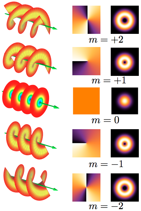
An optical vortex (also known as a photonic quantum vortex, screw dislocation or phase singularity) is a zero of an optical field; a point of zero intensity. The term is also used to describe a beam of light that has such a zero in it. The study of these phenomena is known as singular optics. Explanation In an optical vortex, light is twisted like a corkscrew around its axis of travel. Because of the twisting, the light waves at the axis itself cancel each other out. When projected onto a flat surface, an optical vortex looks like a ring of light, with a dark hole in the center. The vortex is given a number, called the topological charge, according to how many twists the light does in one wavelength. The number is always an integer, and can be positive or negative, depending on the direction of the twist. The higher the number of the twist, the faster the light is spinning around the axis. This spinning carries orbital angular momentum with the wave train, and will induce torque ... [...More Info...] [...Related Items...] OR: [Wikipedia] [Google] [Baidu] |


