|
Restrictive Cardiomyopathy
Restrictive cardiomyopathy (RCM) is a form of cardiomyopathy in which the walls of the heart are rigid (but not thickened). Thus the heart is restricted from stretching and filling with blood properly. It is the least common of the three original subtypes of cardiomyopathy: hypertrophic, dilated, and restrictive. It should not be confused with constrictive pericarditis, a disease which presents similarly but is very different in treatment and prognosis. Signs and symptoms Untreated hearts with RCM often develop the following characteristics: * M or W configuration in an invasive hemodynamic pressure tracing of the RA * Square root sign of part of the invasive hemodynamic pressure tracing Of The LV * Biatrial enlargement * Thickened LV walls (with normal chamber size) * Thickened RV free wall (with normal chamber size) * Elevated right atrial pressure (>12mmHg), * Moderate pulmonary hypertension, * Normal systolic function, * Poor diastolic function, typically Grade III - IV Diast ... [...More Info...] [...Related Items...] OR: [Wikipedia] [Google] [Baidu] |
Micrograph
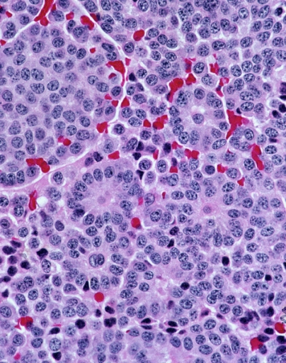
A micrograph or photomicrograph is a photograph or digital image taken through a microscope or similar device to show a magnified image of an object. This is opposed to a macrograph or photomacrograph, an image which is also taken on a microscope but is only slightly magnified, usually less than 10 times. Micrography is the practice or art of using microscopes to make photographs. A micrograph contains extensive details of microstructure. A wealth of information can be obtained from a simple micrograph like behavior of the material under different conditions, the phases found in the system, failure analysis, grain size estimation, elemental analysis and so on. Micrographs are widely used in all fields of microscopy. Types Photomicrograph A light micrograph or photomicrograph is a micrograph prepared using an optical microscope, a process referred to as ''photomicroscopy''. At a basic level, photomicroscopy may be performed simply by connecting a camera to a microscope, th ... [...More Info...] [...Related Items...] OR: [Wikipedia] [Google] [Baidu] |
Primary Hyperoxaluria
Primary hyperoxaluria is a rare condition (autosomal recessive), resulting in increased excretion of oxalate (up to 600 mg a day from normal 50 mg a day), with oxalate stones being common. Signs and symptoms Primary hyperoxaluria is an autosomal recessive disease, meaning both copies of the gene contain the mutation. Both parents must have one copy of this mutated gene to pass it on to their child, but they do not typically show signs or symptoms of the disease. A single kidney stone in children or recurrent stones in adults is often the first warning sign of primary hyperoxaluria. Other symptoms range from recurrent urinary tract infections, severe abdominal pain or pain in the side, blood in the urine, to chronic kidney disease and kidney failure. The age of symptom onset, progression and severity can vary greatly from one person to another, even among members of the same family. Some individuals may have mild cases that go undiagnosed well into adulthood; others may ... [...More Info...] [...Related Items...] OR: [Wikipedia] [Google] [Baidu] |
Hypereosinophilic Syndrome
Hypereosinophilic syndrome is a disease characterized by a persistently elevated eosinophil count (≥ 1500 eosinophils/mm³) in the blood for at least six months without any recognizable cause, with involvement of either the heart, nervous system, or bone marrow. HES is a diagnosis of exclusion, after clonal eosinophilia (such as ''FIP1L1-PDGFRA''-fusion induced hypereosinophelia and leukemia) and reactive eosinophilia (in response to infection, autoimmune disease, atopy, hypoadrenalism, tropical eosinophilia, or cancer) have been ruled out. There are some associations with chronic eosinophilic leukemia as it shows similar characteristics and genetic defects. Last updated: Updated: Oct 4, 2009 by Venkata Samavedi and Emmanuel C Besa If left untreated, HES is progressive and fatal. It is treated with glucocorticoids such as prednisone. The addition of the monoclonal antibody mepolizumab may reduce the dose of glucocorticoids. Signs and symptoms As HES affects many organs at ... [...More Info...] [...Related Items...] OR: [Wikipedia] [Google] [Baidu] |
Carcinoid
A carcinoid (also carcinoid tumor) is a slow-growing type of neuroendocrine tumor originating in the cells of the neuroendocrine system. In some cases, metastasis may occur. Carcinoid tumors of the midgut (jejunum, ileum, appendix, and cecum) are associated with carcinoid syndrome. Carcinoid tumors are the most common malignant tumor of the appendix, but they are most commonly associated with the small intestine, and they can also be found in the rectum and stomach. They are known to grow in the liver, but this finding is usually a manifestation of metastatic disease from a primary carcinoid occurring elsewhere in the body. They have a very slow growth rate compared to most malignant tumors. The median age at diagnosis for all patients with neuroendocrine tumors is 63 years. Signs and symptoms While most carcinoids are asymptomatic through the natural life and are discovered only upon surgery for unrelated reasons (so-called ''coincidental carcinoids''), all carcinoids are ... [...More Info...] [...Related Items...] OR: [Wikipedia] [Google] [Baidu] |
Werner Syndrome
Werner syndrome (WS) or Werner's syndrome, also known as "adult progeria",James, William; Berger, Timothy; Elston, Dirk (2005). ''Andrews' Diseases of the Skin: Clinical Dermatology''. (10th ed.). Saunders. . is a rare, autosomal recessive disorder which is characterized by the appearance of premature aging. Werner syndrome is named after the German scientist Otto Werner. He identified the syndrome in four siblings observed with premature aging, which he explored as the subject of his dissertation of 1904. It has a global incidence rate of less than 1 in 100,000 live births (although incidence in Japan and Sardinia is higher, affecting 1 in 20,000–40,000 and 1 in 50,000, respectively). 1,300 cases had been reported as of 2006. Affected individuals typically grow and develop normally until puberty; the mean age of diagnosis is twenty-four, often realized when the adolescent growth spurt is not observed. The youngest person diagnosed was six years old. The median and mean ages of ... [...More Info...] [...Related Items...] OR: [Wikipedia] [Google] [Baidu] |
Pseudoxanthoma Elasticum
Pseudoxanthoma elasticum (PXE) is a genetic disease that causes mineralization of elastic fibers in some tissues. The most common problems arise in the skin and eyes, and later in blood vessels in the form of premature atherosclerosis. PXE is caused by autosomal recessive mutations in the '' ABCC6'' gene on the short arm of chromosome 16 (16p13.1). Signs and symptoms Usually, pseudoxanthoma elasticum affects the skin first, often in childhood or early adolescence. Small, yellowish papular lesions form and cutaneous laxity mainly affect the neck, axillae (armpits), groin, and flexural creases (the inside parts of the elbows and knees). Skin may become lax and redundant. Many individuals have "oblique mental creases" (horizontal grooves of the chin) PXE first affects the retina through a dimpling of the Bruch membrane (a thin membrane separating the blood vessel-rich layer from the pigmented layer of the retina), that is only visible during ophthalmologic examinations. This is ca ... [...More Info...] [...Related Items...] OR: [Wikipedia] [Google] [Baidu] |
Scleroderma
Scleroderma is a group of autoimmune diseases that may result in changes to the skin, blood vessels, muscles, and internal organs. The disease can be either localized to the skin or involve other organs, as well. Symptoms may include areas of thickened skin, stiffness, feeling tired, and poor blood flow to the fingers or toes with cold exposure. One form of the condition, known as CREST syndrome, classically results in calcium deposits, Raynaud's syndrome, esophageal problems, thickening of the skin of the fingers and toes, and areas of small, dilated blood vessels. The cause is unknown, but it may be due to an abnormal immune response. Risk factors include family history, certain genetic factors, and exposure to silica. The underlying mechanism involves the abnormal growth of connective tissue, which is believed to be the result of the immune system attacking healthy tissues. Diagnosis is based on symptoms, supported by a skin biopsy or blood tests. While no cure is ... [...More Info...] [...Related Items...] OR: [Wikipedia] [Google] [Baidu] |
Diabetic Cardiomyopathy
Diabetic cardiomyopathy is a disorder of the myocardium, heart muscle in people with diabetes mellitus, diabetes. It can lead to inability of the heart to circulate blood through the body effectively, a state known as heart failure(HF),} with accumulation of fluid in the lungs (pulmonary edema) or human leg, legs (peripheral edema). Most heart failure in people with diabetes results from coronary artery disease, and diabetic cardiomyopathy is only said to exist if there is ''no'' coronary artery disease to explain the heart muscle disorder. Signs and symptoms One particularity of diabetic cardiomyopathy is the long latent phase, during which the disease progresses but is completely asymptomatic. In most cases, diabetic cardiomyopathy is detected with concomitant hypertension or coronary artery disease. One of the earliest signs is mild left ventricular diastolic dysfunction with little effect on ventricular filling. Also, the diabetic patient may show subtle signs of diabetic cardi ... [...More Info...] [...Related Items...] OR: [Wikipedia] [Google] [Baidu] |
Niemann–Pick Disease
Niemann–Pick disease is a group of severe inherited metabolic disorders, in which sphingomyelin accumulates in lysosomes in cells (the lysosomes normally degrade material that comes from out of cells). These disorders involve the dysfunctional metabolism of sphingolipids, which are fats found in cell membranes. They can be considered as a kind of sphingolipidosis, which is included in the larger family of lysosomal storage diseases. Signs and symptoms Symptoms are related to the organs in which sphingomyelin accumulates. Enlargement of the liver and spleen (hepatosplenomegaly) may cause reduced appetite, abdominal distension, and pain. Enlargement of the spleen (splenomegaly) may also cause low levels of platelets in the blood (thrombocytopenia). Accumulation of sphingomyelin in the central nervous system (including the cerebellum) results in unsteady gait (ataxia), slurring of speech (dysarthria), and difficulty swallowing (dysphagia). Basal ganglia dysfunction causes abnormal ... [...More Info...] [...Related Items...] OR: [Wikipedia] [Google] [Baidu] |
Mucopolysaccharidosis Type II Hunter Syndrome- Mild Form
Mucopolysaccharidoses are a group of metabolic disorders caused by the absence or malfunctioning of lysosomal enzymes needed to break down molecules called glycosaminoglycans (GAGs). These long chains of sugar carbohydrates occur within the cells that help build bone, cartilage, tendons, corneas, skin and connective tissue. GAGs (formerly called mucopolysaccharides) are also found in the fluids that lubricate joints. Individuals with mucopolysaccharidosis either do not produce enough of one of the eleven enzymes required to break down these sugar chains into simpler molecules, or they produce enzymes that do not work properly. Over time, these GAGs collect in the cells, blood and connective tissues. The result is permanent, progressive cellular damage which affects appearance, physical abilities, organ and system functioning. The mucopolysaccharidoses are part of the lysosomal storage disease family, a group of more than 40 genetic disorders that result when the lysosome organel ... [...More Info...] [...Related Items...] OR: [Wikipedia] [Google] [Baidu] |
Mucopolysaccharidosis Type I Hurler Syndrome
Mucopolysaccharidoses are a group of metabolic disorders caused by the absence or malfunctioning of lysosomal enzymes needed to break down molecules called glycosaminoglycans (GAGs). These long chains of sugar carbohydrates occur within the cells that help build bone, cartilage, tendons, corneas, skin and connective tissue. GAGs (formerly called mucopolysaccharides) are also found in the fluids that lubricate joints. Individuals with mucopolysaccharidosis either do not produce enough of one of the eleven enzymes required to break down these sugar chains into simpler molecules, or they produce enzymes that do not work properly. Over time, these GAGs collect in the cells, blood and connective tissues. The result is permanent, progressive cellular damage which affects appearance, physical abilities, organ and system functioning. The mucopolysaccharidoses are part of the lysosomal storage disease family, a group of more than 40 genetic disorders that result when the lysosome organel ... [...More Info...] [...Related Items...] OR: [Wikipedia] [Google] [Baidu] |
Glycogen Storage Disease
A glycogen storage disease (GSD, also glycogenosis and dextrinosis) is a metabolic disorder caused by an enzyme deficiency affecting glycogen synthesis, glycogen breakdown, or glucose breakdown, typically in muscles and/or liver cells. GSD has two classes of cause: genetic and acquired. Genetic GSD is caused by any inborn error of metabolism (genetically defective enzymes) involved in these processes. In livestock, acquired GSD is caused by intoxication with the alkaloid castanospermine. Types Remarks: * Some GSDs have different forms, e.g. infantile, juvenile, adult (late-onset). * Some GSDs have different subtypes, e.g. GSD1a / GSD1b, GSD9A1 / GSD9A2 / GSD9B / GSD9C / GSD9D. * GSD type 0: Although glycogen synthase deficiency does not result in storage of extra glycogen in the liver, it is often classified with the GSDs as type 0 because it is another defect of glycogen storage and can cause similar problems. * GSD type VIII (GSD 8): In the past it was considered a distin ... [...More Info...] [...Related Items...] OR: [Wikipedia] [Google] [Baidu] |


.jpg)