|
Hypereosinophilic Syndrome
Hypereosinophilic syndrome is a disease characterized by a persistently elevated eosinophil count (≥ 1500 eosinophils/mm³) in the blood for at least six months without any recognizable cause, with involvement of either the heart, nervous system, or bone marrow. HES is a diagnosis of exclusion, after clonal eosinophilia (such as ''FIP1L1-PDGFRA''-fusion induced hypereosinophelia and leukemia) and reactive eosinophilia (in response to infection, autoimmune disease, atopy, hypoadrenalism, tropical eosinophilia, or cancer) have been ruled out. There are some associations with chronic eosinophilic leukemia as it shows similar characteristics and genetic defects. Last updated: Updated: Oct 4, 2009 by Venkata Samavedi and Emmanuel C Besa If left untreated, HES is progressive and fatal. It is treated with glucocorticoids such as prednisone. The addition of the monoclonal antibody mepolizumab may reduce the dose of glucocorticoids. Signs and symptoms As HES affects many organs ... [...More Info...] [...Related Items...] OR: [Wikipedia] [Google] [Baidu] |
Eosinophil
Eosinophils, sometimes called eosinophiles or, less commonly, acidophils, are a variety of white blood cells (WBCs) and one of the immune system components responsible for combating multicellular parasites and certain infections in vertebrates. Along with mast cells and basophils, they also control mechanisms associated with allergy and asthma. They are granulocytes that develop during hematopoiesis in the bone marrow before migrating into blood, after which they are terminally differentiated and do not multiply. They form about 2 to 3% of WBCs. These cells are eosinophilic or " acid-loving" due to their large acidophilic cytoplasmic granules, which show their affinity for acids by their affinity to coal tar dyes: Normally transparent, it is this affinity that causes them to appear brick-red after staining with eosin, a red dye, using the Romanowsky method. The staining is concentrated in small granules within the cellular cytoplasm, which contain many chemical mediato ... [...More Info...] [...Related Items...] OR: [Wikipedia] [Google] [Baidu] |
Lesions
A lesion is any damage or abnormal change in the tissue of an organism, usually caused by disease or trauma. ''Lesion'' is derived from the Latin "injury". Lesions may occur in plants as well as animals. Types There is no designated classification or naming convention for lesions. Since lesions can occur anywhere in the body and the definition of a lesion is so broad, the varieties of lesions are virtually endless. Generally, lesions may be classified by their patterns, their sizes, their locations, or their causes. They can also be named after the person who discovered them. For example, Ghon lesions, which are found in the lungs of those with tuberculosis, are named after the lesion's discoverer, Anton Ghon. The characteristic skin lesions of a varicella zoster virus infection are called ''chickenpox''. Lesions of the teeth are usually called dental caries. Location Lesions are often classified by their tissue types or locations. For example, a "skin lesion" or a " bra ... [...More Info...] [...Related Items...] OR: [Wikipedia] [Google] [Baidu] |
Chromosome 4 (human)
Chromosome 4 is one of the 23 pairs of chromosomes in humans. People normally have two copies of this chromosome. Chromosome 4 spans more than 186 million base pairs (the building material of DNA) and represents between 6 and 6.5 percent of the total DNA in cells. Genomics The chromosome is ~191 megabases in length. In a 2012 paper, 775 protein-encoding genes were identified on this chromosome.Chen LC, Liu MY, Hsiao YC, Choong WK, Wu HY, Hsu WL, Liao PC, Sung TY, Tsai SF, Yu JS, Chen YJ (2012) Decoding the disease-associated proteins encoded in the human chromosome 4. J Proteome Res 211 (27.9%) of these coding sequences did not have any experimental evidence at the protein level, in 2012. 271 appear to be membrane proteins. 54 have been classified as cancer-associated proteins. Genes Number of genes The following are some of the gene count estimates of human chromosome 4. Because researchers use different approaches to genome annotation their predictions of the number of genes o ... [...More Info...] [...Related Items...] OR: [Wikipedia] [Google] [Baidu] |
PDGFRA
PDGFRA, i.e. platelet-derived growth factor receptor A, also termed PDGFRα, i.e. platelet-derived growth factor receptor α, or CD140a i.e. Cluster of Differentiation 140a, is a receptor located on the surface of a wide range of cell types. This receptor binds to certain isoforms of platelet-derived growth factors (PDGFs) and thereby becomes active in stimulating cell signaling pathways that elicit responses such as cellular growth and differentiation. The receptor is critical for the development of certain tissues and organs during embryogenesis and for the maintenance of these tissues and organs, particularly hematologic tissues, throughout life. Mutations in the gene which codes for PDGFRA, i.e. the ''PDGFRA'' gene, are associated with an array of clinically significant neoplasms, notably ones of the clonal hypereosinophilia class of malignancies, as well as gastrointestinal stromal tumors (GISTs). Overall structure This gene encodes a typical receptor tyrosine kinase, whi ... [...More Info...] [...Related Items...] OR: [Wikipedia] [Google] [Baidu] |
Cerebrospinal Fluid
Cerebrospinal fluid (CSF) is a clear, colorless body fluid found within the tissue that surrounds the brain and spinal cord of all vertebrates. CSF is produced by specialised ependymal cells in the choroid plexus of the ventricles of the brain, and absorbed in the arachnoid granulations. There is about 125 mL of CSF at any one time, and about 500 mL is generated every day. CSF acts as a shock absorber, cushion or buffer, providing basic mechanical and immunological protection to the brain inside the skull. CSF also serves a vital function in the cerebral autoregulation of cerebral blood flow. CSF occupies the subarachnoid space (between the arachnoid mater and the pia mater) and the ventricular system around and inside the brain and spinal cord. It fills the ventricles of the brain, cisterns, and sulci, as well as the central canal of the spinal cord. There is also a connection from the subarachnoid space to the bony labyrinth of the inner ear via the per ... [...More Info...] [...Related Items...] OR: [Wikipedia] [Google] [Baidu] |
CT Scan
A computed tomography scan (CT scan; formerly called computed axial tomography scan or CAT scan) is a medical imaging technique used to obtain detailed internal images of the body. The personnel that perform CT scans are called radiographers or radiology technologists. CT scanners use a rotating X-ray tube and a row of detectors placed in a gantry to measure X-ray attenuations by different tissues inside the body. The multiple X-ray measurements taken from different angles are then processed on a computer using tomographic reconstruction algorithms to produce tomographic (cross-sectional) images (virtual "slices") of a body. CT scans can be used in patients with metallic implants or pacemakers, for whom magnetic resonance imaging (MRI) is contraindicated. Since its development in the 1970s, CT scanning has proven to be a versatile imaging technique. While CT is most prominently used in medical diagnosis, it can also be used to form images of non-living objects. The 1979 N ... [...More Info...] [...Related Items...] OR: [Wikipedia] [Google] [Baidu] |
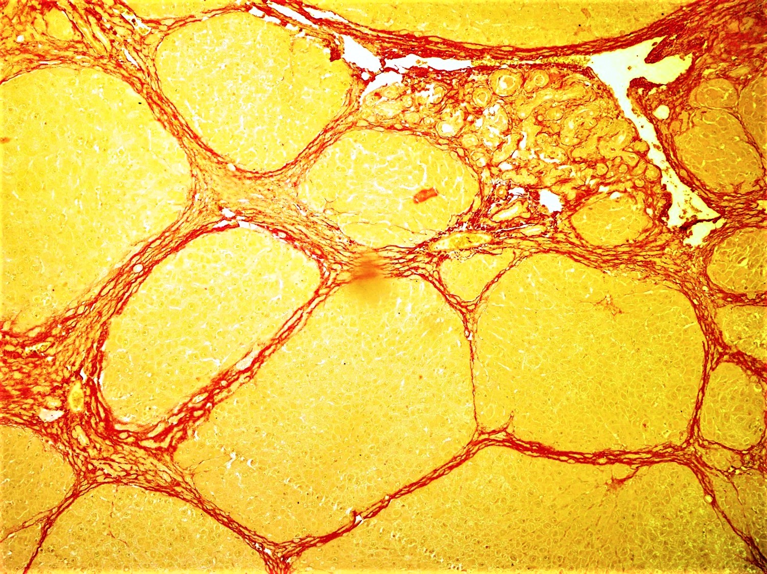
Fibrosis
Fibrosis, also known as fibrotic scarring, is a pathological wound healing in which connective tissue replaces normal parenchymal tissue to the extent that it goes unchecked, leading to considerable tissue remodelling and the formation of permanent scar tissue. Repeated injuries, chronic inflammation and repair are susceptible to fibrosis where an accidental excessive accumulation of extracellular matrix components, such as the collagen is produced by fibroblasts, leading to the formation of a permanent fibrotic scar. In response to injury, this is called scarring, and if fibrosis arises from a single cell line, this is called a fibroma. Physiologically, fibrosis acts to deposit connective tissue, which can interfere with or totally inhibit the normal architecture and function of the underlying organ or tissue. Fibrosis can be used to describe the pathological state of excess deposition of fibrous tissue, as well as the process of connective tissue deposition in healing. Define ... [...More Info...] [...Related Items...] OR: [Wikipedia] [Google] [Baidu] |
Echocardiography
An echocardiography, echocardiogram, cardiac echo or simply an echo, is an ultrasound of the heart. It is a type of medical imaging of the heart, using standard ultrasound or Doppler ultrasound. Echocardiography has become routinely used in the diagnosis, management, and follow-up of patients with any suspected or known heart diseases. It is one of the most widely used diagnostic imaging modalities in cardiology. It can provide a wealth of helpful information, including the size and shape of the heart (internal chamber size quantification), pumping capacity, location and extent of any tissue damage, and assessment of valves. An echocardiogram can also give physicians other estimates of heart function, such as a calculation of the cardiac output, ejection fraction, and diastolic function (how well the heart relaxes). Echocardiography is an important tool in assessing wall motion abnormality in patients with suspected cardiac disease. It is a tool which helps in reaching an ea ... [...More Info...] [...Related Items...] OR: [Wikipedia] [Google] [Baidu] |
Heart Arrhythmia
Arrhythmias, also known as cardiac arrhythmias, heart arrhythmias, or dysrhythmias, are irregularities in the heartbeat, including when it is too fast or too slow. A resting heart rate that is too fast – above 100 beats per minute in adults – is called tachycardia, and a resting heart rate that is too slow – below 60 beats per minute – is called bradycardia. Some types of arrhythmias have no symptoms. Symptoms, when present, may include palpitations or feeling a pause between heartbeats. In more serious cases, there may be lightheadedness, passing out, shortness of breath or chest pain. While most cases of arrhythmia are not serious, some predispose a person to complications such as stroke or heart failure. Others may result in sudden death. Arrhythmias are often categorized into four groups: extra beats, supraventricular tachycardias, ventricular arrhythmias and bradyarrhythmias. Extra beats include premature atrial contractions, premature ventricular contrac ... [...More Info...] [...Related Items...] OR: [Wikipedia] [Google] [Baidu] |
Anaemia
Anemia or anaemia (British English) is a blood disorder in which the blood has a reduced ability to carry oxygen due to a lower than normal number of red blood cells, or a reduction in the amount of hemoglobin. When anemia comes on slowly, the symptoms are often vague, such as tiredness, weakness, shortness of breath, headaches, and a reduced ability to exercise. When anemia is acute, symptoms may include confusion, feeling like one is going to pass out, loss of consciousness, and increased thirst. Anemia must be significant before a person becomes noticeably pale. Symptoms of anemia depend on how quickly hemoglobin decreases. Additional symptoms may occur depending on the underlying cause. Preoperative anemia can increase the risk of needing a blood transfusion following surgery. Anemia can be temporary or long term and can range from mild to severe. Anemia can be caused by blood loss, decreased red blood cell production, and increased red blood cell breakdown. Causes ... [...More Info...] [...Related Items...] OR: [Wikipedia] [Google] [Baidu] |
Ventricle (heart)
A ventricle is one of two large chambers toward the bottom of the heart that collect and expel blood towards the peripheral beds within the body and lungs. The blood pumped by a ventricle is supplied by an atrium (heart), atrium, an adjacent chamber in the upper heart that is smaller than a ventricle. Interventricular means between the ventricles (for example the interventricular septum), while intraventricular means within one ventricle (for example an intraventricular block). In a four-chambered heart, such as that in humans, there are two ventricles that operate in a double circulatory system: the right ventricle pumps blood into the pulmonary circulation to the lungs, and the left ventricle pumps blood into the systemic circulation through the aorta. Structure Ventricles have thicker walls than atria and generate higher blood pressures. The physiological load on the ventricles requiring pumping of blood throughout the body and lungs is much greater than the pressure generated ... [...More Info...] [...Related Items...] OR: [Wikipedia] [Google] [Baidu] |
Hepatosplenomegaly
Hepatosplenomegaly (commonly abbreviated HSM) is the simultaneous enlargement of both the liver (hepatomegaly) and the spleen (splenomegaly). Hepatosplenomegaly can occur as the result of acute viral hepatitis, infectious mononucleosis, and histoplasmosis or it can be the sign of a serious and life-threatening lysosomal storage disease. Systemic venous hypertension can also increase the risk for developing hepatosplenomegaly, which may be seen in those patients with right-sided heart failure. Common causes Rare disorders * Lipoproteinlipase deficiency * Multiple sulfatase deficiency Multiple sulfatase deficiency (MSD), also known as Austin disease, or mucosulfatidosis, is a very rare autosomal recessiveJames, William; Berger, Timothy; Elston, Dirk (2005). ''Andrews' Diseases of the Skin: Clinical Dermatology''. (10th ed.). Sau ... * Osteopetrosis * Adult-onset Still's disease (AOSD) References External links Symptoms and signs: Digestive system and abdomen Med ... [...More Info...] [...Related Items...] OR: [Wikipedia] [Google] [Baidu] |


.png)

