|
Rachischisis
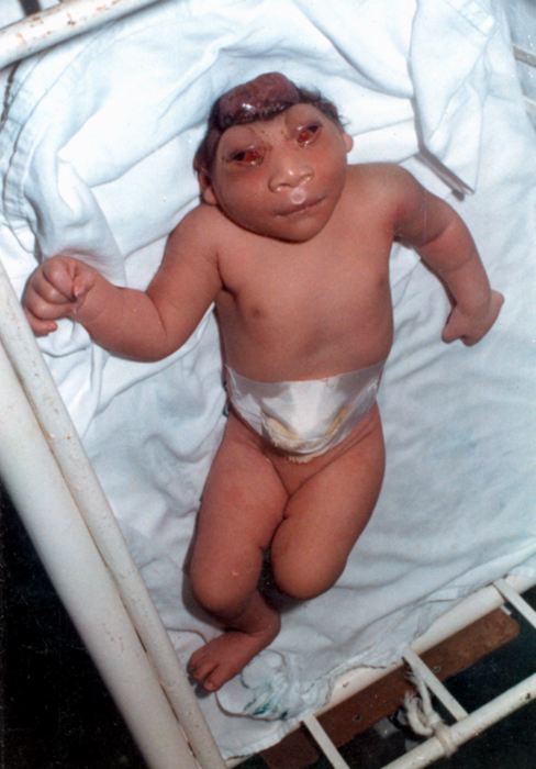
Rachischisis (Greek language, Greek: "rhachis - ῥάχις" - spine, and "schisis - σχίσις" - split) is a developmental birth defect involving the neural tube. This anomaly occurs in utero, when the posterior neuropore of the neural tube fails to close by the 27th intrauterine day. As a consequence the vertebrae overlying the open portion of the spinal cord do not fully form and remain unfused and open, leaving the spinal cord exposed. Patients with rachischisis have motor and sensory deficits, chronic infections, and disturbances in bladder function. This defect often occurs with anencephaly. Craniorachischisis is a variant of rachischisis that occurs when the entire spinal cord and brain are exposed – simultaneous complete rachischisis and anencephaly. It is incompatible with life; affected pregnancies often end in miscarriage or stillbirth. Infants born alive with craniorachischisis die soon after birth. Presentation Interactions with other developmental deformitie ... [...More Info...] [...Related Items...] OR: [Wikipedia] [Google] [Baidu] |
Anencephaly
Anencephaly is the absence of a major portion of the brain, skull, and scalp that occurs during embryonic development. It is a cephalic disorder that results from a neural tube defect that occurs when the rostral (head) end of the neural tube fails to close, usually between the 23rd and 26th day following conception. Strictly speaking, the Greek term translates as "without a brain" (or totally lacking the inside part of the head), but it is accepted that children born with this disorder usually only lack a telencephalon, the largest part of the brain consisting mainly of the cerebral hemispheres, including the neocortex, which is responsible for cognition. The remaining structure is usually covered only by a thin layer of membrane—skin, bone, meninges, etc., are all lacking. With very few exceptions, infants with this disorder do not survive longer than a few hours or days after birth. Signs and symptoms The National Institute of Neurological Disorders and Stroke (NINDS) descr ... [...More Info...] [...Related Items...] OR: [Wikipedia] [Google] [Baidu] |
Spina Bifida
Spina bifida (Latin for 'split spine'; SB) is a birth defect in which there is incomplete closing of the spine and the membranes around the spinal cord during early development in pregnancy. There are three main types: spina bifida occulta, meningocele and myelomeningocele. Meningocele and myelomeningocele may be grouped as spina bifida cystica. The most common location is the lower back, but in rare cases it may be in the middle back or neck. Occulta has no or only mild signs, which may include a hairy patch, dimple, dark spot or swelling on the back at the site of the gap in the spine. Meningocele typically causes mild problems, with a sac of fluid present at the gap in the spine. Myelomeningocele, also known as open spina bifida, is the most severe form. Problems associated with this form include poor ability to walk, impaired bladder or bowel control, accumulation of fluid in the brain (hydrocephalus), a tethered spinal cord and latex allergy. Learning problems are rela ... [...More Info...] [...Related Items...] OR: [Wikipedia] [Google] [Baidu] |
Cranioschisis
Cranioschisis (Greek language, Greek: κρανιον ''kranion'', "skull", and σχίσις ''schisis'', "split"), or dysraphism, is a neural tube defect involving the skull. In this defect, the cranium fails to close completely (especially at the occipital region). Thus, the brain is exposed to the Amnion, amnios and eventually degenerates, causing anencephaly. Craniorachischisis is on the extreme end of the dysraphism spectrum, wherein the entire length of the neural tube fails to close. See also * Rachischisis * Spina bifida References {{Congenital malformations and deformations of musculoskeletal system Congenital disorders of nervous system Congenital disorders of musculoskeletal system ... [...More Info...] [...Related Items...] OR: [Wikipedia] [Google] [Baidu] |
Spina Bifida
Spina bifida (Latin for 'split spine'; SB) is a birth defect in which there is incomplete closing of the spine and the membranes around the spinal cord during early development in pregnancy. There are three main types: spina bifida occulta, meningocele and myelomeningocele. Meningocele and myelomeningocele may be grouped as spina bifida cystica. The most common location is the lower back, but in rare cases it may be in the middle back or neck. Occulta has no or only mild signs, which may include a hairy patch, dimple, dark spot or swelling on the back at the site of the gap in the spine. Meningocele typically causes mild problems, with a sac of fluid present at the gap in the spine. Myelomeningocele, also known as open spina bifida, is the most severe form. Problems associated with this form include poor ability to walk, impaired bladder or bowel control, accumulation of fluid in the brain (hydrocephalus), a tethered spinal cord and latex allergy. Learning problems are rela ... [...More Info...] [...Related Items...] OR: [Wikipedia] [Google] [Baidu] |
Greek Language
Greek ( el, label=Modern Greek, Ελληνικά, Elliniká, ; grc, Ἑλληνική, Hellēnikḗ) is an independent branch of the Indo-European family of languages, native to Greece, Cyprus, southern Italy (Calabria and Salento), southern Albania, and other regions of the Balkans, the Black Sea coast, Asia Minor, and the Eastern Mediterranean. It has the longest documented history of any Indo-European language, spanning at least 3,400 years of written records. Its writing system is the Greek alphabet, which has been used for approximately 2,800 years; previously, Greek was recorded in writing systems such as Linear B and the Cypriot syllabary. The alphabet arose from the Phoenician script and was in turn the basis of the Latin, Cyrillic, Armenian, Coptic, Gothic, and many other writing systems. The Greek language holds a very important place in the history of the Western world. Beginning with the epics of Homer, ancient Greek literature includes many works of lasting impo ... [...More Info...] [...Related Items...] OR: [Wikipedia] [Google] [Baidu] |
Birth Defect
A birth defect, also known as a congenital disorder, is an abnormal condition that is present at birth regardless of its cause. Birth defects may result in disabilities that may be physical, intellectual, or developmental. The disabilities can range from mild to severe. Birth defects are divided into two main types: structural disorders in which problems are seen with the shape of a body part and functional disorders in which problems exist with how a body part works. Functional disorders include metabolic and degenerative disorders. Some birth defects include both structural and functional disorders. Birth defects may result from genetic or chromosomal disorders, exposure to certain medications or chemicals, or certain infections during pregnancy. Risk factors include folate deficiency, drinking alcohol or smoking during pregnancy, poorly controlled diabetes, and a mother over the age of 35 years old. Many are believed to involve multiple factors. Birth defects may be visi ... [...More Info...] [...Related Items...] OR: [Wikipedia] [Google] [Baidu] |
Neural Tube
In the developing chordate (including vertebrates), the neural tube is the embryonic precursor to the central nervous system, which is made up of the brain and spinal cord. The neural groove gradually deepens as the neural fold become elevated, and ultimately the folds meet and coalesce in the middle line and convert the groove into the closed neural tube. In humans, neural tube closure usually occurs by the fourth week of pregnancy (the 28th day after conception). The ectodermal wall of the tube forms the rudiment of the nervous system. The centre of the tube is the ''neural canal''.It is an important structure for the development of fetus's brain and spine Development The neural tube develops in two ways: primary neurulation and secondary neurulation. Primary neurulation divides the ectoderm into three cell types: * The internally located neural tube * The externally located epidermis * The neural crest cells, which develop in the region between the neural tube and epider ... [...More Info...] [...Related Items...] OR: [Wikipedia] [Google] [Baidu] |
Neuropore
Neurulation refers to the folding process in vertebrate embryos, which includes the transformation of the neural plate into the neural tube. The embryo at this stage is termed the neurula. The process begins when the notochord induces the formation of the central nervous system (CNS) by signaling the ectoderm germ layer above it to form the thick and flat neural plate. The neural plate folds in upon itself to form the neural tube, which will later differentiate into the spinal cord and the brain, eventually forming the central nervous system. Computer simulations found that cell wedging and differential proliferation are sufficient for mammalian neurulation. Different portions of the neural tube form by two different processes, called primary and secondary neurulation, in different species. * In primary neurulation, the neural plate creases inward until the edges come in contact and fuse. * In secondary neurulation, the tube forms by hollowing out of the interior of a solid precur ... [...More Info...] [...Related Items...] OR: [Wikipedia] [Google] [Baidu] |
Vertebrae
The spinal column, a defining synapomorphy shared by nearly all vertebrates,Hagfish are believed to have secondarily lost their spinal column is a moderately flexible series of vertebrae (singular vertebra), each constituting a characteristic irregular bone whose complex structure is composed primarily of bone, and secondarily of hyaline cartilage. They show variation in the proportion contributed by these two tissue types; such variations correlate on one hand with the cerebral/caudal rank (i.e., location within the vertebral column, backbone), and on the other with phylogenetic differences among the vertebrate taxon, taxa. The basic configuration of a vertebra varies, but the bone is its ''body'', with the central part of the body constituting the ''centrum''. The upper (closer to) and lower (further from), respectively, the cranium and its central nervous system surfaces of the vertebra body support attachment to the intervertebral discs. The posterior part of a vertebra fo ... [...More Info...] [...Related Items...] OR: [Wikipedia] [Google] [Baidu] |
Acrania
Acrania is a rare congenital disorder that occurs in the human fetus in which the flat bones in the cranial vault are either completely or partially absent. The cerebral hemispheres develop completely but abnormally. The condition is frequently, though not always, associated with anencephaly. The fetus is said to have acrania if it meets the following criteria: the fetus should have a perfectly normal facial bone, a normal cervical column but without the fetal skull and a volume of brain tissue equivalent to at least one-third of the normal brain size. Causes Genetics There are no known family ties in acrania and recurrence rates are extremely low. Not much is known about the exact mechanism involved in acrania. It is hypothesized that, like other developmental malformations, there are multiple origins for acrania. Recent work has identified mutations in the hedgehog acyltransferase (HHAT) gene that have caused acrania along with holoprosencephaly and agnathia. The mutation ... [...More Info...] [...Related Items...] OR: [Wikipedia] [Google] [Baidu] |
Iniencephaly
Iniencephaly is a rare type of cephalic disorder characterised by three common characteristics: a defect to the occipital bone, spina bifida of the cervical vertebrae and retroflexion (backward bending) of the head on the cervical spine. Stillbirth is the most common outcome, with a few rare examples of live birth, after which death invariably occurs within a short time. The disorder was first described by Étienne Geoffroy Saint-Hilaire in 1836. The name is derived from the Ancient Greek word ἰνίον ''inion'', for the occipital bone/nape of the neck. Classifications There are two types of iniencephaly. The more severe group is iniencephaly apertus (open iniencephaly), involving the development of an encephalocele. In the other group, iniencephaly clausus (closed iniencephaly), the encephalocele is absent. Signs and symptoms The affected infant tends to be short, with a disproportionately large head. The fetal head of infants born with iniencephaly are hyperextended whil ... [...More Info...] [...Related Items...] OR: [Wikipedia] [Google] [Baidu] |


