|
RGS2
Regulator of G-protein signaling 2 is a protein that in humans is encoded by the ''RGS2'' gene. It is part of a larger family of RGS proteins that control signalling through G-protein coupled receptors (GPCR). Function RGS2 is thought to have protective effects against myocardial hypertrophy as well as atrial arrhythmias. Increased stimulation of Gs coupled β1-adrenergic receptors and Gq coupled α1-adrenergic receptors in the heart can result in cardiac hypertrophy. In the case of Gq protein coupled receptor (GqPCR) mediated hypertrophy, Gαq will activate the intracellular affectors phospholipase Cβ and rho guanine nucleotide exchange factor to stimulate cell processes which lead to cardiomyocyte hypertrophy. RGS2 functions as a GTPase Activating Protein (GAP) which acts to increase the natural GTPase activity of the Gα subunit. By increasing the GTPase activity of the Gα subunit, RGS2 promotes GTP hydrolysis back to GDP, thus converting the Gα subunit back to its ... [...More Info...] [...Related Items...] OR: [Wikipedia] [Google] [Baidu] |
RGS Protein
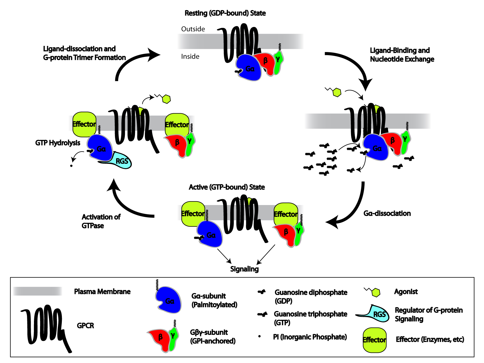
Regulators of G protein signaling (RGS) are protein structural domains or the proteins that contain these domains, that function to activate the GTPase activity of heterotrimeric G-protein G alpha subunit, α-subunits. RGS proteins are multi-functional, GTPase-accelerating proteins that promote GTP hydrolysis by the α-subunit of heterotrimeric G proteins, thereby inactivating the G protein and rapidly switching off G protein-coupled receptor signaling pathways. Upon activation by receptors, G proteins exchange GDP for GTP, are released from the receptor, and dissociate into a free, active GTP-bound α-subunit and G beta-gamma complex, βγ-dimer, both of which activate downstream effectors. The response is terminated upon GTP hydrolysis by the α-subunit (), which can then re-bind the βγ-dimer ( ) and the receptor. RGS proteins markedly reduce the lifespan of GTP-bound α-subunits by stabilising the G protein transition state. Whereas receptors stimulate GTP binding, RGS protein ... [...More Info...] [...Related Items...] OR: [Wikipedia] [Google] [Baidu] |
PRKG1
cGMP-dependent protein kinase 1, alpha isozyme is an enzyme that in humans is encoded by the ''PRKG1'' gene. Interactions PRKG1 has been shown to interact with: * GTF2I, * ITPR1, * MRVI1 Protein MRVI1 is a protein that in humans is encoded by the ''MRVI1'' gene. Function This gene is similar to a mouse putative tumor suppressor gene that is frequently disrupted by mouse AIDS-related virus (MRV). The encoded protein, which is fo ..., * RGS2, and * TNNT1. References Further reading * * * * * * * * * * * * * * * * * * EC 2.7.11 {{gene-10-stub ... [...More Info...] [...Related Items...] OR: [Wikipedia] [Google] [Baidu] |
Gβγ
Heterotrimeric G protein, also sometimes referred to as the ''"large" G proteins'' (as opposed to the subclass of smaller, monomeric small GTPases) are membrane-associated G proteins that form a heterotrimeric complex. The biggest non-structural difference between heterotrimeric and monomeric G protein is that heterotrimeric proteins bind to their cell-surface receptors, called G protein-coupled receptors, directly. These G proteins are made up of ''alpha'' (α), ''beta'' (β) and ''gamma'' (γ) subunits. The alpha subunit is attached to either a GTP or GDP, which serves as an on-off switch for the activation of G-protein. When ligands bind a GPCR, the GPCR acquires GEF (guanine nucleotide exchange factor) ability, which activates the G-protein by exchanging the GDP on the ''alpha'' subunit to GTP. The binding of GTP to the ''alpha'' subunit results in a structural change and its dissociation from the rest of the G-protein. Generally, the ''alpha'' subunit binds membrane-bound ... [...More Info...] [...Related Items...] OR: [Wikipedia] [Google] [Baidu] |
Refractory Period (physiology)
Refractoriness is the fundamental property of any object of autowave nature (especially excitable medium) not to respond on stimuli, if the object stays in the specific ''refractory state''. In common sense, refractory period is the characteristic recovery time, a period that is associated with the motion of the image point on the left branch of the isocline \dot = 0 (for more details, see also Reaction–diffusion and Parabolic partial differential equation). In physiology, a refractory period is a period of time during which an organ or cell is incapable of repeating a particular action, or (more precisely) the amount of time it takes for an excitable membrane to be ready for a second stimulus once it returns to its resting state following an excitation. It most commonly refers to electrically excitable muscle cells or neurons. Absolute refractory period corresponds to depolarization and repolarization, whereas relative refractory period corresponds to hyperpolarization ... [...More Info...] [...Related Items...] OR: [Wikipedia] [Google] [Baidu] |
Voltage-gated Potassium Channel
Voltage-gated potassium channels (VGKCs) are transmembrane channels specific for potassium and sensitive to voltage changes in the cell's membrane potential. During action potentials, they play a crucial role in returning the depolarized cell to a resting state. Classification Alpha subunits Alpha subunits form the actual conductance pore. Based on sequence homology of the hydrophobic transmembrane cores, the alpha subunits of voltage-gated potassium channels are grouped into 12 classes. These are labeled Kvα1-12. The following is a list of the 40 known human voltage-gated potassium channel alpha subunits grouped first according to function and then subgrouped according to the Kv sequence homology classification scheme: Delayed rectifier slowly inactivating or non-inactivating *Kvα1.x - Shaker-related: Kv1.1 (KCNA1), Kv1.2 (KCNA2), Kv1.3 (KCNA3), Kv1.5 (KCNA5), Kv1.6 (KCNA6), Kv1.7 (KCNA7), Kv1.8 ( KCNA10) *Kvα2.x - Shab-related: Kv2.1 ( KCNB1), Kv2.2 ( KCNB2) *Kvα3. ... [...More Info...] [...Related Items...] OR: [Wikipedia] [Google] [Baidu] |
Muscarinic Acetylcholine Receptor M3
The muscarinic acetylcholine receptor, also known as cholinergic/acetylcholine receptor M3, or the muscarinic 3, is a muscarinic acetylcholine receptor encoded by the human gene CHRM3. The M3 muscarinic receptors are located at many places in the body, e.g., smooth muscles, the endocrine glands, the exocrine glands, lungs, pancreas and the brain. In the CNS, they induce emesis. Muscarinic M3 receptors are expressed in regions of the brain that regulate insulin homeostasis, such as the hypothalamus and dorsal vagal complex of the brainstem. These receptors are highly expressed on pancreatic beta cells and are critical regulators of glucose homoestasis by modulating insulin secretion. In general, they cause smooth muscle contraction and increased glandular secretions. They are unresponsive to PTX and CTX. Mechanism Like the M1 muscarinic receptor, M3 receptors are coupled to G proteins of class Gq, which upregulate phospholipase C and, therefore, inositol trisphosphate and ... [...More Info...] [...Related Items...] OR: [Wikipedia] [Google] [Baidu] |
Myocyte
A muscle cell is also known as a myocyte when referring to either a cardiac muscle cell (cardiomyocyte), or a smooth muscle cell as these are both small cells. A skeletal muscle cell is long and threadlike with many nuclei and is called a muscle fiber. Muscle cells (including myocytes and muscle fibers) develop from embryonic precursor cells called myoblasts. Myoblasts fuse to form multinucleated skeletal muscle cells known as syncytia in a process known as myogenesis. Skeletal muscle cells and cardiac muscle cells both contain myofibrils and sarcomeres and form a striated muscle tissue. Cardiac muscle cells form the cardiac muscle in the walls of the heart chambers, and have a single central nucleus. Cardiac muscle cells are joined to neighboring cells by intercalated discs, and when joined in a visible unit they are described as a ''cardiac muscle fiber''. Smooth muscle cells control involuntary movements such as the peristalsis contractions in the esophagus and stoma ... [...More Info...] [...Related Items...] OR: [Wikipedia] [Google] [Baidu] |
Cyclic Adenosine Monophosphate
Cyclic adenosine monophosphate (cAMP, cyclic AMP, or 3',5'-cyclic adenosine monophosphate) is a second messenger important in many biological processes. cAMP is a derivative of adenosine triphosphate (ATP) and used for intracellular signal transduction in many different organisms, conveying the cAMP-dependent pathway. History Earl Sutherland of Vanderbilt University won a Nobel Prize in Physiology or Medicine in 1971 "for his discoveries concerning the mechanisms of the action of hormones", especially epinephrine, via second messengers (such as cyclic adenosine monophosphate, cyclic AMP). Synthesis Cyclic AMP is synthesized from ATP by adenylate cyclase located on the inner side of the plasma membrane and anchored at various locations in the interior of the cell. Adenylate cyclase is ''activated'' by a range of signaling molecules through the activation of adenylate cyclase stimulatory G ( Gs)-protein-coupled receptors. Adenylate cyclase is ''inhibited'' by agonists of adenyl ... [...More Info...] [...Related Items...] OR: [Wikipedia] [Google] [Baidu] |
Adenyl Cyclase
Adenylate cyclase (EC 4.6.1.1, also commonly known as adenyl cyclase and adenylyl cyclase, abbreviated AC) is an enzyme with systematic name ATP diphosphate-lyase (cyclizing; 3′,5′-cyclic-AMP-forming). It catalyzes the following reaction: :ATP = 3′,5′-cyclic AMP + diphosphate It has key regulatory roles in essentially all cells. It is the most polyphyletic known enzyme: six distinct classes have been described, all catalyzing the same reaction but representing unrelated gene families with no known sequence or structural homology. The best known class of adenylyl cyclases is class III or AC-III (Roman numerals are used for classes). AC-III occurs widely in eukaryotes and has important roles in many human tissues. All classes of adenylyl cyclase catalyse the conversion of adenosine triphosphate (ATP) to 3',5'-cyclic AMP (cAMP) and pyrophosphate.Magnesium ions are generally required and appear to be closely involved in the enzymatic mechanism. The cAMP produced by AC ... [...More Info...] [...Related Items...] OR: [Wikipedia] [Google] [Baidu] |
Protein
Proteins are large biomolecules and macromolecules that comprise one or more long chains of amino acid residues. Proteins perform a vast array of functions within organisms, including catalysing metabolic reactions, DNA replication, responding to stimuli, providing structure to cells and organisms, and transporting molecules from one location to another. Proteins differ from one another primarily in their sequence of amino acids, which is dictated by the nucleotide sequence of their genes, and which usually results in protein folding into a specific 3D structure that determines its activity. A linear chain of amino acid residues is called a polypeptide. A protein contains at least one long polypeptide. Short polypeptides, containing less than 20–30 residues, are rarely considered to be proteins and are commonly called peptides. The individual amino acid residues are bonded together by peptide bonds and adjacent amino acid residues. The sequence of amino acid ... [...More Info...] [...Related Items...] OR: [Wikipedia] [Google] [Baidu] |
ARHGEF1
Rho guanine nucleotide exchange factor 1 is a protein that in humans is encoded by the ''ARHGEF1'' gene. This protein is also called RhoGEF1 or p115-RhoGEF. Function Rho guanine nucleotide exchange factor 1 is guanine nucleotide exchange factor (GEF) for the RhoA small GTPase protein. Rho is a small GTPase protein that is inactive when bound to the guanine nucleotide GDP. But when acted on by Rho GEF proteins such as RhoGEF1, this GDP is released and replaced by GTP, leading to the active state of Rho. In this active, GTP-bound conformation, Rho can bind to and activate specific effector proteins and enzymes to regulate cellular functions. In particular, active Rho is a major regulator of the cell actin cytoskeleton. RhoGEF1 is a member of a group of four RhoGEF proteins known to be activated by G protein coupled receptors coupled to the G12 and G13 heterotrimeric G proteins. The others are ARHGEF11 (also known as PDZ-RhoGEF), ARHGEF12 (also known as LARG) and AKAP13 ... [...More Info...] [...Related Items...] OR: [Wikipedia] [Google] [Baidu] |


