|
Gβγ
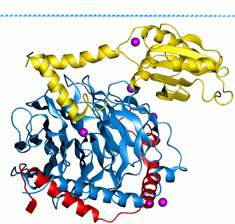
Heterotrimeric G protein, also sometimes referred to as the ''"large" G proteins'' (as opposed to the subclass of smaller, monomeric small GTPases) are membrane-associated G proteins that form a heterotrimeric complex. The biggest non-structural difference between heterotrimeric and monomeric G protein is that heterotrimeric proteins bind to their cell-surface receptors, called G protein-coupled receptors, directly. These G proteins are made up of ''alpha'' (α), ''beta'' (β) and ''gamma'' (γ) subunits. The alpha subunit is attached to either a GTP or GDP, which serves as an on-off switch for the activation of G-protein. When ligands bind a GPCR, the GPCR acquires GEF (guanine nucleotide exchange factor) ability, which activates the G-protein by exchanging the GDP on the ''alpha'' subunit to GTP. The binding of GTP to the ''alpha'' subunit results in a structural change and its dissociation from the rest of the G-protein. Generally, the ''alpha'' subunit binds membrane-bound ... [...More Info...] [...Related Items...] OR: [Wikipedia] [Google] [Baidu] |
Gq Alpha Subunit
Gq protein alpha subunit is a family of heterotrimeric G protein alpha subunits. This family is also commonly called the Gq/11 (Gq/G11) family or Gq/11/14/15 family to include closely related family members. G alpha subunits may be referred to as Gq alpha, Gαq, or Gqα. Gq proteins couple to G protein-coupled receptors to activate beta-type phospholipase C (PLC-β) enzymes. PLC-β in turn hydrolyzes phosphatidylinositol 4,5-bisphosphate (PIP2) to diacyl glycerol (DAG) and inositol trisphosphate (IP3). IP3 acts as a second messenger to release stored calcium into the cytoplasm, while DAG acts as a second messenger that activates protein kinase C (PKC). Family members In humans, there are four distinct proteins in the Gq alpha subunit family: * Gαq is encoded by the gene GNAQ. * Gα11 is encoded by the gene GNA11. * Gα14 is encoded by the gene GNA14. * Gα15 is encoded by the gene GNA15. Function The general function of Gq is to activate intracellular signaling p ... [...More Info...] [...Related Items...] OR: [Wikipedia] [Google] [Baidu] |
GPCR Cycle
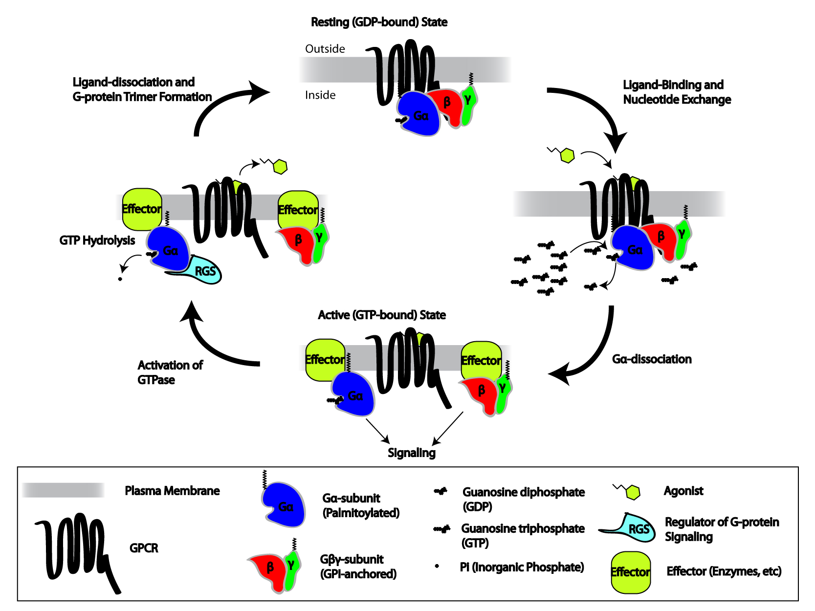
G protein-coupled receptors (GPCRs), also known as seven-(pass)-transmembrane domain receptors, 7TM receptors, heptahelical receptors, serpentine receptors, and G protein-linked receptors (GPLR), form a large group of evolutionarily-related proteins that are cell surface receptors that detect molecules outside the cell and activate cellular responses. Coupling with G proteins, they are called seven-transmembrane receptors because they pass through the cell membrane seven times. Text was copied from this source, which is available under Attribution 2.5 Generic (CC BY 2.5) license. Ligands can bind either to extracellular N-terminus and loops (e.g. glutamate receptors) or to the binding site within transmembrane helices (Rhodopsin-like family). They are all activated by agonists although a spontaneous auto-activation of an empty receptor can also be observed. G protein-coupled receptors are found only in eukaryotes, including yeast, choanoflagellates, and ... [...More Info...] [...Related Items...] OR: [Wikipedia] [Google] [Baidu] |
G Protein-coupled Receptor
G protein-coupled receptors (GPCRs), also known as seven-(pass)-transmembrane domain receptors, 7TM receptors, heptahelical receptors, serpentine receptors, and G protein-linked receptors (GPLR), form a large group of evolutionarily-related proteins that are cell surface receptors that detect molecules outside the cell and activate cellular responses. Coupling with G proteins, they are called seven-transmembrane receptors because they pass through the cell membrane seven times. Text was copied from this source, which is available under Attribution 2.5 Generic (CC BY 2.5) license. Ligands can bind either to extracellular N-terminus and loops (e.g. glutamate receptors) or to the binding site within transmembrane helices (Rhodopsin-like family). They are all activated by agonists although a spontaneous auto-activation of an empty receptor can also be observed. G protein-coupled receptors are found only in eukaryotes, including yeast, choanoflagellates, and ... [...More Info...] [...Related Items...] OR: [Wikipedia] [Google] [Baidu] |
Gi Alpha Subunit
Gi protein alpha subunit is a family of heterotrimeric G protein alpha subunits. This family is also commonly called the Gi/o (Gi /Go ) family or Gi/o/z/t family to include closely related family members. G alpha subunits may be referred to as Gi alpha, Gαi, or Giα. Family members There are four distinct subtypes of alpha subunits in the Gi/o/z/t alpha subunit family that define four families of heterotrimeric G proteins: * Gi proteins: Gi1α, Gi2α, and Gi3α * Go protein: Goα (in mouse there is alternative splicing to generate Go1α and Go2α) * Gz protein: Gzα * Transducins (Gt proteins): Gt1α, Gt2α, Gt3α Giα proteins Gi1α Gi1α is encoded by the gene GNAI1. Gi2α Gi2α is encoded by the gene GNAI2. Gi3α Gi3α is encoded by the gene GNAI3. Goα protein Go1α is encoded by the gene GNAO1. Gzα protein Gzα is encoded by the gene GNAZ. Transducin proteins Gt1α Transducin/Gt1α is encoded by the gene GNAT1. Gt2α Transducin 2/Gt ... [...More Info...] [...Related Items...] OR: [Wikipedia] [Google] [Baidu] |
Gs Alpha Subunit
The Gs alpha subunit (Gαs, Gsα) is a subunit of the heterotrimeric G protein Gs that stimulates the cAMP-dependent pathway by activating adenylyl cyclase. Gsα is a GTPase that functions as a cellular signaling protein. Gsα is the founding member of one of the four families of heterotrimeric G proteins, defined by the alpha subunits they contain: the Gαs family, Gαi/Gαo family, Gαq family, and Gα12/Gα13 family. The Gs-family has only two members: the other member is Golf, named for its predominant expression in the olfactory system. In humans, Gsα is encoded by the GNAS complex locus, while Golfα is encoded by the GNAL gene. Function The general function of Gs is to activate intracellular signaling pathways in response to activation of cell surface G protein-coupled receptors (GPCRs). GPCRs function as part of a three-component system of receptor-transducer-effector. The transducer in this system is a heterotrimeric G protein, composed of three subunits: a Gα ... [...More Info...] [...Related Items...] OR: [Wikipedia] [Google] [Baidu] |
Gustducin
Gustducin is a G protein associated with taste and the gustatory system, found in some taste receptor cells. Research on the discovery and isolation of gustducin is recent. It is known to play a large role in the transduction of bitter, sweet and umami stimuli. Its pathways (especially for detecting bitter stimuli) are many and diverse. An intriguing feature of gustducin is its similarity to transducin. These two G proteins have been shown to be structurally and functionally similar, leading researchers to believe that the sense of taste evolved in a similar fashion to the sense of sight. Gustducin is a heterotrimeric protein composed of the products of the GNAT3 (α-subunit), GNB1 (β-subunit) and GNG13 (γ-subunit). Discovery Gustducin was discovered in 1992 when degenerate oligonucleotide primers were synthesized and mixed with a taste tissue cDNA library. The DNA products were amplified by the polymerase chain reaction method, and eight positive clones were shown to ... [...More Info...] [...Related Items...] OR: [Wikipedia] [Google] [Baidu] |
Transducin
Transducin (Gt) is a protein naturally expressed in vertebrate retina rods and cones and it is very important in vertebrate phototransduction. It is a type of heterotrimeric G-protein with different α subunits in rod and cone photoreceptors. Light leads to conformational changes in rhodopsin, which in turn leads to the activation of transducin. Transducin activates phosphodiesterase, which results in the breakdown of cyclic guanosine monophosphate (cGMP). The intensity of the flash response is directly proportional to the number of transducin activated. Function in phototransduction Transducin is activated by metarhodopsin II, a conformational change in rhodopsin caused by the absorption of a photon by the rhodopsin moiety retinal. The light causes isomerization of retinal from 11-cis to all-trans. Isomerization causes a change in the opsin to become metarhodopsin II. When metarhodopsin activates transducin, the guanosine diphosphate (GDP) bound to the α subunit (Tα) is exc ... [...More Info...] [...Related Items...] OR: [Wikipedia] [Google] [Baidu] |
G Protein
G proteins, also known as guanine nucleotide-binding proteins, are a family of proteins that act as molecular switches inside cells, and are involved in transmitting signals from a variety of stimuli outside a cell to its interior. Their activity is regulated by factors that control their ability to bind to and hydrolyze guanosine triphosphate (GTP) to guanosine diphosphate (GDP). When they are bound to GTP, they are 'on', and, when they are bound to GDP, they are 'off'. G proteins belong to the larger group of enzymes called GTPases. There are two classes of G proteins. The first function as monomeric small GTPases (small G-proteins), while the second function as heterotrimeric G protein complexes. The latter class of complexes is made up of '' alpha'' (α), ''beta'' (β) and ''gamma'' (γ) subunits. In addition, the beta and gamma subunits can form a stable dimeric complex referred to as the beta-gamma complex . Heterotrimeric G proteins located within the cell are activ ... [...More Info...] [...Related Items...] OR: [Wikipedia] [Google] [Baidu] |
G-Protein
G proteins, also known as guanine nucleotide-binding proteins, are a family of proteins that act as molecular switches inside cells, and are involved in transmitting signals from a variety of stimuli outside a cell to its interior. Their activity is regulated by factors that control their ability to bind to and hydrolyze guanosine triphosphate (GTP) to guanosine diphosphate (GDP). When they are bound to GTP, they are 'on', and, when they are bound to GDP, they are 'off'. G proteins belong to the larger group of enzymes called GTPases. There are two classes of G proteins. The first function as monomeric small GTPases (small G-proteins), while the second function as heterotrimeric G protein complexes. The latter class of complexes is made up of ''alpha'' (α), ''beta'' (β) and ''gamma'' (γ) subunits. In addition, the beta and gamma subunits can form a stable dimeric complex referred to as the beta-gamma complex . Heterotrimeric G proteins located within the cell are activa ... [...More Info...] [...Related Items...] OR: [Wikipedia] [Google] [Baidu] |
GNAT1
Guanine nucleotide-binding protein G(t) subunit alpha-1 is a protein that in humans is encoded by the ''GNAT1'' gene. Transducin is a 3-subunit guanine nucleotide-binding protein (G protein) which stimulates the coupling of rhodopsin and cGMP-phosphodiesterase A phosphodiesterase (PDE) is an enzyme that breaks a phosphodiester bond. Usually, ''phosphodiesterase'' refers to cyclic nucleotide phosphodiesterases, which have great clinical significance and are described below. However, there are many oth ... during visual impulses. The transducin alpha subunits in rods and cones are encoded by separate genes. This gene encodes the alpha subunit in rods. Alternative splicing of this gene results in two transcript variants. References Further reading * * * * * * * * * * * * * * * * External links * {{gene-3-stub ... [...More Info...] [...Related Items...] OR: [Wikipedia] [Google] [Baidu] |
GNAT2
Guanine nucleotide-binding protein G(t) subunit alpha-2 is a protein that in humans is encoded by the ''GNAT2'' gene. Function Transducin is a 3-subunit guanine nucleotide-binding protein (G protein) which stimulates the coupling of rhodopsin and cGMP-phosphodiesterase during visual impulses. The transducin alpha subunits in rods and cones A cone is a three-dimensional geometric shape that tapers smoothly from a flat base (frequently, though not necessarily, circular) to a point called the apex or vertex. A cone is formed by a set of line segments, half-lines, or lines conn ... are encoded by separate genes. This gene encodes the alpha subunit in cones. References Further reading * * * * * * * * * * * * * * * * External links GeneReviews/NIH/NCBI/UW entry on Achromatopsia OMIM entries on Achromatopsia {{gene-1-stub ... [...More Info...] [...Related Items...] OR: [Wikipedia] [Google] [Baidu] |
Phosphodiesterase
A phosphodiesterase (PDE) is an enzyme that breaks a phosphodiester bond. Usually, ''phosphodiesterase'' refers to cyclic nucleotide phosphodiesterases, which have great clinical significance and are described below. However, there are many other families of phosphodiesterases, including phospholipases C and D, autotaxin, sphingomyelin phosphodiesterase, DNases, RNases, and restriction endonucleases (which all break the phosphodiester backbone of DNA or RNA), as well as numerous less-well-characterized small-molecule phosphodiesterases. The cyclic nucleotide phosphodiesterases comprise a group of enzymes that degrade the phosphodiester bond in the second messenger molecules cAMP and cGMP. They regulate the localization, duration, and amplitude of cyclic nucleotide signaling within subcellular domains. PDEs are therefore important regulators of signal transduction mediated by these second messenger molecules. History These multiple forms (isoforms or subtypes) of phosphodies ... [...More Info...] [...Related Items...] OR: [Wikipedia] [Google] [Baidu] |


