|
Riga–Fede Disease
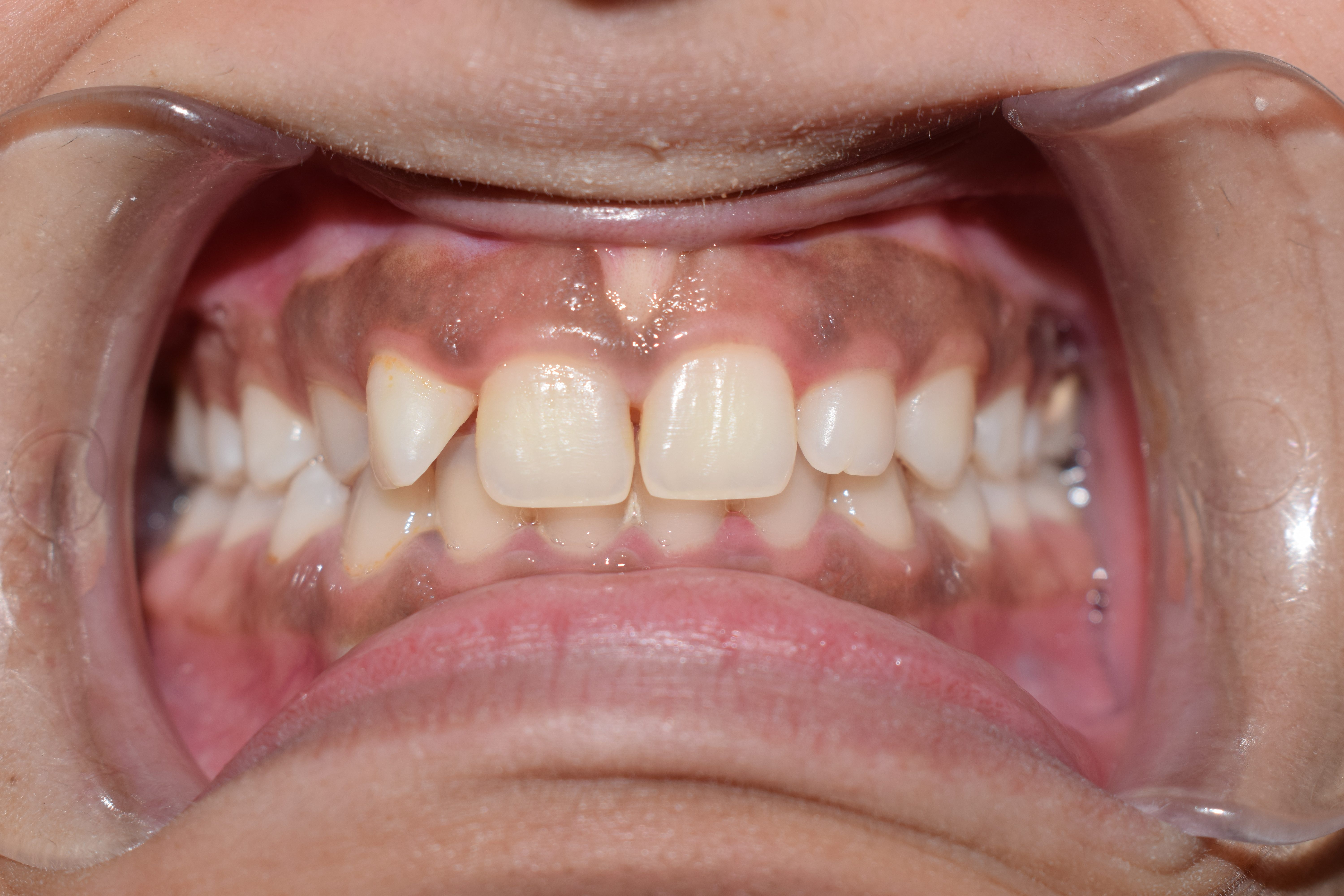
Riga–Fede disease (RFD) is a rare and benign mucosal condition, characterized by a tongue ulcer that is frequently brought on by traumatizing injuries sustained from repeatedly moving the tongue back and forth over the mandibular anterior incisors. Signs and symptoms Clinically, it typically manifests as a tongue-localized ulcer (60 percent of lesions), though it can also affect the lip, palate, gingiva, vestibular mucosa, and floor of the mouth. RFD may not cause any symptoms or may occasionally be accompanied by pain. Causes RFD is most frequently linked to the emergence of the natal-neonatal teeth in newborns or the primary lower incisor in older infants. Diagnosis Histopathologically speaking, RFD is characterized by mucosal areas that are ulcerated, granulation tissue present, and a mixed inflammatory infiltrate that is abundant in mast cells, macrophages, lymphocytes, and eosinophils Eosinophils, sometimes called eosinophiles or, less commonly, acidophils, are a variet ... [...More Info...] [...Related Items...] OR: [Wikipedia] [Google] [Baidu] |
Tongue
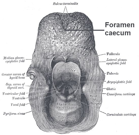
The tongue is a muscular organ (anatomy), organ in the mouth of a typical tetrapod. It manipulates food for mastication and swallowing as part of the digestive system, digestive process, and is the primary organ of taste. The tongue's upper surface (dorsum) is covered by taste buds housed in numerous lingual papillae. It is sensitive and kept moist by saliva and is richly supplied with nerves and blood vessels. The tongue also serves as a natural means of oral hygiene, cleaning the teeth. A major function of the tongue is the enabling of speech in humans and animal communication, vocalization in other animals. The human tongue is divided into two parts, an oral cavity, oral part at the front and a pharynx, pharyngeal part at the back. The left and right sides are also separated along most of its length by a vertical section of connective tissue, fibrous tissue (the lingual septum) that results in a groove, the median sulcus, on the tongue's surface. There are two groups of muscle ... [...More Info...] [...Related Items...] OR: [Wikipedia] [Google] [Baidu] |
Mandible
In anatomy, the mandible, lower jaw or jawbone is the largest, strongest and lowest bone in the human facial skeleton. It forms the lower jaw and holds the lower tooth, teeth in place. The mandible sits beneath the maxilla. It is the only movable bone of the skull (discounting the ossicles of the middle ear). It is connected to the temporal bones by the temporomandibular joints. The bone is formed prenatal development, in the fetus from a fusion of the left and right mandibular prominences, and the point where these sides join, the mandibular symphysis, is still visible as a faint ridge in the midline. Like other symphyses in the body, this is a midline articulation where the bones are joined by fibrocartilage, but this articulation fuses together in early childhood.Illustrated Anatomy of the Head and Neck, Fehrenbach and Herring, Elsevier, 2012, p. 59 The word "mandible" derives from the Latin word ''mandibula'', "jawbone" (literally "one used for chewing"), from ''wikt:mandere ... [...More Info...] [...Related Items...] OR: [Wikipedia] [Google] [Baidu] |
Gingiva
The gums or gingiva (plural: ''gingivae'') consist of the mucosal tissue that lies over the mandible and maxilla inside the mouth. Gum health and disease can have an effect on general health. Structure The gums are part of the soft tissue lining of the mouth. They surround the teeth and provide a seal around them. Unlike the soft tissue linings of the lips and cheeks, most of the gums are tightly bound to the underlying bone which helps resist the friction of food passing over them. Thus when healthy, it presents an effective barrier to the barrage of periodontal insults to deeper tissue. Healthy gums are usually coral pink in light skinned people, and may be naturally darker with melanin pigmentation. Changes in color, particularly increased redness, together with swelling and an increased tendency to bleed, suggest an inflammation that is possibly due to the accumulation of bacterial plaque. Overall, the clinical appearance of the tissue reflects the underlying histology, b ... [...More Info...] [...Related Items...] OR: [Wikipedia] [Google] [Baidu] |
Neonatal Teeth
Natal teeth are teeth that are present above the gumline (have already erupted) at birth, and neonatal teeth are teeth that emerge through the gingiva during the first month of life (the neonatal period). The incidence of neonatal teeth varies considerably, between 1:700 and 1:30,000 depending on the type of study; the highest prevalence is found in the only study that relies on personal examination of patients. Natal teeth, and neonatal teeth, can be the baby's normal deciduous teeth, sprouting prematurely. These should be preserved, if possible. Alternately, they could be supernumary teeth, extra teeth, not part of the normal allotment of teeth. Signs and symptoms Most often natal teeth are mandibular central incisors. They have little root structure and are attached to the end of the gum by soft tissue and are often mobile. Causes Most of the time, natal teeth are not related to a medical condition. However, sometimes they may be associated with: * Ellis–van Creveld syn ... [...More Info...] [...Related Items...] OR: [Wikipedia] [Google] [Baidu] |
Macrophage
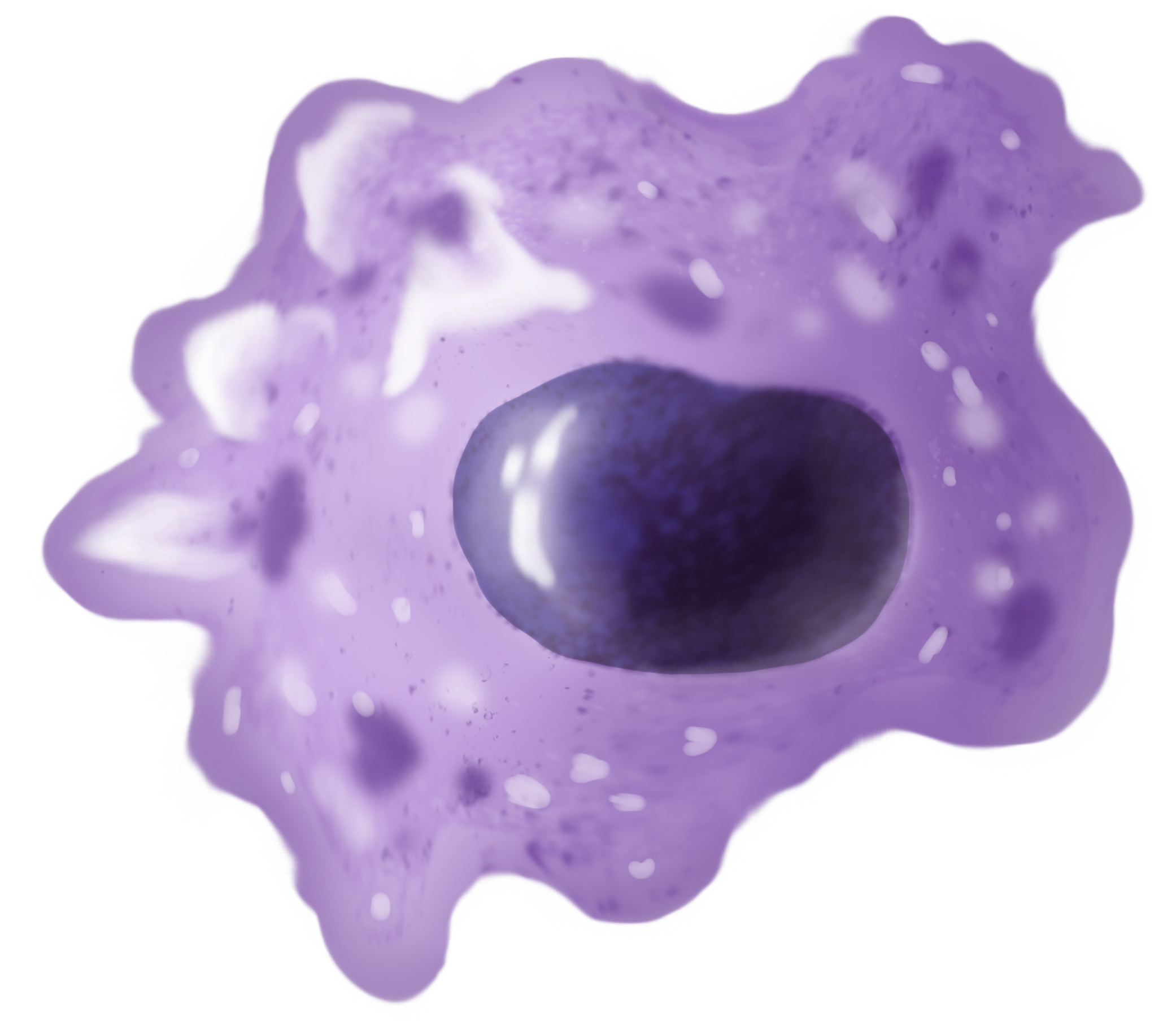
Macrophages (abbreviated as M φ, MΦ or MP) ( el, large eaters, from Greek ''μακρός'' (') = large, ''φαγεῖν'' (') = to eat) are a type of white blood cell of the immune system that engulfs and digests pathogens, such as cancer cells, microbes, cellular debris, and foreign substances, which do not have proteins that are specific to healthy body cells on their surface. The process is called phagocytosis, which acts to defend the host against infection and injury. These large phagocytes are found in essentially all tissues, where they patrol for potential pathogens by amoeboid movement. They take various forms (with various names) throughout the body (e.g., histiocytes, Kupffer cells, alveolar macrophages, microglia, and others), but all are part of the mononuclear phagocyte system. Besides phagocytosis, they play a critical role in nonspecific defense (innate immunity) and also help initiate specific defense mechanisms (adaptive immunity) by recruiting other immune ... [...More Info...] [...Related Items...] OR: [Wikipedia] [Google] [Baidu] |
Lymphocyte
A lymphocyte is a type of white blood cell (leukocyte) in the immune system of most vertebrates. Lymphocytes include natural killer cells (which function in cell-mediated, cytotoxic innate immunity), T cells (for cell-mediated, cytotoxic adaptive immunity), and B cells (for humoral, antibody-driven adaptive immunity). They are the main type of cell found in lymph, which prompted the name "lymphocyte". Lymphocytes make up between 18% and 42% of circulating white blood cells. Types The three major types of lymphocyte are T cells, B cells and natural killer (NK) cells. Lymphocytes can be identified by their large nucleus. T cells and B cells T cells (thymus cells) and B cells ( bone marrow- or bursa-derived cells) are the major cellular components of the adaptive immune response. T cells are involved in cell-mediated immunity, whereas B cells are primarily responsible for humoral immunity (relating to antibodies). The function of T cells and B cells is to recognize sp ... [...More Info...] [...Related Items...] OR: [Wikipedia] [Google] [Baidu] |
Eosinophil
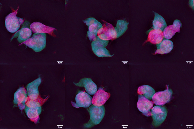
Eosinophils, sometimes called eosinophiles or, less commonly, acidophils, are a variety of white blood cells (WBCs) and one of the immune system components responsible for combating multicellular parasites and certain infections in vertebrates. Along with mast cells and basophils, they also control mechanisms associated with allergy and asthma. They are granulocytes that develop during hematopoiesis in the bone marrow before migrating into blood, after which they are terminally differentiated and do not multiply. They form about 2 to 3% of WBCs. These cells are eosinophilic or "acid-loving" due to their large acidophilic cytoplasmic granules, which show their affinity for acids by their affinity to coal tar dyes: Normally transparent, it is this affinity that causes them to appear brick-red after staining with eosin, a red dye, using the Romanowsky method. The staining is concentrated in small granules within the cellular cytoplasm, which contain many chemical mediators, such ... [...More Info...] [...Related Items...] OR: [Wikipedia] [Google] [Baidu] |
Dental Extraction
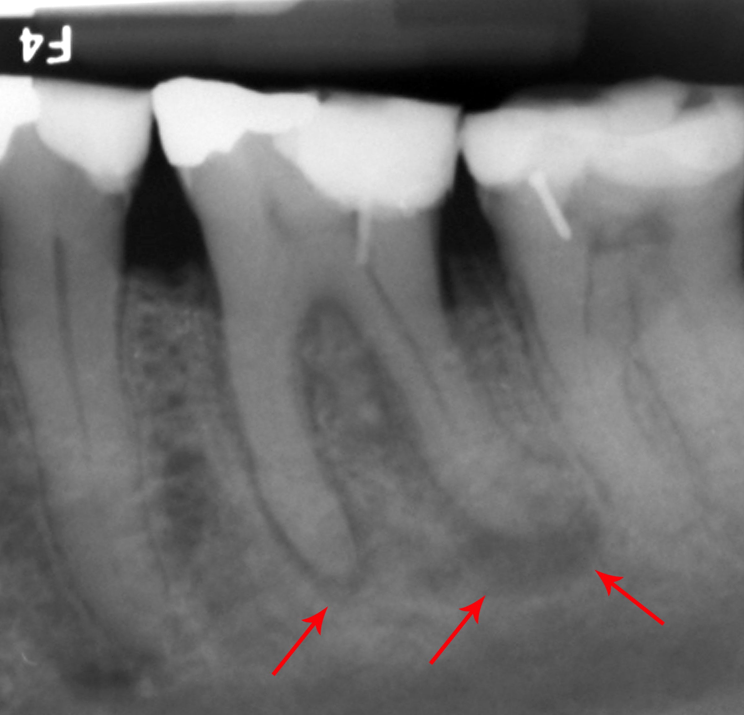
A dental extraction (also referred to as tooth extraction, exodontia, exodontics, or informally, tooth pulling) is the removal of teeth from the dental alveolus (socket) in the alveolar bone. Extractions are performed for a wide variety of reasons, but most commonly to remove teeth which have become unrestorable through tooth decay, periodontal disease, or dental trauma, especially when they are associated with toothache. Sometimes impacted wisdom teeth (wisdom teeth that are stuck and unable to grow normally into the mouth) cause recurrent infections of the gum (pericoronitis), and may be removed when other conservative treatments have failed (cleaning, antibiotics and operculectomy). In orthodontics, if the teeth are crowded, healthy teeth may be extracted (often bicuspids) to create space so the rest of the teeth can be straightened. Procedure Extractions could be categorized into non-surgical (simple) and surgical, depending on the type of tooth to be removed and other fac ... [...More Info...] [...Related Items...] OR: [Wikipedia] [Google] [Baidu] |
Oral Mucosal Pathology
The word oral may refer to: Relating to the mouth * Relating to the mouth, the first portion of the alimentary canal that primarily receives food and liquid **Oral administration of medicines ** Oral examination (also known as an oral exam or oral test), a practice in many schools and disciplines in which an examiner poses questions to the student in spoken form ** Oral hygiene, practices involved in cleaning the mouth and preventing disease **Oral medication **Oral rehydration therapy, a simple treatment for dehydration associated with diarrhea **Oral sex, sexual activity involving the stimulation of genitalia by use of the mouth, tongue, teeth or throat. **Oral stage, a human development phase in Freudian developmental psychology **Oral tradition, cultural material and tradition transmitted orally from one generation to another **Oralism, the education of deaf students through oral language by using lip reading, and mimicking of mouth shapes and breathing patterns **Speech commu ... [...More Info...] [...Related Items...] OR: [Wikipedia] [Google] [Baidu] |
Tongue Disorders
The tongue is a muscular organ in the mouth of a typical tetrapod. It manipulates food for mastication and swallowing as part of the digestive process, and is the primary organ of taste. The tongue's upper surface (dorsum) is covered by taste buds housed in numerous lingual papillae. It is sensitive and kept moist by saliva and is richly supplied with nerves and blood vessels. The tongue also serves as a natural means of cleaning the teeth. A major function of the tongue is the enabling of speech in humans and vocalization in other animals. The human tongue is divided into two parts, an oral part at the front and a pharyngeal part at the back. The left and right sides are also separated along most of its length by a vertical section of fibrous tissue (the lingual septum) that results in a groove, the median sulcus, on the tongue's surface. There are two groups of muscles of the tongue. The four intrinsic muscles alter the shape of the tongue and are not attached to bone. The f ... [...More Info...] [...Related Items...] OR: [Wikipedia] [Google] [Baidu] |

