|
Renal Lobe
The renal lobe is a portion of a kidney consisting of a renal pyramid and the renal cortex above it. In humans, on average there are 7 to 18 renal lobes. It is visible without a microscope, though it is easier to see in humans than in other animals. It is composed of many renal lobules, which are not visible without a microscope. See also * Renal capsule * Renal medulla The renal medulla (Latin: ''medulla renis'' 'marrow of the kidney') is the innermost part of the kidney. The renal medulla is split up into a number of sections, known as the renal pyramids. Blood enters into the kidney via the renal artery, which ... References External links * Kidney anatomy {{Portal bar, Anatomy ... [...More Info...] [...Related Items...] OR: [Wikipedia] [Google] [Baidu] |
Renal Pyramids
The renal medulla (Latin: ''medulla renis'' 'marrow of the kidney') is the innermost part of the kidney. The renal medulla is split up into a number of sections, known as the renal pyramids. Blood enters into the kidney via the renal artery, which then splits up to form the segmental arteries which then branch to form interlobar arteries. The interlobar arteries each in turn branch into arcuate arteries, which in turn branch to form interlobular arteries, and these finally reach the glomeruli. At the glomerulus the blood reaches a highly disfavourable pressure gradient and a large exchange surface area, which forces the serum portion of the blood out of the vessel and into the renal tubules. Flow continues through the renal tubules, including the proximal tubule, the loop of Henle, through the distal tubule and finally leaves the kidney by means of the collecting duct, leading to the renal pelvis, the dilated portion of the ureter. The renal medulla contains the structures of t ... [...More Info...] [...Related Items...] OR: [Wikipedia] [Google] [Baidu] |
Renal Sinus
The renal sinus is a cavity within the kidney which is occupied by the renal pelvis, renal calyces, blood vessels, nerves and fat. The renal hilum extends into a large cavity within the kidney occupied by the renal vessels, minor renal calyces, major renal calyces, renal pelvis and some adipose tissue Adipose tissue (also known as body fat or simply fat) is a loose connective tissue composed mostly of adipocytes. It also contains the stromal vascular fraction (SVF) of cells including preadipocytes, fibroblasts, Blood vessel, vascular endothel .... Additional images File:Slide3iii.JPG, Renal sinus File:Slide22iii.JPG, Renal sinus References Kidney anatomy {{genitourinary-stub ... [...More Info...] [...Related Items...] OR: [Wikipedia] [Google] [Baidu] |
Renal Lobules
A cortical lobule (or renal lobule) is a part of a renal lobe. It consists of the nephrons grouped around a single medullary ray, and draining into a single collecting duct. Its near identical parallel is the rectal The rectum (: rectums or recta) is the final straight portion of the large intestine in humans and some other mammals, and the Gastrointestinal tract, gut in others. Before expulsion through the anus or cloaca, the rectum stores the feces te ... lobe, which is present in the majority of mammals. References External links * Kidney anatomy {{Portal bar, Anatomy ... [...More Info...] [...Related Items...] OR: [Wikipedia] [Google] [Baidu] |
Microscope
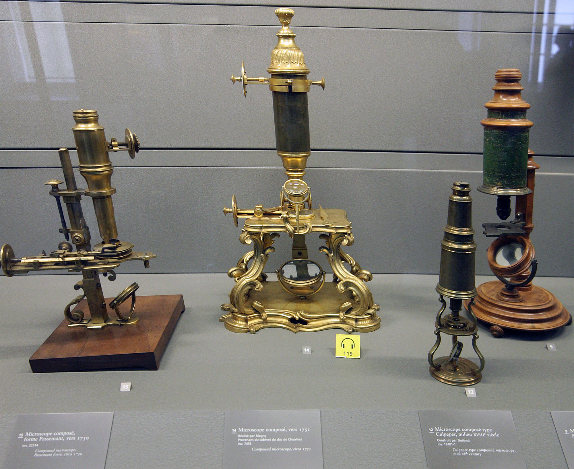
A microscope () is a laboratory equipment, laboratory instrument used to examine objects that are too small to be seen by the naked eye. Microscopy is the science of investigating small objects and structures using a microscope. Microscopic means being invisible to the eye unless aided by a microscope. There are many types of microscopes, and they may be grouped in different ways. One way is to describe the method an instrument uses to interact with a sample and produce images, either by sending a beam of light or electrons through a sample in its optical path, by detecting fluorescence, photon emissions from a sample, or by scanning across and a short distance from the surface of a sample using a probe. The most common microscope (and the first to be invented) is the optical microscope, which uses lenses to refract visible light that passed through a microtome, thinly sectioned sample to produce an observable image. Other major types of microscopes are the fluorescence micro ... [...More Info...] [...Related Items...] OR: [Wikipedia] [Google] [Baidu] |
Renal Cortex
The renal cortex is the outer portion of the kidney between the renal capsule and the renal medulla. In the adult, it forms a continuous smooth outer zone with a number of projections ( cortical columns) that extend down between the pyramids. It contains the renal corpuscles and the renal tubules except for parts of the loop of Henle which descend into the renal medulla. It also contains blood vessels and cortical collecting ducts. The renal cortex is the part of the kidney where ultrafiltration occurs. Erythropoietin is produced in the renal cortex. Additional images File:Njuren.gif, Kidney File:Kidney-Cortex.JPG, Microscopic cross section of the renal cortex File:Kidney_cd10_ihc.jpg, CD10 immunohistochemical staining of normal kidney In humans, the kidneys are two reddish-brown bean-shaped blood-filtering organ (anatomy), organs that are a multilobar, multipapillary form of mammalian kidneys, usually without signs of external lobulation. They are located on the le ... [...More Info...] [...Related Items...] OR: [Wikipedia] [Google] [Baidu] |
Renal Pyramid
The renal medulla (Latin: ''medulla renis'' 'marrow of the kidney') is the innermost part of the kidney. The renal medulla is split up into a number of sections, known as the renal pyramids. Blood enters into the kidney via the renal artery, which then splits up to form the segmental arteries which then branch to form interlobar arteries. The interlobar arteries each in turn branch into arcuate arteries, which in turn branch to form interlobular arteries, and these finally reach the glomerulus (kidney), glomeruli. At the glomerulus the blood reaches a highly disfavourable pressure gradient and a large exchange surface area, which forces the serum (blood), serum portion of the blood out of the vessel and into the renal tubules. Flow continues through the renal tubules, including the proximal tubule, the loop of Henle, through the distal tubule and finally leaves the kidney by means of the collecting duct, leading to the renal pelvis, the dilated portion of the ureter. The renal med ... [...More Info...] [...Related Items...] OR: [Wikipedia] [Google] [Baidu] |
Kidney
In humans, the kidneys are two reddish-brown bean-shaped blood-filtering organ (anatomy), organs that are a multilobar, multipapillary form of mammalian kidneys, usually without signs of external lobulation. They are located on the left and right in the retroperitoneal space, and in adult humans are about in length. They receive blood from the paired renal artery, renal arteries; blood exits into the paired renal veins. Each kidney is attached to a ureter, a tube that carries excreted urine to the urinary bladder, bladder. The kidney participates in the control of the volume of various body fluids, fluid osmolality, Acid-base homeostasis, acid-base balance, various electrolyte concentrations, and removal of toxins. Filtration occurs in the glomerulus (kidney), glomerulus: one-fifth of the blood volume that enters the kidneys is filtered. Examples of substances reabsorbed are solute-free water, sodium, bicarbonate, glucose, and amino acids. Examples of substances secreted are hy ... [...More Info...] [...Related Items...] OR: [Wikipedia] [Google] [Baidu] |
Interlobar Veins
The interlobar veins are veins of the renal circulation The renal circulation supplies the blood to the kidneys via the renal artery, renal arteries, left and right, which branch directly from the abdominal aorta. Despite their relatively small size, the kidneys receive approximately 20% of the cardiac ... which drain the renal lobes. They collect blood from the arcuate veins. The interlobar veins unite to form a renal vein. Each interlobar vein passes along the edge of the renal pyramids. References External links * - "Renal Vasculature: Efferent Arterioles & Peritubular Capillaries" * - "Urinary System: neonatal kidney, vasculature" Kidney anatomy Thoracic veins {{circulatory-stub ... [...More Info...] [...Related Items...] OR: [Wikipedia] [Google] [Baidu] |
Interlobar Arteries
The interlobar arteries are vessels of the renal circulation The renal circulation supplies the blood to the kidneys via the renal artery, renal arteries, left and right, which branch directly from the abdominal aorta. Despite their relatively small size, the kidneys receive approximately 20% of the cardiac ... which supply the renal lobes. The interlobar arteries branch from the lobar arteries which branch from the segmental arteries, from the renal artery. They give rise to arcuate arteries. References External links * - "Renal Vasculature: Efferent Arterioles & Peritubular Capillaries" * - "Urinary System: neonatal kidney, vasculature" Diagram at eku.edu* * Kidney anatomy {{circulatory-stub ... [...More Info...] [...Related Items...] OR: [Wikipedia] [Google] [Baidu] |
Urinary System
The human urinary system, also known as the urinary tract or renal system, consists of the kidneys, ureters, urinary bladder, bladder, and the urethra. The purpose of the urinary system is to eliminate waste from the body, regulate blood volume and blood pressure, control levels of Electrolyte, electrolytes and Metabolite, metabolites, and regulate Acid–base homeostasis, blood pH. The urinary tract is the body's drainage system for the eventual removal of urine. The kidneys have an extensive blood supply via the Renal artery, renal arteries which leave the kidneys via the renal vein. Each kidney consists of functional units called nephrons. Following filtration of blood and further processing, waste (in the form of urine) exits the kidney via the ureters, tubes made of smooth muscle fibres that propel urine towards the urinary bladder, where it is stored and subsequently expelled through the urethra during urination. The female and male urinary system are very similar, differin ... [...More Info...] [...Related Items...] OR: [Wikipedia] [Google] [Baidu] |
Renal Column
The renal columns, Bertin columns, or columns of Bertin, a.k.a. columns of Bertini are extensions of the renal cortex in between the renal pyramids. They allow the cortex to be better anchored. (Cortical extensions into the medullary space.) Each column consists of lines of blood vessels and urinary tubes and a fibrous material. A hypertrophied renal column (or ''renal pseudotumor'') may be differentiated from an actual renal tumor with the help of a DMSA scan. The scan will show the area as one with normal activity if it is a pseudotumor or will show decreased uptake if it is a cystic or solid renal mass. See also * Renal pyramids * Renal papilla * Renal medulla The renal medulla (Latin: ''medulla renis'' 'marrow of the kidney') is the innermost part of the kidney. The renal medulla is split up into a number of sections, known as the renal pyramids. Blood enters into the kidney via the renal artery, which ... Additional images File:Slide12iii.JPG, Renal column File:Slide1 ... [...More Info...] [...Related Items...] OR: [Wikipedia] [Google] [Baidu] |
Renal Papilla
In humans, the kidneys are two reddish-brown bean-shaped blood-filtering organs that are a multilobar, multipapillary form of mammalian kidneys, usually without signs of external lobulation. They are located on the left and right in the retroperitoneal space, and in adult humans are about in length. They receive blood from the paired renal arteries; blood exits into the paired renal veins. Each kidney is attached to a ureter, a tube that carries excreted urine to the bladder. The kidney participates in the control of the volume of various body fluids, fluid osmolality, acid-base balance, various electrolyte concentrations, and removal of toxins. Filtration occurs in the glomerulus: one-fifth of the blood volume that enters the kidneys is filtered. Examples of substances reabsorbed are solute-free water, sodium, bicarbonate, glucose, and amino acids. Examples of substances secreted are hydrogen, ammonium, potassium and uric acid. The nephron is the structural and func ... [...More Info...] [...Related Items...] OR: [Wikipedia] [Google] [Baidu] |

