|
RESOLFT
RESOLFT, an acronym for REversible Saturable OpticaL Fluorescence Transitions, denotes a group of optical fluorescence microscopy techniques with very high resolution. Using standard far field visible light optics a resolution far below the diffraction limit down to molecular scales can be obtained. With conventional microscopy techniques, it is not possible to distinguish features that are located at distances less than about half the wavelength used (i.e. about 200 nm for visible light). This diffraction limit is based on the wave nature of light. In conventional microscopes the limit is determined by the used wavelength and the numerical aperture of the optical system. The RESOLFT concept surmounts this limit by temporarily switching the molecules to a state in which they cannot send a (fluorescence-) signal upon illumination. This concept is different from for example electron microscopy where instead the used wavelength is much smaller. Working principle RESOLFT micro ... [...More Info...] [...Related Items...] OR: [Wikipedia] [Google] [Baidu] |
RESOLFT Principle
RESOLFT, an acronym for REversible Saturable OpticaL Fluorescence Transitions, denotes a group of optical fluorescence microscopy techniques with very high resolution. Using standard far field visible light optics a resolution far below the diffraction limit down to molecular scales can be obtained. With conventional microscopy techniques, it is not possible to distinguish features that are located at distances less than about half the wavelength used (i.e. about 200 nm for visible light). This diffraction limit is based on the wave nature of light. In conventional microscopes the limit is determined by the used wavelength and the numerical aperture of the optical system. The RESOLFT concept surmounts this limit by temporarily switching the molecules to a state in which they cannot send a (fluorescence-) signal upon illumination. This concept is different from for example electron microscopy where instead the used wavelength is much smaller. Working principle RESOLFT microsco ... [...More Info...] [...Related Items...] OR: [Wikipedia] [Google] [Baidu] |
4Pi Microscope
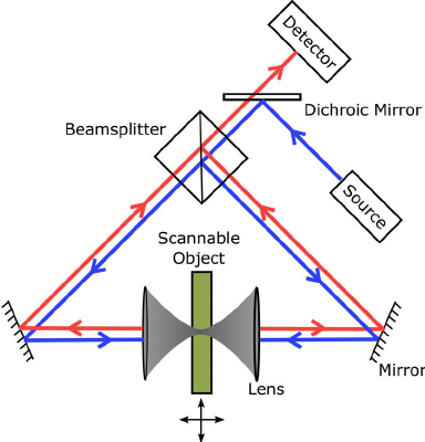
A 4Pi microscope is a laser scanning fluorescence microscope with an improved optical axis, axial Optical resolution, resolution. With it the typical range of the axial resolution of 500–700 nm can be improved to 100–150 nm, which corresponds to an almost spherical focal spot with 5–7 times less volume than that of standard confocal microscopy. Working principle The improvement in resolution is achieved by using two opposing objective lenses, which both are focused to the same geometrical location. Also the difference in optical path length through each of the two objective lenses is carefully aligned to be minimal. By this method, molecules residing in the common focal area of both objectives can be illuminated coherently from both sides and the reflected or emitted light can also be collected coherently, i.e. coherent superposition of emitted light on the detector is possible. The solid angle \Omega that is used for illumination and detection is increased and appro ... [...More Info...] [...Related Items...] OR: [Wikipedia] [Google] [Baidu] |
GSD Microscopy
Ground state depletion microscopy (GSD Microscopy) is an implementation of the RESOLFT concept. The method was proposed in 1995 and experimentally demonstrated in 2007. It is the second concept to overcome the diffraction barrier in far-field optical microscopy published by Stefan Hell. Using nitrogen-vacancy centers in diamonds a resolution of up to 7.8 nm was achieved in 2009. This is far below the diffraction limit (~200 nm). Principle In GSD microscopy, fluorescent markers are used. In one condition, the marker can freely be excited from ground state and returns spontaneously via emission of a fluorescence photon. However, if light of appropriate wavelength is additionally applied the dye can be excited to a long-lived dark state, i.e. a state where no fluorescence occurs. As long as the molecule is in the long-lived dark state (e.g. a triplet state In quantum mechanics, a triplet is a quantum state of a system with a spin of quantum number =1, such that there a ... [...More Info...] [...Related Items...] OR: [Wikipedia] [Google] [Baidu] |
STED Microscopy
Stimulated emission depletion (STED) microscopy is one of the techniques that make up super-resolution microscopy. It creates super-resolution images by the selective deactivation of fluorophores, minimizing the area of illumination at the focal point, and thus enhancing the achievable resolution for a given system. It was developed by Stefan W. Hell and Jan Wichmann in 1994, and was first experimentally demonstrated by Hell and Thomas Klar in 1999. Hell was awarded the Nobel Prize in Chemistry in 2014 for its development. In 1986, V.A. Okhonin (Institute of Biophysics, USSR Academy of Sciences, Siberian Branch, Krasnoyarsk) had patented the STED idea. This patent was unknown to Hell and Wichmann in 1994. STED microscopy is one of several types of super resolution microscopy techniques that have recently been developed to bypass the diffraction limit of light microscopy to increase resolution. STED is a deterministic functional technique that exploits the non-linear response of ... [...More Info...] [...Related Items...] OR: [Wikipedia] [Google] [Baidu] |
Confocal Microscopy
Confocal microscopy, most frequently confocal laser scanning microscopy (CLSM) or laser confocal scanning microscopy (LCSM), is an optical imaging technique for increasing optical resolution and contrast of a micrograph by means of using a spatial pinhole to block out-of-focus light in image formation. Capturing multiple two-dimensional images at different depths in a sample enables the reconstruction of three-dimensional structures (a process known as optical sectioning) within an object. This technique is used extensively in the scientific and industrial communities and typical applications are in life sciences, semiconductor inspection and materials science. Light travels through the sample under a conventional microscope as far into the specimen as it can penetrate, while a confocal microscope only focuses a smaller beam of light at one narrow depth level at a time. The CLSM achieves a controlled and highly limited depth of field. Basic concept The principle of co ... [...More Info...] [...Related Items...] OR: [Wikipedia] [Google] [Baidu] |
Green Fluorescent Protein
The green fluorescent protein (GFP) is a protein that exhibits bright green fluorescence when exposed to light in the blue to ultraviolet range. The label ''GFP'' traditionally refers to the protein first isolated from the jellyfish ''Aequorea victoria'' and is sometimes called ''avGFP''. However, GFPs have been found in other organisms including corals, sea anemones, zoanithids, copepods and lancelets. The GFP from ''A. victoria'' has a major excitation peak at a wavelength of 395 nm and a minor one at 475 nm. Its emission peak is at 509 nm, which is in the lower green portion of the visible spectrum. The fluorescence quantum yield (QY) of GFP is 0.79. The GFP from the sea pansy (''Renilla reniformis'') has a single major excitation peak at 498 nm. GFP makes for an excellent tool in many forms of biology due to its ability to form an internal chromophore without requiring any accessory cofactors, gene products, or enzymes / substrates other than mo ... [...More Info...] [...Related Items...] OR: [Wikipedia] [Google] [Baidu] |
Stimulated Emission
Stimulated emission is the process by which an incoming photon of a specific frequency can interact with an excited atomic electron (or other excited molecular state), causing it to drop to a lower energy level. The liberated energy transfers to the electromagnetic field, creating a new photon with a frequency, polarization, and direction of travel that are all identical to the photons of the incident wave. This is in contrast to spontaneous emission, which occurs at a characteristic rate for each of the atoms/oscillators in the upper energy state regardless of the external electromagnetic field. According to the American Physical Society, the first person to correctly predict the phenomenon of stimulated emission was Albert Einstein in a series of papers starting in 1916, culminating in what is now called the Einstein B Coefficient. Einstein's work became the theoretical foundation of the MASER and LASER. The process is identical in form to atomic absorption in which the energ ... [...More Info...] [...Related Items...] OR: [Wikipedia] [Google] [Baidu] |
Ernst Karl Abbe
Ernst Karl Abbe HonFRMS (23 January 1840 – 14 January 1905) was a German physicist, optical scientist, entrepreneur, and social reformer. Together with Otto Schott and Carl Zeiss, he developed numerous optical instruments. He was also a co-owner of Carl Zeiss AG, a German manufacturer of scientific microscopes, astronomical telescopes, planetariums, and other advanced optical systems. Personal life Abbe was born 23 January 1840 in Eisenach, Saxe-Weimar-Eisenach, to Georg Adam Abbe and Elisabeth Christina Barchfeldt. He came from a humble home – his father was a foreman in a spinnery. Supported by his father's employer, Abbe was able to attend secondary school and to obtain the general qualification for university entrance with fairly good grades, at the Eisenach Gymnasium, which he graduated from in 1857. By the time he left school, his scientific talent and his strong will had already become obvious. Thus, in spite of the family's strained financial situation, his father ... [...More Info...] [...Related Items...] OR: [Wikipedia] [Google] [Baidu] |
Confocal Laser Scanning Microscopy
Confocal microscopy, most frequently confocal laser scanning microscopy (CLSM) or laser confocal scanning microscopy (LCSM), is an optical imaging technique for increasing optical resolution and contrast of a micrograph by means of using a spatial pinhole to block out-of-focus light in image formation. Capturing multiple two-dimensional images at different depths in a sample enables the reconstruction of three-dimensional structures (a process known as optical sectioning) within an object. This technique is used extensively in the scientific and industrial communities and typical applications are in life sciences, semiconductor inspection and materials science. Light travels through the sample under a conventional microscope as far into the specimen as it can penetrate, while a confocal microscope only focuses a smaller beam of light at one narrow depth level at a time. The CLSM achieves a controlled and highly limited depth of field. Basic concept The principle of ... [...More Info...] [...Related Items...] OR: [Wikipedia] [Google] [Baidu] |
Far Field
The near field and far field are regions of the electromagnetic (EM) field around an object, such as a transmitting antenna, or the result of radiation scattering off an object. Non-radiative ''near-field'' behaviors dominate close to the antenna or scattering object, while electromagnetic radiation ''far-field'' behaviors dominate at greater distances. Far-field E (electric) and B (magnetic) field strength decreases as the distance from the source increases, resulting in an inverse-square law for the radiated ''power'' intensity of electromagnetic radiation. By contrast, near-field E and B strength decrease more rapidly with distance: the radiative field decreases by the inverse-distance squared, the reactive field by an inverse-cube law, resulting in a diminished power in the parts of the electric field by an inverse fourth-power and sixth-power, respectively. The rapid drop in power contained in the near-field ensures that effects due to the near-field essentially vanish a ... [...More Info...] [...Related Items...] OR: [Wikipedia] [Google] [Baidu] |
Microscopy
Microscopy is the technical field of using microscopes to view objects and areas of objects that cannot be seen with the naked eye (objects that are not within the resolution range of the normal eye). There are three well-known branches of microscopy: optical, electron, and scanning probe microscopy, along with the emerging field of X-ray microscopy. Optical microscopy and electron microscopy involve the diffraction, reflection, or refraction of electromagnetic radiation/electron beams interacting with the specimen, and the collection of the scattered radiation or another signal in order to create an image. This process may be carried out by wide-field irradiation of the sample (for example standard light microscopy and transmission electron microscopy) or by scanning a fine beam over the sample (for example confocal laser scanning microscopy and scanning electron microscopy). Scanning probe microscopy involves the interaction of a scanning probe with the surface of the objec ... [...More Info...] [...Related Items...] OR: [Wikipedia] [Google] [Baidu] |
Superresolution Microscopy 02
Super-resolution imaging (SR) is a class of techniques that enhance (increase) the resolution of an imaging system. In optical SR the diffraction limit of systems is transcended, while in geometrical SR the resolution of digital imaging sensors is enhanced. In some radar and sonar imaging applications (e.g. magnetic resonance imaging (MRI), high-resolution computed tomography), subspace decomposition-based methods (e.g. MUSIC) and compressed sensing-based algorithms (e.g., SAMV) are employed to achieve SR over standard periodogram algorithm. Super-resolution imaging techniques are used in general image processing and in super-resolution microscopy. Basic concepts Because some of the ideas surrounding super-resolution raise fundamental issues, there is need at the outset to examine the relevant physical and information-theoretical principles: * Diffraction limit: The detail of a physical object that an optical instrument can reproduce in an image has limits that are mandated b ... [...More Info...] [...Related Items...] OR: [Wikipedia] [Google] [Baidu] |


