|
REC8
Meiotic recombination protein REC8 homolog is a protein that in humans is encoded by the ''REC8'' gene. Rec8 is a meiosis-specific component of the cohesin complex that binds sister chromatids in preparation for the two divisions of meiosis. Rec8 is sequentially removed from sister chromatids. It is removed from the arms of chromosomes in the first division - separating homologous chromosomes from each other. However, Rec8 is maintained at centromeres so that sister chromatids are kept joined until anaphase of meiosis II, at which point removal of remaining cohesin leads to the separation of sister chromatids. Function This gene encodes a member of the kleisin family of SMC (structural maintenance of chromosome) protein partners. The protein localizes to the axial elements of chromosomes during meiosis in both oocytes and spermatocytes. REC8 protein appears to participate with other cohesins STAG3, SMC1ß and SMC3 in sister chromatid cohesion throughout the whole meiotic proc ... [...More Info...] [...Related Items...] OR: [Wikipedia] [Google] [Baidu] |
Establishment Of Sister Chromatid Cohesion
Sister chromatid cohesion refers to the process by which sister chromatids are paired and held together during certain phases of the cell cycle. Establishment of sister chromatid cohesion is the process by which chromatin-associated cohesin protein becomes competent to physically bind together the sister chromatids. In general, cohesion is established during S phase as DNA is replicated, and is lost when chromosomes segregate during mitosis and meiosis. Some studies have suggested that cohesion aids in aligning the kinetochores during mitosis by forcing the kinetochores to face opposite cell poles. Cohesin loading Cohesin first associates with the chromosomes during G1 phase. The cohesin ring is composed of two SMC (structural maintenance of chromosomes) proteins and two additional Scc proteins. Cohesin may originally interact with chromosomes via the ATPase domains of the SMC proteins. In yeast, the loading of cohesin on the chromosomes depends on proteins Scc2 and Scc4. Cohesin ... [...More Info...] [...Related Items...] OR: [Wikipedia] [Google] [Baidu] |
Cohesin
Cohesin is a protein complex that mediates sister chromatid cohesion, homologous recombination, and DNA looping. Cohesin is formed of SMC3, SMC1, SCC1 and SCC3 ( SA1 or SA2 in humans). Cohesin holds sister chromatids together after DNA replication until anaphase when removal of cohesin leads to separation of sister chromatids. The complex forms a ring-like structure and it is believed that sister chromatids are held together by entrapment inside the cohesin ring. Cohesin is a member of the SMC family of protein complexes which includes Condensin, MukBEF and SMC-ScpAB. Cohesin was separately discovered in budding yeast by Douglas Koshland and Kim Nasmyth. Structure Cohesin is a multi-subunit protein complex, made up of SMC1, SMC3, RAD21 and SCC3 (SA1 or SA2). SMC1 and SMC3 are members of the Structural Maintenance of Chromosomes (SMC) family. SMC proteins have two main structural characteristics: an ATP-binding cassette-like 'head' domain with ATPase activity (form ... [...More Info...] [...Related Items...] OR: [Wikipedia] [Google] [Baidu] |
SMC1B
Structural maintenance of chromosomes protein 1B (SMC-1B) is a protein that in humans is encoded by the ''SMC1B'' gene. SMC-1B belongs to a family of proteins required for chromatid cohesion and DNA recombination during meiosis and mitosis. SMC1ß protein appears to participate with other cohesins REC8, STAG3 and SMC3 in sister-chromatid cohesion throughout the whole meiotic process in human oocyte An oocyte (, ), oöcyte, or ovocyte is a female gametocyte or germ cell involved in reproduction. In other words, it is an immature ovum, or egg cell. An oocyte is produced in a female fetus in the ovary during female gametogenesis. The female ...s. References Further reading * * * * * * * {{Nucleus ... [...More Info...] [...Related Items...] OR: [Wikipedia] [Google] [Baidu] |
SMC3
Structural maintenance of chromosomes protein 3 (SMC3) is a protein that in humans is encoded by the SMC3 gene. SMC3 is a subunit of the Cohesin complex which mediates sister chromatid cohesion, homologous recombination and DNA looping. Cohesin is formed of SMC3, SMC1, RAD21 and either SA1 or SA2. In humans, SMC3 is present in all cohesin complexes whereas there are multiple paralogs for the other subunits. SMC3 is a member of the SMC protein family. Members of this family are key regulators of DNA repair, chromosome condensation and chromosome segregation. Structure and interactions The domain organisation of SMC proteins is evolutionarily conserved and is composed of an N-terminal Walker A motif, coiled-coil, "hinge", coiled-coil and a C-terminal Walker B motif. The protein folds back on itself to form a rod-shaped molecule with a heterodimerisation "hinge" domain at one end and an ABC-type ATPase "head" at the other. These globular domains are separated by a ~50 ... [...More Info...] [...Related Items...] OR: [Wikipedia] [Google] [Baidu] |
SGOL2
Shugoshin 2 (Shugoshin-2), also known as Shugoshin-like 2, is a protein which in humans is encoded by the ''SGO2'' gene. Function Shugoshin-2 is one of the two mammalian orthologs of the Shugoshin/Mei-S322 family of proteins that regulate sister chromatid cohesion by protecting the integrity of a multiprotein complex named cohesin. This protective system is essential for faithful chromosome segregation during mitosis and meiosis, which is the physical basis of Mendelian inheritance. Model organisms Model organisms have been used in the study of SGO2 function. A conditional knockout mouse line, called ''Sgol2tm1a(EUCOMM)Wtsi'' was generated as part of the International Knockout Mouse Consortium program — a high-throughput mutagenesis project to generate and distribute animal models of disease to interested scientists — at the Wellcome Trust Sanger Institute. Male and female animals underwent a standardized phenotypic screen to determine the effects of deletion. Twenty tw ... [...More Info...] [...Related Items...] OR: [Wikipedia] [Google] [Baidu] |
STAG3 (gene)
Stromal antigen 3 is a protein that in humans is encoded by the STAG3 gene. STAG3 protein is a component of a cohesin complex that regulates the separation of sister chromatids specifically during meiosis. STAG3 appears to participate in sister-chromatid cohesion throughout the meiotic process in human oocytes. A homozygous 1-bp deletion inducing a frameshift mutation in STAG3 causes premature ovarian failure Primary ovarian insufficiency (POI) (also called premature ovarian insufficiency, premature menopause, and premature ovarian failure) is the partial or total loss of reproductive and hormonal function of the ovaries before age 40 because of fol .... References Further reading * * * * * * * Genes on human chromosome 7 {{gene-7-stub ... [...More Info...] [...Related Items...] OR: [Wikipedia] [Google] [Baidu] |
Separase
Separase, also known as separin, is a cysteine protease responsible for triggering anaphase by hydrolysing cohesin, which is the protein responsible for binding sister chromatids during the early stage of anaphase. In humans, separin is encoded by the ''ESPL1'' gene. History In ''S. cerevisiae'', separase is encoded by the ''esp1'' gene. Esp1 was discovered by Kim Nasmyth and coworkers in 1998. In 2021, structures of human separase were determined in complex with either securin or CDK1-cyclin B1-CKS1 using cryo-EM by scientists of the University of Geneva. Function Stable cohesion between sister chromatids before anaphase and their timely separation during anaphase are critical for cell division and chromosome inheritance. In vertebrates, sister chromatid cohesion is released in 2 steps via distinct mechanisms. The first step involves phosphorylation of STAG1 or STAG2 in the cohesin complex. The second step involves cleavage of the cohesin subunit SCC1 ( RAD21) by separase, ... [...More Info...] [...Related Items...] OR: [Wikipedia] [Google] [Baidu] |
Protein Phosphatase 2
Protein phosphatase 2 (PP2), also known as PP2A, is an enzyme that in humans is encoded by the ''PPP2CA'' gene. The PP2A heterotrimeric protein phosphatase is ubiquitously expressed, accounting for a large fraction of phosphatase activity in eukaryotic cells. Its serine/threonine phosphatase activity has a broad substrate specificity and diverse cellular functions. Among the targets of PP2A are proteins of oncogenic signaling cascades, such as Raf, MEK, and AKT, where PP2A may act as a tumor suppressor. Structure and function PP2A consists of a dimeric core enzyme composed of the structural A and catalytic C subunits, and a regulatory B subunit. When the PP2A catalytic C subunit associates with the A and B subunits several species of holoenzymes are produced with distinct functions and characteristics. The A subunit, a founding member of the HEAT repeat protein family (huntington-elongation-A subunit-TOR), is the scaffold required for the formation of the heterotrimeric co ... [...More Info...] [...Related Items...] OR: [Wikipedia] [Google] [Baidu] |
Spindle Checkpoint
The spindle checkpoint, also known as the metaphase-to-anaphase transition, the spindle assembly checkpoint (SAC), the metaphase checkpoint, or the mitotic checkpoint, is a cell cycle checkpoint during mitosis or meiosis that prevents the separation of the duplicated chromosomes (anaphase) until each chromosome is properly attached to the spindle. To achieve proper segregation, the two kinetochores on the sister chromatids must be attached to opposite spindle poles (bipolar orientation). Only this pattern of attachment will ensure that each daughter cell receives one copy of the chromosome. The defining biochemical feature of this checkpoint is the stimulation of the anaphase-promoting complex by M-phase cyclin-CDK complexes, which in turn causes the proteolytic destruction of cyclins and proteins that hold the sister chromatids together. Overview and importance The beginning of metaphase is characterized by the connection of the microtubules to the kinetochores of the chrom ... [...More Info...] [...Related Items...] OR: [Wikipedia] [Google] [Baidu] |
Protein
Proteins are large biomolecules and macromolecules that comprise one or more long chains of amino acid residues. Proteins perform a vast array of functions within organisms, including catalysing metabolic reactions, DNA replication, responding to stimuli, providing structure to cells and organisms, and transporting molecules from one location to another. Proteins differ from one another primarily in their sequence of amino acids, which is dictated by the nucleotide sequence of their genes, and which usually results in protein folding into a specific 3D structure that determines its activity. A linear chain of amino acid residues is called a polypeptide. A protein contains at least one long polypeptide. Short polypeptides, containing less than 20–30 residues, are rarely considered to be proteins and are commonly called peptides. The individual amino acid residues are bonded together by peptide bonds and adjacent amino acid residues. The sequence of amino acid residue ... [...More Info...] [...Related Items...] OR: [Wikipedia] [Google] [Baidu] |
Oocyte
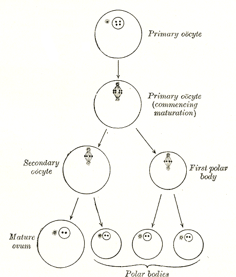
An oocyte (, ), oöcyte, or ovocyte is a female gametocyte or germ cell involved in reproduction. In other words, it is an immature ovum, or egg cell. An oocyte is produced in a female fetus in the ovary during female gametogenesis. The female germ cells produce a primordial germ cell (PGC), which then undergoes mitosis, forming oogonia. During oogenesis, the oogonia become primary oocytes. An oocyte is a form of genetic material that can be collected for cryoconservation. Formation The formation of an oocyte is called oocytogenesis, which is a part of oogenesis. Oogenesis results in the formation of both primary oocytes during fetal period, and of secondary oocytes after it as part of ovulation. Characteristics Cytoplasm Oocytes are rich in cytoplasm, which contains yolk granules to nourish the cell early in development. Nucleus During the primary oocyte stage of oogenesis, the nucleus is called a germinal vesicle. The only normal human type of secondary oocyte has t ... [...More Info...] [...Related Items...] OR: [Wikipedia] [Google] [Baidu] |
_and_SMC1_(green)_(PDB_2WD5)_from_mice_(Kurze_et_al._2009).png)


