|
Quadratojugal Boss
The quadratojugal is a skull bone present in many vertebrates, including some living reptiles and amphibians. Anatomy and function In animals with a quadratojugal bone, it is typically found connected to the jugal (cheek) bone from the front and the squamosal bone from above. It is usually positioned at the rear lower corner of the cranium. Many modern tetrapods lack a quadratojugal bone as it has been lost or fused to other bones. Modern examples of tetrapods without a quadratojugal include salamanders, mammals, birds, and squamates (lizards and snakes). In tetrapods with a quadratojugal bone, it often forms a portion of the jaw joint. Developmentally, the quadratojugal bone is a dermal bone in the temporal series, forming the original braincase. The squamosal and quadratojugal bones together form the cheek region and may provide muscular attachments for facial muscles. In reptiles and amphibians In most modern reptiles and amphibians, the quadratojugal is a prominen ... [...More Info...] [...Related Items...] OR: [Wikipedia] [Google] [Baidu] |
Skull
The skull is a bone protective cavity for the brain. The skull is composed of four types of bone i.e., cranial bones, facial bones, ear ossicles and hyoid bone. However two parts are more prominent: the cranium and the mandible. In humans, these two parts are the neurocranium and the viscerocranium ( facial skeleton) that includes the mandible as its largest bone. The skull forms the anterior-most portion of the skeleton and is a product of cephalisation—housing the brain, and several sensory structures such as the eyes, ears, nose, and mouth. In humans these sensory structures are part of the facial skeleton. Functions of the skull include protection of the brain, fixing the distance between the eyes to allow stereoscopic vision, and fixing the position of the ears to enable sound localisation of the direction and distance of sounds. In some animals, such as horned ungulates (mammals with hooves), the skull also has a defensive function by providing the mount (on the front ... [...More Info...] [...Related Items...] OR: [Wikipedia] [Google] [Baidu] |
Infratemporal Fenestra
An infratemporal fenestra, also called the lateral temporal fenestra or simply temporal fenestra, is an opening in the skull behind the orbit in some animals. It is ventrally bordered by a zygomatic arch. An opening in front of the eye sockets, conversely, is called an antorbital fenestra. Both of these openings reduce the weight of the skull. Infratemporal fenestrae are commonly (although not universally) seen in the fossilized skulls of dinosaurs. Synapsids, including mammals, have one temporal fenestra, while sauropsids Sauropsida ("lizard faces") is a clade of amniotes, broadly equivalent to the class Reptilia. Sauropsida is the sister taxon to Synapsida, the other clade of amniotes which includes mammals as its only modern representatives. Although early synap ..., the birds and reptiles, have two. References {{ref list Dinosaur anatomy Foramina of the skull ... [...More Info...] [...Related Items...] OR: [Wikipedia] [Google] [Baidu] |
Eusthenopteron Quadratojugal
''Eusthenopteron'' (from el, εὖ , 'good', el, σθένος , 'strength', and el, πτερόν 'wing' or 'fin') is a genus of prehistoric sarcopterygian (often called lobe-finned fishes) which has attained an iconic status from its close relationships to tetrapods. Early depictions of this animal show it emerging onto land; however, paleontologists now widely agree that it was a strictly aquatic animal.M. Laurin, F. J. Meunier, D. Germain, and M. Lemoine 2007A microanatomical and histological study of the paired fin skeleton of the Devonian sarcopterygian ''Eusthenopteron foordi'' ''Journal of Paleontology'' 81: 143–153. The genus ''Eusthenopteron'' is known from several species that lived during the Late Devonian period, about 385 million years ago. ''Eusthenopteron'' was first described by J. F. Whiteaves in 1881, as part of a large collection of fishes from Miguasha, Quebec. Some 2,000 ''Eusthenopteron'' specimens have been collected from Miguasha, one of which was t ... [...More Info...] [...Related Items...] OR: [Wikipedia] [Google] [Baidu] |
Middle Ear
The middle ear is the portion of the ear medial to the eardrum, and distal to the oval window of the cochlea (of the inner ear). The mammalian middle ear contains three ossicles, which transfer the vibrations of the eardrum into waves in the fluid and membranes of the inner ear. The hollow space of the middle ear is also known as the tympanic cavity and is surrounded by the tympanic part of the temporal bone. The auditory tube (also known as the Eustachian tube or the pharyngotympanic tube) joins the tympanic cavity with the nasal cavity (nasopharynx), allowing pressure to equalize between the middle ear and throat. The primary function of the middle ear is to efficiently transfer acoustic energy from compression waves in air to fluid–membrane waves within the cochlea. Structure Ossicles The middle ear contains three tiny bones known as the ossicles: '' malleus'', '' incus'', and ''stapes''. The ossicles were given their Latin names for their distinctive shapes; they ar ... [...More Info...] [...Related Items...] OR: [Wikipedia] [Google] [Baidu] |
Ossicles
The ossicles (also called auditory ossicles) are three bones in either middle ear that are among the smallest bones in the human body. They serve to transmit sounds from the air to the fluid-filled labyrinth (cochlea). The absence of the auditory ossicles would constitute a moderate-to-severe hearing loss. The term "ossicle" literally means "tiny bone". Though the term may refer to any small bone throughout the body, it typically refers to the malleus, incus, and stapes (hammer, anvil, and stirrup) of the middle ear. Structure The ossicles are, in order from the eardrum to the inner ear (from superficial to deep): the malleus, incus, and stapes, terms that in Latin are translated as "the hammer, anvil, and stirrup". * The malleus ( la, "hammer") articulates with the incus through the incudomalleolar joint and is attached to the tympanic membrane (eardrum), from which vibrational sound pressure motion is passed. * The incus ( la, "anvil") is connected to both the other bones. * ... [...More Info...] [...Related Items...] OR: [Wikipedia] [Google] [Baidu] |
Incus
The ''incus'' (plural incudes) or anvil is a bone in the middle ear. The anvil-shaped small bone is one of three ossicles in the middle ear. The ''incus'' receives vibrations from the ''malleus'', to which it is connected laterally, and transmits these to the ''stapes'' medially. The ''incus'' is so-called because of its resemblance to an anvil ( la, Incus). Structure The incus is the second of the ossicles, three bones in the middle ear which act to transmit sound. It is shaped like an anvil, and has a long and short crus extending from the body, which articulates with the malleus. The short crus attaches to the posterior ligament of the incus. The long crus articulates with the stirrup at the lenticular process. The superior ligament of the incus attaches at the body of the incus to the roof of the tympanic cavity. Function Vibrations in the middle ear are received via the tympanic membrane. The malleus, resting on the membrane, conveys vibrations to the incus. This in tu ... [...More Info...] [...Related Items...] OR: [Wikipedia] [Google] [Baidu] |
Stapes
The ''stapes'' or stirrup is a bone in the middle ear of humans and other animals which is involved in the conduction of sound vibrations to the inner ear. This bone is connected to the oval window by its annular ligament, which allows the footplate to transmit sound energy through the oval window into the inner ear. The ''stapes'' is the smallest and lightest bone in the human body, and is so-called because of its resemblance to a stirrup ( la, Stapes). Structure The ''stapes'' is the third bone of the three ossicles in the middle ear and the smallest in the human body. It measures roughly , greater along the head-base span. It rests on the oval window, to which it is connected by an annular ligament and articulates with the ''incus'', or anvil through the incudostapedial joint. They are connected by anterior and posterior limbs ( la, crura). Development The ''stapes'' develops from the second pharyngeal arch during the sixth to eighth week of embryological life. The ce ... [...More Info...] [...Related Items...] OR: [Wikipedia] [Google] [Baidu] |
Mammaliaformes
Mammaliaformes ("mammalian forms") is a clade that contains the crown group mammals and their closest Extinction, extinct relatives; the group adaptive radiation, radiated from earlier probainognathian cynodonts. It is defined as the clade originating from the most recent common ancestor of Morganucodonta and the crown group mammals; the latter is the clade originating with the most recent common ancestor of extant Monotremata, Marsupialia, and Placentalia. Besides Morganucodonta and the crown group mammals, Mammaliaformes includes Docodonta and ''Hadrocodium'' as well as the Triassic ''Tikitherium'', the earliest known member of the group. Mammaliaformes is a term of phylogenetic nomenclature. In contrast, the assignment of organisms to Mammalia has traditionally been founded on traits and, on this basis, Mammalia is slightly more inclusive than Mammaliaformes. In particular, trait-based taxonomy generally includes ''Adelobasileus'' and ''Sinoconodon'' in Mammalia, though they fa ... [...More Info...] [...Related Items...] OR: [Wikipedia] [Google] [Baidu] |
Cranial Kinesis
Cranial kinesis is the term for significant movement of skull bones relative to each other in addition to movement at the joint between the upper and lower jaw. It is usually taken to mean relative movement between the upper jaw and the braincase. Most vertebrates have some form of kinetic skull. Cranial kinesis, or lack thereof, is usually linked to feeding. Animals which must exert powerful bite forces, such as crocodiles, often have rigid skulls with little or no kinesis, for maximum strength. Animals which swallow large prey whole (snakes), which grip awkwardly shaped food items (parrots eating nuts), or, most often, which feed in the water via suction feeding often have very kinetic skulls, frequently with numerous mobile joints. In the case of mammals, which have akinetic skulls (except for perhaps hares), the lack of kinesis is most likely to be related to the secondary palate, which prevents relative movement. This in turn is a consequence of the need to be able to create ... [...More Info...] [...Related Items...] OR: [Wikipedia] [Google] [Baidu] |
Squamosal Bone
The squamosal is a skull bone found in most reptiles, amphibians, and birds. In fishes, it is also called the pterotic bone. In most tetrapods, the squamosal and quadratojugal bones form the cheek series of the skull. The bone forms an ancestral component of the dermal roof and is typically thin compared to other skull bones. The squamosal bone lies ventral to the temporal series and otic notch, and is bordered anteriorly by the postorbital. Posteriorly, the squamosal articulates with the quadrate and pterygoid bones. The squamosal is bordered anteroventrally by the jugal and ventrally by the quadratojugal. Function in reptiles In reptiles, the quadrate and articular bones of the skull articulate to form the jaw joint. The squamosal bone lies anterior to the quadrate bone. Anatomy in synapsids Non-mammalian synapsids In non-mammalian synapsids, the jaw is composed of four bony elements and referred to as a quadro-articular jaw because the joint is between the articular an ... [...More Info...] [...Related Items...] OR: [Wikipedia] [Google] [Baidu] |
Postorbital Bone
The ''postorbital'' is one of the bones in vertebrate skulls which forms a portion of the dermal skull roof and, sometimes, a ring about the orbit. Generally, it is located behind the postfrontal and posteriorly to the orbital fenestra. In some vertebrates, the postorbital is fused with the postfrontal to create a postorbitofrontal. Birds have a separate postorbital as an embryo, but the bone fuses with the frontal Front may refer to: Arts, entertainment, and media Films * ''The Front'' (1943 film), a 1943 Soviet drama film * ''The Front'', 1976 film Music * The Front (band), an American rock band signed to Columbia Records and active in the 1980s and e ... before it hatches. References * Roemer, A. S. 1956. ''Osteology of the Reptiles''. University of Chicago Press. 772 pp. Skull {{Vertebrate anatomy-stub ... [...More Info...] [...Related Items...] OR: [Wikipedia] [Google] [Baidu] |
Caecilian
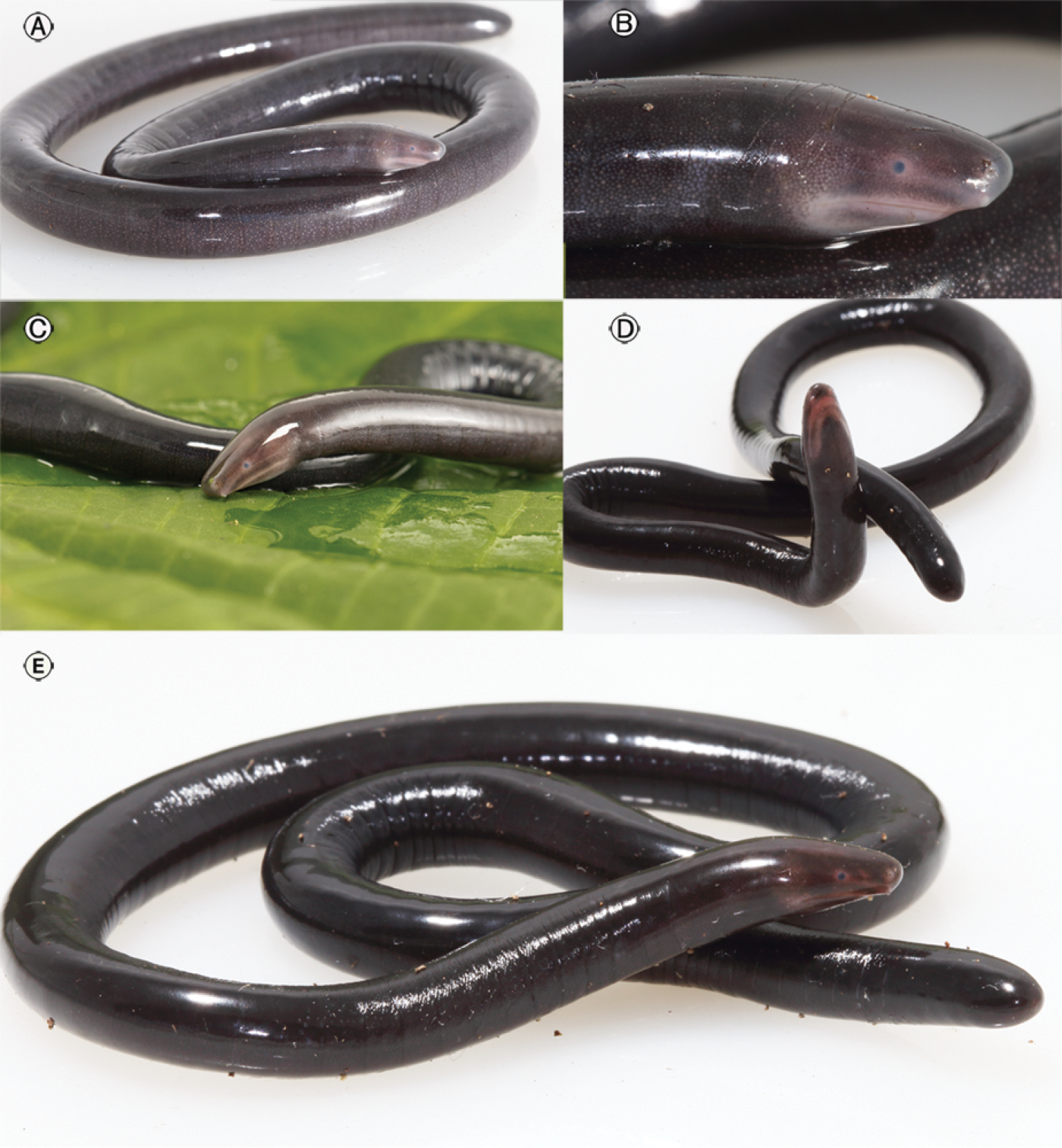
Caecilians (; ) are a group of limbless, vermiform or serpentine amphibians. They mostly live hidden in the ground and in stream substrates, making them the least familiar order of amphibians. Caecilians are mostly distributed in the tropics of South and Central America, Africa, and southern Asia. Their diet consists of small subterranean creatures such as earthworms. All modern caecilians and their closest fossil relatives are grouped as a clade, Apoda , within the larger group Gymnophiona , which also includes more primitive extinct caecilian-like amphibians. The name derives from the Greek words γυμνος (''gymnos'', naked) and οφις (''ophis'', snake), as the caecilians were originally thought to be related to snakes. The body is cylindrical dark brown or bluish black in colour. The skin is slimy and bears grooves or ringlike markings. Description Caecilians completely lack limbs, making the smaller species resemble worms, while the larger species, with lengths up ... [...More Info...] [...Related Items...] OR: [Wikipedia] [Google] [Baidu] |


