|
Posterior Cruciate Ligament Injury
The function of the posterior cruciate ligament (PCL) is to prevent the femur from sliding off the anterior edge of the tibia and to prevent the tibia from displacing posterior to the femur. Common causes of PCL injuries are direct blows to the flexed knee, such as the knee hitting the dashboard in a car accident or falling hard on the knee, both instances displacing the tibia posterior to the femur. Surgery to repair the posterior cruciate ligament is controversial due to its placement and technical difficulty. The posterior drawer test is one of the tests used by doctors and physiotherapists to detect injury to the PCL. An additional test of posterior cruciate ligament injury is the ''posterior sag test'', where, in contrast to the drawer test, no active force is applied. Rather, the person lies supine with the leg held by another person so that the hip is flexed to 90 degrees and the knee 90 degrees. The main parameter in this test is ''step-off'', which is the shortest distance ... [...More Info...] [...Related Items...] OR: [Wikipedia] [Google] [Baidu] |
Posterior Cruciate Ligament
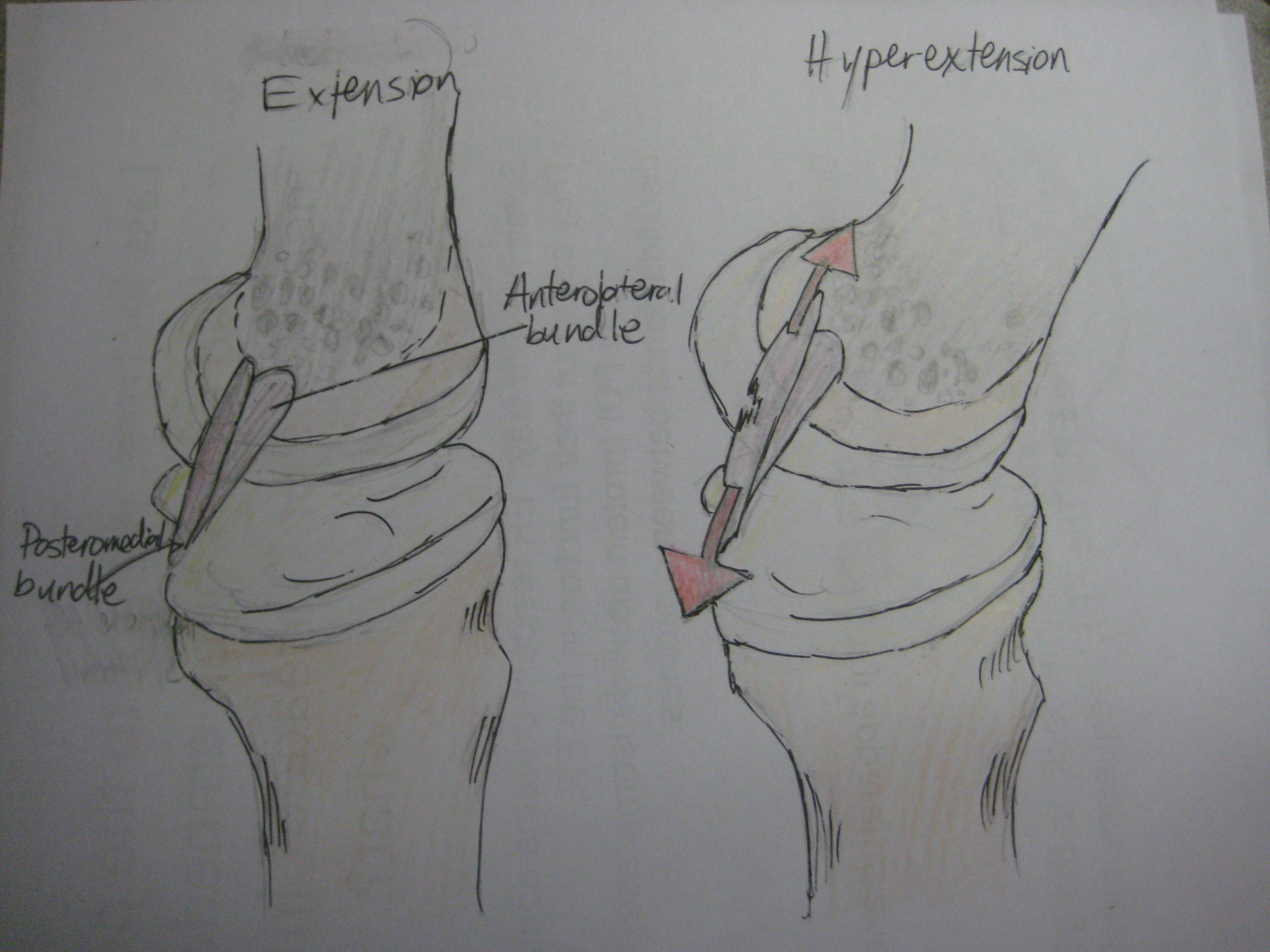
The posterior cruciate ligament (PCL) is a ligament in each knee of humans and various other animals. It works as a counterpart to the anterior cruciate ligament (ACL). It connects the posterior intercondylar area of the tibia to the medial condyle of the femur. This configuration allows the PCL to resist forces pushing the tibia posteriorly relative to the femur. The PCL and ACL are intracapsular ligaments because they lie deep within the knee joint. They are both isolated from the fluid-filled synovial cavity, with the synovial membrane wrapped around them. The PCL gets its name by attaching to the posterior portion of the tibia. The PCL, ACL, MCL, and LCL are the four main ligaments of the knee in primates. Structure The PCL is located within the knee joint where it stabilizes the articulating bones, particularly the femur and the tibia, during movement. It originates from the lateral edge of the medial femoral condyle and the roof of the intercondyle notch then stretches ... [...More Info...] [...Related Items...] OR: [Wikipedia] [Google] [Baidu] |
Ligaments
A ligament is the fibrous connective tissue that connects bones to other bones. It is also known as ''articular ligament'', ''articular larua'', ''fibrous ligament'', or ''true ligament''. Other ligaments in the body include the: * Peritoneal ligament: a fold of peritoneum or other membranes. * Fetal remnant ligament: the remnants of a fetal tubular structure. * Periodontal ligament: a group of fibers that attach the cementum of teeth to the surrounding alveolar bone. Ligaments are similar to tendons and fasciae as they are all made of connective tissue. The differences among them are in the connections that they make: ligaments connect one bone to another bone, tendons connect muscle to bone, and fasciae connect muscles to other muscles. These are all found in the skeletal system of the human body. Ligaments cannot usually be regenerated naturally; however, there are periodontal ligament stem cells located near the periodontal ligament which are involved in the adult regener ... [...More Info...] [...Related Items...] OR: [Wikipedia] [Google] [Baidu] |
Ligament
A ligament is the fibrous connective tissue that connects bones to other bones. It is also known as ''articular ligament'', ''articular larua'', ''fibrous ligament'', or ''true ligament''. Other ligaments in the body include the: * Peritoneal ligament: a fold of peritoneum or other membranes. * Fetal remnant ligament: the remnants of a fetal tubular structure. * Periodontal ligament: a group of fibers that attach the cementum of teeth to the surrounding alveolar bone. Ligaments are similar to tendons and fasciae as they are all made of connective tissue. The differences among them are in the connections that they make: ligaments connect one bone to another bone, tendons connect muscle to bone, and fasciae connect muscles to other muscles. These are all found in the skeletal system of the human body. Ligaments cannot usually be regenerated naturally; however, there are periodontal ligament stem cells located near the periodontal ligament which are involved in the adult regener ... [...More Info...] [...Related Items...] OR: [Wikipedia] [Google] [Baidu] |
Anterior Cruciate Ligament
The anterior cruciate ligament (ACL) is one of a pair of cruciate ligaments (the other being the posterior cruciate ligament) in the human knee. The two ligaments are also called "cruciform" ligaments, as they are arranged in a crossed formation. In the quadruped stifle joint (analogous to the knee), based on its anatomical position, it is also referred to as the cranial cruciate ligament. The term cruciate translates to cross. This name is fitting because the ACL crosses the posterior cruciate ligament to form an “X”. It is composed of strong, fibrous material and assists in controlling excessive motion. This is done by limiting mobility of the joint. The anterior cruciate ligament is one of the four main ligaments of the knee, providing 85% of the restraining force to anterior tibial displacement at 30 and 90° of knee flexion. The ACL is the most injured ligament of the four located in the knee. Structure The ACL originates from deep within the notch of the distal fe ... [...More Info...] [...Related Items...] OR: [Wikipedia] [Google] [Baidu] |
Ligaments
A ligament is the fibrous connective tissue that connects bones to other bones. It is also known as ''articular ligament'', ''articular larua'', ''fibrous ligament'', or ''true ligament''. Other ligaments in the body include the: * Peritoneal ligament: a fold of peritoneum or other membranes. * Fetal remnant ligament: the remnants of a fetal tubular structure. * Periodontal ligament: a group of fibers that attach the cementum of teeth to the surrounding alveolar bone. Ligaments are similar to tendons and fasciae as they are all made of connective tissue. The differences among them are in the connections that they make: ligaments connect one bone to another bone, tendons connect muscle to bone, and fasciae connect muscles to other muscles. These are all found in the skeletal system of the human body. Ligaments cannot usually be regenerated naturally; however, there are periodontal ligament stem cells located near the periodontal ligament which are involved in the adult regener ... [...More Info...] [...Related Items...] OR: [Wikipedia] [Google] [Baidu] |
Prone Knee Flexion
Prone position () is a body position in which the person lies flat with the chest down and the back up. In anatomical terms of location, the dorsal side is up, and the ventral side is down. The supine position is the 180° contrast. Etymology The word ''prone'', meaning "naturally inclined to something, apt, liable," has been recorded in English since 1382; the meaning "lying face-down" was first recorded in 1578, but is also referred to as "lying down" or "going prone." ''Prone'' derives from the Latin ', meaning "bent forward, inclined to," from the adverbial form of the prefix ''pro-'' "forward." Both the original, literal, and the derived figurative sense were used in Latin, but the figurative is older in English. Anatomy In anatomy, the prone position is a position of the body lying face down. It is opposed to the supine position which is face up. Using the terms defined in the anatomical position, the ventral side is down, and the dorsal side is up. Concerning th ... [...More Info...] [...Related Items...] OR: [Wikipedia] [Google] [Baidu] |
Hamstring Muscles
In human anatomy, a hamstring () is any one of the three posterior thigh muscles in between the hip and the knee (from medial to lateral: semimembranosus, semitendinosus and biceps femoris). The hamstrings are susceptible to injury. In quadrupeds, the hamstring is the single large tendon found behind the knee or comparable area. Criteria The common criteria of any hamstring muscles are: # Muscles should originate from ischial tuberosity. # Muscles should be inserted over the knee joint, in the tibia or in the fibula. # Muscles will be innervated by the tibial nerve, tibial branch of the sciatic nerve. # Muscle will participate in flexion of the knee joint and extension of the hip joint. Those muscles which fulfill all of the four criteria are called true hamstrings. The adductor magnus reaches only up to the adductor tubercle of the femur, but it is included amongst the hamstrings because the tibial collateral ligament of the knee joint morphologically is the degenerated tendon ... [...More Info...] [...Related Items...] OR: [Wikipedia] [Google] [Baidu] |
Injury
An injury is any physiological damage to living tissue caused by immediate physical stress. An injury can occur intentionally or unintentionally and may be caused by blunt trauma, penetrating trauma, burning, toxic exposure, asphyxiation, or overexertion. Injuries can occur in any part of the body, and different symptoms are associated with different injuries. Treatment of a major injury is typically carried out by a health professional and varies greatly depending on the nature of the injury. Traffic collisions are the most common cause of accidental injury and injury-related death among humans. Injuries are distinct from chronic conditions, psychological trauma, infections, or medical procedures, though injury can be a contributing factor to any of these. Several major health organizations have established systems for the classification and description of human injuries. Occurrence Injuries may be intentional or unintentional. Intentional injuries may be acts of ... [...More Info...] [...Related Items...] OR: [Wikipedia] [Google] [Baidu] |
Quadriceps
The quadriceps femoris muscle (, also called the quadriceps extensor, quadriceps or quads) is a large muscle group that includes the four prevailing muscles on the front of the thigh. It is the sole extensor muscle of the knee, forming a large fleshy mass which covers the front and sides of the femur. The name derives . Structure Parts The quadriceps femoris muscle is subdivided into four separate muscles (the 'heads'), with the first superficial to the other three over the femur (from the trochanters to the condyles): *The rectus femoris muscle occupies the middle of the thigh, covering most of the other three quadriceps muscles. It originates on the ilium. It is named for its straight course. *The vastus lateralis muscle is on the ''lateral side'' of the femur (i.e. on the outer side of the thigh). *The vastus medialis muscle is on the ''medial side'' of the femur (i.e. on the inner part thigh). *The vastus intermedius muscle lies between vastus lateralis and vastus mediali ... [...More Info...] [...Related Items...] OR: [Wikipedia] [Google] [Baidu] |
Range Of Motion
Range of motion (or ROM), is the linear or angular distance that a moving object may normally travel while properly attached to another. It is also called range of travel (or ROT), particularly when talking about mechanical devices and in mechanical engineering fields. For example, a sound volume control knob. As used in the biomedical field and by weightlifters, range of motion refers to the distance and direction a joint can move between the flexed position and the extended position. The act of attempting to increase this distance through therapeutic exercises (range of motion therapy—stretching from flexion to extension for physiological gain) is also sometimes called range of motion. Measuring range of motion Each specific joint has a normal range of motion that is expressed in degrees. The reference values for the normal ROM in individuals differ slightly depending on age and gender. For example, as an individual ages, they typically lose a small amount of ROM. Analog ... [...More Info...] [...Related Items...] OR: [Wikipedia] [Google] [Baidu] |
Posterolateral Knee Injuries
Posterolateral corner injuries (PLC injuries) of the knee are injuries to a complex area formed by the interaction of multiple structures. Injuries to the posterolateral corner can be debilitating to the person and require recognition and treatment to avoid long term consequences. Injuries to the PLC often occur in combination with other ligamentous injuries to the knee; most commonly the anterior cruciate ligament (ACL) and posterior cruciate ligament (PCL). As with any injury, an understanding of the anatomy and functional interactions of the posterolateral corner is important to diagnosing and treating the injury. Signs and symptoms Patients often complain of pain and instability at the joint. With concurrent Nerve injury, nerve injuries, patients may experience numbness, tingling and weakness of the Ankle dorsiflexion, ankle dorsiflexors and great toe extensors, or a footdrop. Complications Follow-up studies by Levy et al. and Stannard at al. both examined failure rates for ... [...More Info...] [...Related Items...] OR: [Wikipedia] [Google] [Baidu] |


