|
Post-traumatic Epilepsy
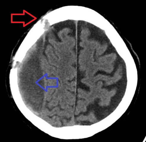
Post-traumatic epilepsy (PTE) is a form of acquired epilepsy that results from brain damage caused by physical trauma to the brain (traumatic brain injury, abbreviated TBI). A person with PTE experiences repeated post-traumatic seizures (PTS, seizures that result from TBI) more than a week after the initial injury. PTE is estimated to constitute 5% of all cases of epilepsy and over 20% of cases of acquired epilepsy (in which seizures are caused by an identifiable organic brain condition). It is not known who will develop epilepsy after TBI and who will not. However, the likelihood that a person will develop PTE is influenced by the severity and type of injury; for example penetrating injuries and those that involve bleeding within the brain confer a higher risk. The onset of PTE can occur within a short time of the physical trauma that causes it, or months or years after. People with head trauma may remain at a higher risk for post-traumatic seizures than the general pop ... [...More Info...] [...Related Items...] OR: [Wikipedia] [Google] [Baidu] |
Epilepsy
Epilepsy is a group of non-communicable neurological disorders characterized by recurrent epileptic seizures. Epileptic seizures can vary from brief and nearly undetectable periods to long periods of vigorous shaking due to abnormal electrical activity in the brain. These episodes can result in physical injuries, either directly such as broken bones or through causing accidents. In epilepsy, seizures tend to recur and may have no immediate underlying cause. Isolated seizures that are provoked by a specific cause such as poisoning are not deemed to represent epilepsy. People with epilepsy may be treated differently in various areas of the world and experience varying degrees of social stigma due to the alarming nature of their symptoms. The underlying mechanism of epileptic seizures is excessive and abnormal neuronal activity in the cortex of the brain which can be observed in the electroencephalogram (EEG) of an individual. The reason this occurs in most cases of epilepsy is u ... [...More Info...] [...Related Items...] OR: [Wikipedia] [Google] [Baidu] |
Level Of Consciousness
An altered level of consciousness is any measure of arousal other than normal. Level of consciousness (LOC) is a measurement of a person's arousability and responsiveness to stimuli from the environment. A mildly depressed level of consciousness or alertness may be classed as lethargy; someone in this state can be aroused with little difficulty. People who are obtunded have a more depressed level of consciousness and cannot be fully aroused. Those who are not able to be aroused from a sleep-like state are said to be stuporous. Coma is the inability to make any purposeful response. Scales such as the Glasgow coma scale have been designed to measure the level of consciousness. An altered level of consciousness can result from a variety of factors, including alterations in the chemical environment of the brain (e.g. exposure to poisons or intoxicants), insufficient oxygen or blood flow in the brain, and excessive pressure within the skull. Prolonged unconsciousness is unde ... [...More Info...] [...Related Items...] OR: [Wikipedia] [Google] [Baidu] |
Status Epilepticus
Status epilepticus (SE), or status seizure, is a single seizure lasting more than 5 minutes or 2 or more seizures within a 5-minute period without the person returning to normal between them. Previous definitions used a 30-minute time limit. The seizures can be of the tonic–clonic type, with a regular pattern of contraction and extension of the arms and legs, or of types that do not involve contractions, such as absence seizures or complex partial seizures. Status epilepticus is a life-threatening medical emergency, particularly if treatment is delayed. Status epilepticus may occur in those with a history of epilepsy as well as those with an underlying problem of the brain. These underlying brain problems may include trauma, infections, or strokes, among others. Diagnosis often involves checking the blood sugar, imaging of the head, a number of blood tests, and an electroencephalogram. Psychogenic nonepileptic seizures may present similarly to status epilepticus. Other conditio ... [...More Info...] [...Related Items...] OR: [Wikipedia] [Google] [Baidu] |
Frontal Lobe
The frontal lobe is the largest of the four major lobes of the brain in mammals, and is located at the front of each cerebral hemisphere (in front of the parietal lobe and the temporal lobe). It is parted from the parietal lobe by a groove between tissues called the central sulcus and from the temporal lobe by a deeper groove called the lateral sulcus (Sylvian fissure). The most anterior rounded part of the frontal lobe (though not well-defined) is known as the frontal pole, one of the three poles of the cerebrum. The frontal lobe is covered by the frontal cortex. The frontal cortex includes the premotor cortex, and the primary motor cortex – parts of the motor cortex. The front part of the frontal cortex is covered by the prefrontal cortex. There are four principal gyri in the frontal lobe. The precentral gyrus is directly anterior to the central sulcus, running parallel to it and contains the primary motor cortex, which controls voluntary movements of specific body parts ... [...More Info...] [...Related Items...] OR: [Wikipedia] [Google] [Baidu] |
Cerebral Edema
Cerebral edema is excess accumulation of fluid (edema) in the intracellular or extracellular spaces of the brain. This typically causes impaired nerve function, increased pressure within the skull, and can eventually lead to direct compression of brain tissue and blood vessels. Symptoms vary based on the location and extent of edema and generally include headaches, nausea, vomiting, seizures, drowsiness, visual disturbances, dizziness, and in severe cases, coma and death. Cerebral edema is commonly seen in a variety of brain injuries including ischemic stroke, subarachnoid hemorrhage, traumatic brain injury, subdural, epidural, or intracerebral hematoma, hydrocephalus, brain cancer, brain infections, low blood sodium levels, high altitude, and acute liver failure. Diagnosis is based on symptoms and physical examination findings and confirmed by serial neuroimaging ( computed tomography scans and magnetic resonance imaging). The treatment of cerebral edema depends on the ... [...More Info...] [...Related Items...] OR: [Wikipedia] [Google] [Baidu] |
Intracranial Hematoma
Intracranial hemorrhage (ICH), also known as intracranial bleed, is bleeding within the skull. Subtypes are intracerebral bleeds ( intraventricular bleeds and intraparenchymal bleeds), subarachnoid bleeds, epidural bleeds, and subdural bleeds. More often than not it ends in a lethal outcome. Intracerebral bleeding affects 2.5 per 10,000 people each year. Signs and symptoms Intracranial hemorrhage is a serious medical emergency because the buildup of blood within the skull can lead to increases in intracranial pressure, which can crush delicate brain tissue or limit its blood supply. Severe increases in intracranial pressure (ICP) can cause brain herniation, in which parts of the brain are squeezed past structures in the skull. Causes Trauma is the most common cause of intracranial hemorrhage. It can cause epidural hemorrhage, subdural hemorrhage, and subarachnoid hemorrhage. Other condition such as hemorrhagic parenchymal contusion and cerebral microhemorrhages can also b ... [...More Info...] [...Related Items...] OR: [Wikipedia] [Google] [Baidu] |
Subdural Haematoma
A subdural hematoma (SDH) is a type of bleeding in which a collection of blood—usually but not always associated with a traumatic brain injury—gathers between the inner layer of the dura mater and the arachnoid mater of the meninges surrounding the brain. It usually results from tears in bridging veins that cross the subdural space. Subdural hematomas may cause an increase in the pressure inside the skull, which in turn can cause compression of and damage to delicate brain tissue. Acute subdural hematomas are often life-threatening. Chronic subdural hematomas have a better prognosis if properly managed. In contrast, epidural hematomas are usually caused by tears in arteries, resulting in a build-up of blood between the dura mater and the skull. The third type of brain hemorrhage, known as a subarachnoid hemorrhage, causes bleeding into the subarachnoid space between the arachnoid mater and the pia mater. __TOC__ Signs and symptoms The symptoms of a subdural hematoma hav ... [...More Info...] [...Related Items...] OR: [Wikipedia] [Google] [Baidu] |
Intracerebral Hematoma
Intracerebral hemorrhage (ICH), also known as cerebral bleed, intraparenchymal bleed, and hemorrhagic stroke, or haemorrhagic stroke, is a sudden bleeding into the tissues of the brain, into its ventricles, or into both. It is one kind of bleeding within the skull and one kind of stroke. Symptoms can include headache, one-sided weakness, vomiting, seizures, decreased level of consciousness, and neck stiffness. Often, symptoms get worse over time. Fever is also common. Causes include brain trauma, aneurysms, arteriovenous malformations, and brain tumors. The biggest risk factors for spontaneous bleeding are high blood pressure and amyloidosis. Other risk factors include alcoholism, low cholesterol, blood thinners, and cocaine use. Diagnosis is typically by CT scan. Other conditions that may present similarly include ischemic stroke. Treatment should typically be carried out in an intensive care unit. Guidelines recommend decreasing the blood pressure to a systolic of 140&nbs ... [...More Info...] [...Related Items...] OR: [Wikipedia] [Google] [Baidu] |
Penetrating Head Trauma
A penetrating head injury, or open head injury, is a head injury in which the dura mater, the outer layer of the meninges, is breached.University of Vermont College of Medicine"Neuropathology: Trauma to the CNS."Accessed through web archive on August 8, 2007. Penetrating injury can be caused by high-velocity projectiles or objects of lower velocity such as knives, or bone fragments from a skull fracture that are driven into the brain. Head injuries caused by penetrating trauma are serious medical emergencies and may cause permanent disability or death. A penetrating head injury involves "a wound in which an object breaches the cranium but does not exit it." In contrast, a perforating head injury is a wound in which the object passes through the head and leaves an exit wound.Vinas FC and Pilitsis J. 2006"Penetrating Head Trauma."Emedicine.com. Retrieved on February 6, 2007. Cause In penetrating injury from high-velocity missiles, injuries may occur not only from initial lacera ... [...More Info...] [...Related Items...] OR: [Wikipedia] [Google] [Baidu] |
Depressed Skull Fracture
A skull fracture is a break in one or more of the eight bones that form the cranial portion of the skull, usually occurring as a result of blunt force trauma. If the force of the impact is excessive, the bone may fracture at or near the site of the impact and cause damage to the underlying structures within the skull such as the membranes, blood vessels, and brain. While an uncomplicated skull fracture can occur without associated physical or neurological damage and is in itself usually not clinically significant, a fracture in healthy bone indicates that a substantial amount of force has been applied and increases the possibility of associated injury. Any significant blow to the head results in a concussion, with or without loss of consciousness. A fracture in conjunction with an overlying laceration that tears the epidermis and the meninges, or runs through the paranasal sinuses and the middle ear structures, bringing the outside environment into contact with the cranial cavit ... [...More Info...] [...Related Items...] OR: [Wikipedia] [Google] [Baidu] |
National Society For Epilepsy
The Epilepsy Society (formerly known as the National Society for Epilepsy) is the largest medical charity in the field of epilepsy in the United Kingdom, providing services for people with epilepsy for over 100 years. Based in Chalfont St Peter, Buckinghamshire, UK, its stated mission is "to enhance the quality of life of people affected by epilepsy by promoting research, education and public awareness and by delivering specialist medical care and support services." The Epilepsy Society has close partnerships with the National Hospital for Neurology and Neurosurgery (NHNN) and the UCL Institute of Neurology, both located in Queen Square, London. Services Epilepsy Society is a leading epilepsy medical charity supporting all people affected by epilepsy. The services provided by the charity include: * Residential care for over 100 adults within care homes at the Chalfont Centre and also in supported living accommodation. [...More Info...] [...Related Items...] OR: [Wikipedia] [Google] [Baidu] |
Haptoglobin
Haptoglobin (abbreviated as Hp) is the protein that in humans is encoded by the ''HP'' gene. In blood plasma, haptoglobin binds with high affinity to ''free'' hemoglobin released from erythrocytes, and thereby inhibits its deleterious oxidative activity. Compared to Hp, hemopexin binds to ''free'' heme. The haptoglobin-hemoglobin complex will then be removed by the reticuloendothelial system (mostly the spleen). In clinical settings, the haptoglobin assay is used to screen for and monitor intravascular hemolytic anemia. In intravascular hemolysis, free hemoglobin will be released into circulation and hence haptoglobin will bind the hemoglobin. This causes a decline in haptoglobin levels. Function Hemoglobin that has been released into the blood plasma by damaged red blood cells has harmful effects. The ''HP'' gene encodes a preproprotein that is processed to yield both alpha and beta chains, which subsequently combines as a tetramer to produce haptoglobin. Haptoglobin function ... [...More Info...] [...Related Items...] OR: [Wikipedia] [Google] [Baidu] |


