|
Pineocytoma
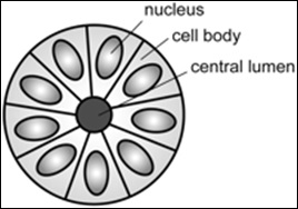
Pineocytoma, is a benign, slowly growing tumor of the pineal gland. Unlike the similar condition pineal gland cyst, it is uncommon. Diagnosis Pineocytomas are diagnosed from tissue, i.e. a brain biopsy.They consist of: * Cytopathology, cytologically benign cells (with nucleus (cell), nuclei of uniform size, regular nuclear membranes, and light chromatin) and, * have the characteristic Pineocytomatous/neurocytic pseudorosettes, pineocytomatous/neurocytic rosettes, which is an irregular circular/flower-like arrangement of cells with a large meshwork of fibers (neuropil) at the centre. Pineocytomatous/neurocytic rosettes are superficially similar to Homer Wright rosettes; however, they differ from Homer Wright rosettes as they have (1) more neuropil at centre of the rosette and, (2) the edge of neuropil meshwork irregular/undulating. Management See also * Pineal gland References External links {{Endocrine gland neoplasia Endocrine neoplasia Brain tumor ... [...More Info...] [...Related Items...] OR: [Wikipedia] [Google] [Baidu] |
Homer Wright Rosette
In histopathology, a palisade is a single layer of relatively long cells, arranged loosely perpendicular to a surface and parallel to each other. A rosette is a palisade in a halo or spoke-and-wheel arrangement, surrounding a central core or hub. A pseudorosette is a perivascular radial arrangement of neoplastic cells around a small blood vessel. Rosette A ''rosette'' is a cell formation in a halo or spoke-and-wheel arrangement, surrounding a central core or hub. The central hub may consist of an empty-appearing lumen or a space filled with cytoplasmic processes. The cytoplasm of each of the cells in the rosette is often wedge-shaped with the apex directed toward the central core: the nuclei of the cells participating in the rosette are peripherally positioned and form a ring or halo around the hub. Pathogenesis Rosettes may be considered primary or secondary manifestations of tumor architecture. Primary rosettes form as a characteristic growth pattern of a given tumor type wh ... [...More Info...] [...Related Items...] OR: [Wikipedia] [Google] [Baidu] |
Pineocytomatous/neurocytic Pseudorosettes
In histopathology, a palisade is a single layer of relatively long cells, arranged loosely perpendicular to a surface and parallel to each other. A rosette is a palisade in a halo or spoke-and-wheel arrangement, surrounding a central core or hub. A pseudorosette is a perivascular radial arrangement of neoplastic cells around a small blood vessel. Rosette A ''rosette'' is a cell formation in a halo or spoke-and-wheel arrangement, surrounding a central core or hub. The central hub may consist of an empty-appearing lumen or a space filled with cytoplasmic processes. The cytoplasm of each of the cells in the rosette is often wedge-shaped with the apex directed toward the central core: the nuclei of the cells participating in the rosette are peripherally positioned and form a ring or halo around the hub. Pathogenesis Rosettes may be considered primary or secondary manifestations of tumor architecture. Primary rosettes form as a characteristic growth pattern of a given tumor type wh ... [...More Info...] [...Related Items...] OR: [Wikipedia] [Google] [Baidu] |
Micrograph
A micrograph or photomicrograph is a photograph or digital image taken through a microscope or similar device to show a magnified image of an object. This is opposed to a macrograph or photomacrograph, an image which is also taken on a microscope but is only slightly magnified, usually less than 10 times. Micrography is the practice or art of using microscopes to make photographs. A micrograph contains extensive details of microstructure. A wealth of information can be obtained from a simple micrograph like behavior of the material under different conditions, the phases found in the system, failure analysis, grain size estimation, elemental analysis and so on. Micrographs are widely used in all fields of microscopy. Types Photomicrograph A light micrograph or photomicrograph is a micrograph prepared using an optical microscope, a process referred to as ''photomicroscopy''. At a basic level, photomicroscopy may be performed simply by connecting a camera to a microscope, th ... [...More Info...] [...Related Items...] OR: [Wikipedia] [Google] [Baidu] |
HPS Stain
In histology, the HPS stain, or hematoxylin phloxine saffron stain, is a way of marking tissues. HPS is similar to H&E, the standard bearer in histology. However, it differentiates between the most common connective tissue (collagen) and muscle and cytoplasm by staining the former yellow and the latter two pink, . polysciences.com. Accessed 6 December 2009. unlike an H&E stain, which stains all three pink. HPS stained sections are more expensive than H&E stained sections, primarily due to the cost of saffron. See also *Histopathology
Histopathology (compound of three Greek words: ''histos'' "tissue", πάθος ''pathos'' "suffering ...
[...More Info...] [...Related Items...] OR: [Wikipedia] [Google] [Baidu] |
Tumor
A neoplasm () is a type of abnormal and excessive growth of tissue. The process that occurs to form or produce a neoplasm is called neoplasia. The growth of a neoplasm is uncoordinated with that of the normal surrounding tissue, and persists in growing abnormally, even if the original trigger is removed. This abnormal growth usually forms a mass, when it may be called a tumor. ICD-10 classifies neoplasms into four main groups: benign neoplasms, in situ neoplasms, malignant neoplasms, and neoplasms of uncertain or unknown behavior. Malignant neoplasms are also simply known as cancers and are the focus of oncology. Prior to the abnormal growth of tissue, as neoplasia, cells often undergo an abnormal pattern of growth, such as metaplasia or dysplasia. However, metaplasia or dysplasia does not always progress to neoplasia and can occur in other conditions as well. The word is from Ancient Greek 'new' and 'formation, creation'. Types A neoplasm can be benign, potentially m ... [...More Info...] [...Related Items...] OR: [Wikipedia] [Google] [Baidu] |
Pineal Gland
The pineal gland, conarium, or epiphysis cerebri, is a small endocrine gland in the brain of most vertebrates. The pineal gland produces melatonin, a serotonin-derived hormone which modulates sleep, sleep patterns in both circadian rhythm, circadian and Season, seasonal cycles. The shape of the gland resembles a pine cone, which gives it its name. The pineal gland is located in the epithalamus, near the center of the brain, between the two cerebral hemisphere, hemispheres, tucked in a groove where the two halves of the thalamus join. The pineal gland is one of the neuroendocrinology, neuroendocrine Circumventricular organs, secretory circumventricular organs in which capillaries are mostly Vascular permeability, permeable to solutes in the blood. Nearly all vertebrate species possess a pineal gland. The most important exception is a primitive vertebrate, the hagfish. Even in the hagfish, however, there may be a "pineal equivalent" structure in the dorsal diencephalon. The lanc ... [...More Info...] [...Related Items...] OR: [Wikipedia] [Google] [Baidu] |
Pineal Gland Cyst
A pineal gland cyst is a usually benign (non-malignant) cyst in the pineal gland, a small endocrine gland in the brain. Historically, these fluid-filled bodies appeared on of magnetic resonance imaging (MRI) brain scans, but were more frequently diagnosed at death, seen in of autopsies. A 2007 study by Pu ''et al''. found a frequency of 23% in brain scans (with a mean diameter of 4.3 mm). Despite the pineal gland being in the center of the brain, due to recent advancements in endoscopic medicine, endoscopic brain surgery to drain and/or remove the cyst can be done with the patient spending 5-10 nights in the hospital, and being fully recovered in weeks, rather than a year, as is the case with open-skull brain surgery. The National Organization for Rare Disorders states that pineal cysts larger than 5.0 mm are "rare findings" and are possibly symptomatic. If narrowing of the cerebral aqueduct occurs, many neurological symptoms may exist, including headaches, vertigo, na ... [...More Info...] [...Related Items...] OR: [Wikipedia] [Google] [Baidu] |
Brain Biopsy
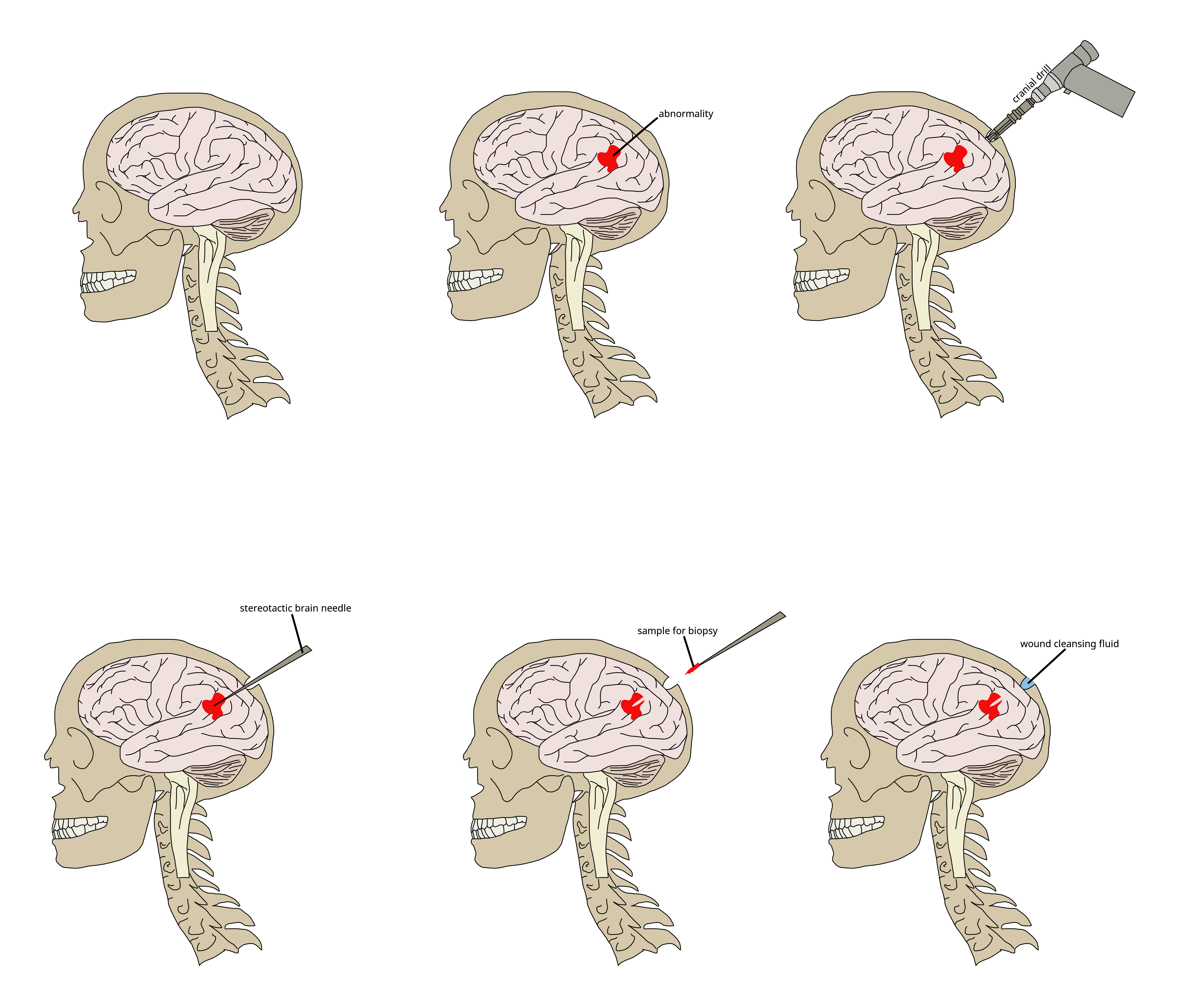
Brain biopsy is the removal of a small piece of brain tissue for the diagnosis of abnormalities of the brain. It is used to diagnose tumors, infection, inflammation, and other brain disorders. By examining the tissue sample under a microscope, the biopsy sample provides information about the appropriate diagnosis and treatment. Indications Given the potential risks surrounding the procedure, cerebral biopsy is indicated only if other diagnostic approaches (e.g. magnetic resonance imaging) have been insufficient in showing the cause of symptoms, and if it is felt that the benefits of histological diagnosis will influence the treatment plan. If the person has a brain tumor, biopsy is 95% sensitive. The procedure can also be valuable in people who are immunocompromised and who have evidence of brain lesions that could be caused by opportunistic infection An opportunistic infection is an infection caused by pathogens (bacteria, fungi, parasites or viruses) that take advantage of an ... [...More Info...] [...Related Items...] OR: [Wikipedia] [Google] [Baidu] |
Cytopathology
Cytopathology (from Greek , ''kytos'', "a hollow"; , ''pathos'', "fate, harm"; and , '' -logia'') is a branch of pathology that studies and diagnoses diseases on the cellular level. The discipline was founded by George Nicolas Papanicolaou in 1928. Cytopathology is generally used on samples of free cells or tissue fragments, in contrast to histopathology, which studies whole tissues. Cytopathology is frequently, less precisely, called "cytology", which means "the study of cells". Cytopathology is commonly used to investigate diseases involving a wide range of body sites, often to aid in the diagnosis of cancer but also in the diagnosis of some infectious diseases and other inflammatory conditions. For example, a common application of cytopathology is the Pap smear, a screening tool used to detect precancerous cervical lesions that may lead to cervical cancer. Cytopathologic tests are sometimes called smear tests because the samples may be smeared across a glass microscope slid ... [...More Info...] [...Related Items...] OR: [Wikipedia] [Google] [Baidu] |
Nucleus (cell)
The cell nucleus (pl. nuclei; from Latin or , meaning ''kernel'' or ''seed'') is a membrane-bound organelle found in eukaryotic cells. Eukaryotic cells usually have a single nucleus, but a few cell types, such as mammalian red blood cells, have no nuclei, and a few others including osteoclasts have many. The main structures making up the nucleus are the nuclear envelope, a double membrane that encloses the entire organelle and isolates its contents from the cellular cytoplasm; and the nuclear matrix, a network within the nucleus that adds mechanical support. The cell nucleus contains nearly all of the cell's genome. Nuclear DNA is often organized into multiple chromosomes – long stands of DNA dotted with various proteins, such as histones, that protect and organize the DNA. The genes within these chromosomes are structured in such a way to promote cell function. The nucleus maintains the integrity of genes and controls the activities of the cell by regulating gene express ... [...More Info...] [...Related Items...] OR: [Wikipedia] [Google] [Baidu] |
Nuclear Membrane
The nuclear envelope, also known as the nuclear membrane, is made up of two lipid bilayer membranes that in eukaryotic cells surround the nucleus, which encloses the genetic material. The nuclear envelope consists of two lipid bilayer membranes: an inner nuclear membrane and an outer nuclear membrane. The space between the membranes is called the perinuclear space. It is usually about 10–50 nm wide. The outer nuclear membrane is continuous with the endoplasmic reticulum membrane. The nuclear envelope has many nuclear pores that allow materials to move between the cytosol and the nucleus. Intermediate filament proteins called lamins form a structure called the nuclear lamina on the inner aspect of the inner nuclear membrane and give structural support to the nucleus. Structure The nuclear envelope is made up of two lipid bilayer membranes, an inner nuclear membrane and an outer nuclear membrane. These membranes are connected to each other by nuclear pores. Two sets of in ... [...More Info...] [...Related Items...] OR: [Wikipedia] [Google] [Baidu] |
Chromatin
Chromatin is a complex of DNA and protein found in eukaryotic cells. The primary function is to package long DNA molecules into more compact, denser structures. This prevents the strands from becoming tangled and also plays important roles in reinforcing the DNA during cell division, preventing DNA damage, and regulating gene expression and DNA replication. During mitosis and meiosis, chromatin facilitates proper segregation of the chromosomes in anaphase; the characteristic shapes of chromosomes visible during this stage are the result of DNA being coiled into highly condensed chromatin. The primary protein components of chromatin are histones. An octamer of two sets of four histone cores (Histone H2A, Histone H2B, Histone H3, and Histone H4) bind to DNA and function as "anchors" around which the strands are wound.Maeshima, K., Ide, S., & Babokhov, M. (2019). Dynamic chromatin organization without the 30-nm fiber. ''Current opinion in cell biology, 58,'' 95–104. https://doi.o ... [...More Info...] [...Related Items...] OR: [Wikipedia] [Google] [Baidu] |


