|
Pharynx
The pharynx (: pharynges) is the part of the throat behind the human mouth, mouth and nasal cavity, and above the esophagus and trachea (the tubes going down to the stomach and the lungs respectively). It is found in vertebrates and invertebrates, though its structure varies across species. The pharynx carries food to the esophagus and air to the larynx. The flap of cartilage called the epiglottis stops food from entering the larynx. In humans, the pharynx is part of the Digestion, digestive system and the conducting zone of the respiratory system. (The conducting zone—which also includes the nostrils of the Human nose, nose, the larynx, trachea, bronchus, bronchi, and bronchioles—filters, warms, and moistens air and conducts it into the lungs). The human pharynx is conventionally divided into three sections: the nasopharynx, oropharynx, and laryngopharynx (hypopharynx). In humans, two sets of pharyngeal muscles form the pharynx and determine the shape of its lumen (anatomy), ... [...More Info...] [...Related Items...] OR: [Wikipedia] [Google] [Baidu] [Amazon] |
Digestive System
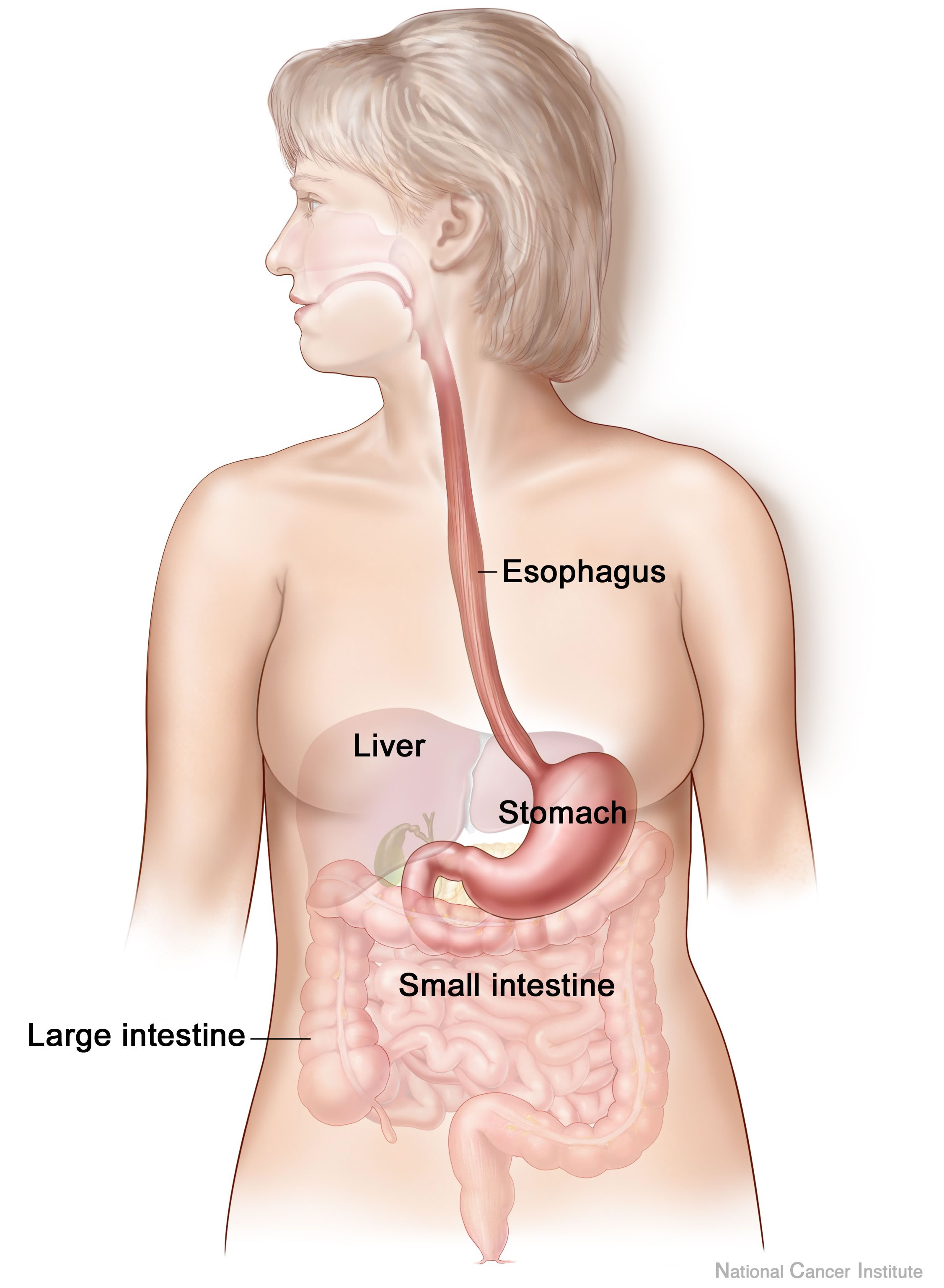
The human digestive system consists of the gastrointestinal tract plus the accessory organs of digestion (the tongue, salivary glands, pancreas, liver, and gallbladder). Digestion involves the breakdown of food into smaller and smaller components, until they can be absorbed and assimilated into the body. The process of digestion has three stages: the cephalic phase, the gastric phase, and the intestinal phase. The first stage, the cephalic phase of digestion, begins with secretions from gastric glands in response to the sight and smell of food, and continues in the human mouth, mouth with the mechanical breakdown of food by chewing, and the chemical breakdown by digestive enzymes in the saliva. Saliva contains amylase, and lingual lipase, secreted by the salivary glands, and serous glands on the tongue. Chewing mixes the food with saliva to produce a Bolus (digestion), bolus to be Swallowing, swallowed down the esophagus to enter the stomach. The second stage, the gastric phase ... [...More Info...] [...Related Items...] OR: [Wikipedia] [Google] [Baidu] [Amazon] |
Pharyngeal Branches Of Ascending Pharyngeal Artery
The ascending pharyngeal artery is an artery of the neck that supplies the pharynx. Its named branches are the inferior tympanic artery, pharyngeal artery, and posterior meningeal artery. inferior tympanic artery, and the meningeal branches (including the posterior meningeal artery). Anatomy The ascending pharyngeal artery is a long and slender vessel. It is deeply seated in the neck, beneath the other branches of the external carotid and under the stylopharyngeus muscle. It lies just superior to the bifurcation of the common carotid arteries. Origin It is the smallest and first medial branch of proximal external carotid artery, arising from the medial surface of the artery. Typically the ascending thyroid artery arises from the external carotid before the ascending pharyngeal, but in variant anatomy the thyroid may arise earlier from the bifurcation or common carotid. Course and relations The artery ascends vertically in between the internal carotid artery and t ... [...More Info...] [...Related Items...] OR: [Wikipedia] [Google] [Baidu] [Amazon] |
Esophagus
The esophagus (American English), oesophagus (British English), or œsophagus (Œ, archaic spelling) (American and British English spelling differences#ae and oe, see spelling difference) all ; : ((o)e)(œ)sophagi or ((o)e)(œ)sophaguses), colloquially known also as the food pipe, food tube, or gullet, is an Organ (anatomy), organ in vertebrates through which food passes, aided by Peristalsis, peristaltic contractions, from the Human pharynx, pharynx to the stomach. The esophagus is a :wiktionary:fibromuscular, fibromuscular tube, about long in adults, that travels behind the trachea and human heart, heart, passes through the Thoracic diaphragm, diaphragm, and empties into the uppermost region of the stomach. During swallowing, the epiglottis tilts backwards to prevent food from going down the larynx and lungs. The word ''esophagus'' is from Ancient Greek οἰσοφάγος (oisophágos), from οἴσω (oísō), future form of φέρω (phérō, "I carry") + ἔφαγον ( ... [...More Info...] [...Related Items...] OR: [Wikipedia] [Google] [Baidu] [Amazon] |
Lung
The lungs are the primary Organ (biology), organs of the respiratory system in many animals, including humans. In mammals and most other tetrapods, two lungs are located near the Vertebral column, backbone on either side of the heart. Their function in the respiratory system is to extract oxygen from the atmosphere and transfer it into the bloodstream, and to release carbon dioxide from the bloodstream into the atmosphere, in a process of gas exchange. Respiration is driven by different muscular systems in different species. Mammals, reptiles and birds use their musculoskeletal systems to support and foster breathing. In early tetrapods, air was driven into the lungs by the pharyngeal muscles via buccal pumping, a mechanism still seen in amphibians. In humans, the primary muscle that drives breathing is the Thoracic diaphragm, diaphragm. The lungs also provide airflow that makes Animal communication#Auditory, vocalisation including speech possible. Humans have two lungs, a ri ... [...More Info...] [...Related Items...] OR: [Wikipedia] [Google] [Baidu] [Amazon] |
Ascending Palatine Artery
The ascending palatine artery is an artery is a branch of the facial artery which ascends along the neck before splitting into two terminal branches; one branch supplies the soft palate, and the other supplies the palatine tonsil and pharyngotympanic tube. Structure Origin The ascending palatine artery arises from the proximal facial artery (close to the facial artery's origin). Course It passes superior-ward between the styloglossus muscle and stylopharyngeus muscle to reach the side of the pharynx. It ascends along the side of the pharynx between the superior pharyngeal constrictor and the medial pterygoid muscle to near the base of the skull. Near the levator veli palatini muscle, the artery splits into its two terminal branches. Branches One terminal branch passes along the levator veli palatini muscle, winding around the superior border of the superior pharyngeal constrictor to provide arterial supply to the soft palate and anastomose with the greater palatine ... [...More Info...] [...Related Items...] OR: [Wikipedia] [Google] [Baidu] [Amazon] |
Epiglottis
The epiglottis (: epiglottises or epiglottides) is a leaf-shaped flap in the throat that prevents food and water from entering the trachea and the lungs. It stays open during breathing, allowing air into the larynx. During swallowing, it closes to prevent aspiration of food into the lungs, forcing the swallowed liquids or food to go along the esophagus toward the stomach instead. It is thus the valve that diverts passage to either the trachea or the esophagus. The epiglottis is made of elastic cartilage covered with a mucous membrane, attached to the entrance of the larynx. It projects upwards and backwards behind the tongue and the hyoid bone. The epiglottis may be inflamed in a condition called epiglottitis, which is most commonly due to the vaccine-preventable bacterium ''Haemophilus influenzae''. Dysfunction may cause the inhalation of food, called aspiration, which may lead to pneumonia or airway obstruction. The epiglottis is also an important landmark for intubation ... [...More Info...] [...Related Items...] OR: [Wikipedia] [Google] [Baidu] [Amazon] |
Larynx
The larynx (), commonly called the voice box, is an organ (anatomy), organ in the top of the neck involved in breathing, producing sound and protecting the trachea against food aspiration. The opening of larynx into pharynx known as the laryngeal inlet is about 4–5 centimeters in diameter. The larynx houses the vocal cords, and manipulates pitch (music), pitch and sound pressure, volume, which is essential for phonation. It is situated just below where the tract of the pharynx splits into the trachea and the esophagus. The word 'larynx' (: larynges) comes from the Ancient Greek word ''lárunx'' ʻlarynx, gullet, throatʼ. Structure The triangle-shaped larynx consists largely of cartilages that are attached to one another, and to surrounding structures, by muscles or by fibrous and elastic tissue components. The larynx is lined by a respiratory epithelium, ciliated columnar epithelium except for the vocal folds. The laryngeal cavity, cavity of the larynx extends from its tria ... [...More Info...] [...Related Items...] OR: [Wikipedia] [Google] [Baidu] [Amazon] |
Pharyngeal Plexus Of Vagus Nerve
The pharyngeal plexus is a nerve plexus located upon the outer surface of the pharynx. It contains a motor component (derived from the vagus nerve (cranial nerve X)), a sensory component (derived from the glossopharyngeal nerve (cranial nerve IX)), and sympathetic component (derived from the superior cervical ganglion). The plexus provides motor innervation to most muscles of the soft palate (all but the tensor veli palatini muscle) and most muscles of the pharynx (all but the stylopharyngeus muscle). The larynx meanwhile receives motor innervation from the vagus nerve (CN X) via its external branch of the superior laryngeal nerve and its recurrent laryngeal nerve, and ''not'' through the pharyngeal plexus. Anatomy The pharyngeal plexus occurs upon the outer surface of the pharynx - especially superficial to the middle pharyngeal constrictor muscle. Afferents It has the following components: * Motor – pharyngeal branch of vagus nerve (CN X) which arises from the ... [...More Info...] [...Related Items...] OR: [Wikipedia] [Google] [Baidu] [Amazon] |
Pharyngeal Muscles
The pharyngeal muscles are a group of muscles that form the pharynx, which is posterior to the oral cavity, determining the shape of its lumen, and affecting its sound properties as the primary resonating cavity. The pharyngeal muscles (involuntary skeletal) push food into the esophagus. There are two muscular layers of the pharynx: the outer circular layer and the inner longitudinal layer. The outer circular layer includes: * Superior constrictor muscle * Middle constrictor muscle * Inferior constrictor muscle During swallowing, these muscles constrict to propel a bolus downwards (an involuntary process). The inner longitudinal layer includes: * Stylopharyngeus muscle * Salpingopharyngeus muscle * Palatopharyngeus muscle During swallowing, these muscles act to shorten and widen the pharynx. They are innervated by the pharyngeal branch of the vagus nerve (CN X) with the exception of the stylopharyngeus muscle which is innervated by the glossopharyngeal nerve The gl ... [...More Info...] [...Related Items...] OR: [Wikipedia] [Google] [Baidu] [Amazon] |
Bronchus
A bronchus ( ; : bronchi, ) is a passage or airway in the lower respiratory tract that conducts air into the lungs. The first or primary bronchi to branch from the trachea at the carina are the right main bronchus and the left main bronchus. These are the widest bronchi, and enter the right lung, and the left lung at each hilum. The main bronchi branch into narrower secondary bronchi or lobar bronchi, and these branch into narrower tertiary bronchi or segmental bronchi. Further divisions of the segmental bronchi are known as 4th order, 5th order, and 6th order segmental bronchi, or grouped together as subsegmental bronchi. The bronchi, when too narrow to be supported by cartilage, are known as bronchioles. No gas exchange takes place in the bronchi. Structure The trachea (windpipe) divides at the carina into two main or primary bronchi, the left bronchus and the right bronchus. The carina of the trachea is located at the level of the sternal angle and the fifth thoracic ver ... [...More Info...] [...Related Items...] OR: [Wikipedia] [Google] [Baidu] [Amazon] |
Human Nose
The human nose is the first organ of the respiratory system. It is also the principal organ in the olfactory system. The shape of the nose is determined by the nasal bones and the nasal cartilages, including the nasal septum, which separates the nostrils and divides the nasal cavity into two. The nose has an important function in breathing. The nasal mucosa lining the nasal cavity and the paranasal sinuses carries out the necessary conditioning of inhaled air by warming and moistening it. Nasal conchae, shell-like bones in the walls of the cavities, play a major part in this process. Filtering of the air by nasal hair in the nostrils prevents large particles from entering the lungs. Sneezing is a reflex to expel unwanted particles from the nose that irritate the mucosal lining. Sneezing can Transmission (medicine), transmit infections, because aerosols are created in which the Respiratory droplets, droplets can harbour pathogens. Another major function of the nose is olfactio ... [...More Info...] [...Related Items...] OR: [Wikipedia] [Google] [Baidu] [Amazon] |
Bronchiole
The bronchioles ( ) are the smaller branches of the bronchial airways in the lower respiratory tract. They include the terminal bronchioles, and finally the respiratory bronchioles that mark the start of the respiratory zone delivering air to the gas exchanging units of the alveoli. The bronchioles no longer contain the cartilage that is found in the bronchi, or glands in their submucosa. Structure The pulmonary lobule is the portion of the lung ventilated by one bronchiole. Bronchioles are approximately 1 mm or less in diameter and their walls consist of ciliated cuboidal epithelium and a layer of smooth muscle. Bronchioles divide into even smaller bronchioles, called ''terminal'', which are 0.5 mm or less in diameter. Terminal bronchioles in turn divide into smaller respiratory bronchioles which divide into alveolar ducts. Terminal bronchioles mark the end of the conducting division of air flow in the respiratory system while respiratory bronchioles are t ... [...More Info...] [...Related Items...] OR: [Wikipedia] [Google] [Baidu] [Amazon] |


