|
Parinaud Syndrome
Parinaud's syndrome is an inability to move the eyes up and down. It is caused by compression of the vertical gaze center at the rostral interstitial nucleus of medial longitudinal fasciculus (riMLF). The eyes lose the ability to move upward and down. It is a group of abnormalities of eye movement and pupil dysfunction. It is caused by lesions of the upper brain stem and is named for Henri Parinaud (1844–1905), considered to be the father of French ophthalmology. Signs and symptoms Parinaud's syndrome is a cluster of abnormalities of eye movement and pupil dysfunction, characterized by: * Paralysis of upwards gaze: Downward gaze is usually preserved. This vertical palsy is supranuclear, so doll's head maneuver should elevate the eyes, but eventually all upward gaze mechanisms fail. * Pseudo- Argyll Robertson pupils: Accommodative paresis ensues, and pupils become mid-dilated and show light-near dissociation. * Convergence-retraction nystagmus: Attempts at upward gaze often ... [...More Info...] [...Related Items...] OR: [Wikipedia] [Google] [Baidu] |
Rostral Interstitial Nucleus Of Medial Longitudinal Fasciculus
The rostral interstitial nucleus of medial longitudinal fasciculus (riMLF) is a portion of the medial longitudinal fasciculus which controls Gaze (physiology), vertical gaze. They project to the vestibular nuclei. External links * https://uni-tuebingen.de/uni/knl/Vilis/originof.htm * http://anatomy.umc.edu/faculty/lynch.html * https://web.archive.org/web/20091021004541/http://isc.temple.edu/neuroanatomy/lab/atlas/papc/ Brainstem {{Neuroanatomy-stub ... [...More Info...] [...Related Items...] OR: [Wikipedia] [Google] [Baidu] |
Esotropia
Esotropia is a form of strabismus in which one or both eyes turns inward. The condition can be constantly present, or occur intermittently, and can give the affected individual a "cross-eyed" appearance. It is the opposite of exotropia and usually involves more severe axis deviation than esophoria. Esotropia is sometimes erroneously called "lazy eye", which describes the condition of amblyopia; a reduction in vision of one or both eyes that is not the result of any pathology of the eye and cannot be resolved by the use of corrective lenses. Amblyopia can, however, arise as a result of esotropia occurring in childhood: In order to relieve symptoms of diplopia or double vision, the child's brain will ignore or "suppress" the image from the esotropic eye, which when allowed to continue untreated will lead to the development of amblyopia. Treatment options for esotropia include glasses to correct refractive errors (see accommodative esotropia below), the use of prisms and/or orthopti ... [...More Info...] [...Related Items...] OR: [Wikipedia] [Google] [Baidu] |
Pineal Gland
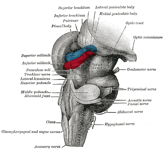
The pineal gland, conarium, or epiphysis cerebri, is a small endocrine gland in the brain of most vertebrates. The pineal gland produces melatonin, a serotonin-derived hormone which modulates sleep, sleep patterns in both circadian rhythm, circadian and Season, seasonal cycles. The shape of the gland resembles a pine cone, which gives it its name. The pineal gland is located in the epithalamus, near the center of the brain, between the two cerebral hemisphere, hemispheres, tucked in a groove where the two halves of the thalamus join. The pineal gland is one of the neuroendocrinology, neuroendocrine Circumventricular organs, secretory circumventricular organs in which capillaries are mostly Vascular permeability, permeable to solutes in the blood. Nearly all vertebrate species possess a pineal gland. The most important exception is a primitive vertebrate, the hagfish. Even in the hagfish, however, there may be a "pineal equivalent" structure in the dorsal diencephalon. The lanc ... [...More Info...] [...Related Items...] OR: [Wikipedia] [Google] [Baidu] |
Brain Tumors
A brain tumor occurs when abnormal cells form within the brain. There are two main types of tumors: malignant tumors and benign (non-cancerous) tumors. These can be further classified as primary tumors, which start within the brain, and secondary tumors, which most commonly have spread from tumors located outside the brain, known as brain metastasis tumors. All types of brain tumors may produce symptoms that vary depending on the size of the tumor and the part of the brain that is involved. Where symptoms exist, they may include headaches, seizures, problems with vision, vomiting and mental changes. Other symptoms may include difficulty walking, speaking, with sensations, or unconsciousness. The cause of most brain tumors is unknown. Uncommon risk factors include exposure to vinyl chloride, Epstein–Barr virus, ionizing radiation, and inherited syndromes such as neurofibromatosis, tuberous sclerosis, and von Hippel-Lindau Disease. Studies on mobile phone exposure have not s ... [...More Info...] [...Related Items...] OR: [Wikipedia] [Google] [Baidu] |
Cranial Nerve
Cranial nerves are the nerves that emerge directly from the brain (including the brainstem), of which there are conventionally considered twelve pairs. Cranial nerves relay information between the brain and parts of the body, primarily to and from regions of the head and neck, including the special senses of vision, taste, smell, and hearing. The cranial nerves emerge from the central nervous system above the level of the first vertebra of the vertebral column. Each cranial nerve is paired and is present on both sides. There are conventionally twelve pairs of cranial nerves, which are described with Roman numerals I–XII. Some considered there to be thirteen pairs of cranial nerves, including cranial nerve zero. The numbering of the cranial nerves is based on the order in which they emerge from the brain and brainstem, from front to back. The terminal nerves (0), olfactory nerves (I) and optic nerves (II) emerge from the cerebrum, and the remaining ten pairs arise from t ... [...More Info...] [...Related Items...] OR: [Wikipedia] [Google] [Baidu] |
Oculomotor
The oculomotor nerve, also known as the third cranial nerve, cranial nerve III, or simply CN III, is a cranial nerve that enters the orbit (anatomy), orbit through the superior orbital fissure and innervates extraocular muscles that enable most eye movement, movements of the eye and that raise the eyelid. The nerve also contains fibers that innervate the intrinsic eye muscles that enable pupillary constriction and accommodation (ability to focus on near objects as in reading). The oculomotor nerve is derived from the Basal plate (neural tube), basal plate of the embryonic midbrain. Cranial nerves trochlear nerve, IV and abducens, VI also participate in control of Eye movement (sensory), eye movement. Structure The oculomotor nerve originates from the third nerve nucleus (neuroanatomy), nucleus at the level of the superior colliculus in the midbrain. The third nerve nucleus is located ventral to the cerebral aqueduct, on the pre-aqueductal grey matter. The fibers from the two ... [...More Info...] [...Related Items...] OR: [Wikipedia] [Google] [Baidu] |
Superior Colliculus
In neuroanatomy, the superior colliculus () is a structure lying on the roof of the mammalian midbrain. In non-mammalian vertebrates, the homologous structure is known as the optic tectum, or optic lobe. The adjective form ''tectal'' is commonly used for both structures. In mammals, the superior colliculus forms a major component of the midbrain. It is a paired structure and together with the paired inferior colliculi forms the corpora quadrigemina. The superior colliculus is a layered structure, with a pattern that is similar to all mammals. The layers can be grouped into the superficial layers ( stratum opticum and above) and the deeper remaining layers. Neurons in the superficial layers receive direct input from the retina and respond almost exclusively to visual stimuli. Many neurons in the deeper layers also respond to other modalities, and some respond to stimuli in multiple modalities. The deeper layers also contain a population of motor-related neurons, capable of activat ... [...More Info...] [...Related Items...] OR: [Wikipedia] [Google] [Baidu] |
Mesencephalic
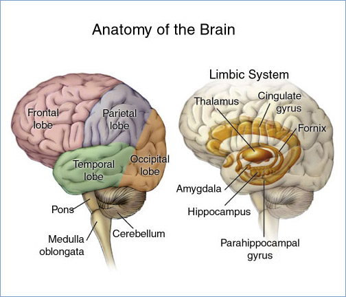
The midbrain or mesencephalon is the forward-most portion of the brainstem and is associated with vision, hearing, motor control, sleep and wakefulness, arousal (alertness), and temperature regulation. The name comes from the Greek ''mesos'', "middle", and ''enkephalos'', "brain". Structure The principal regions of the midbrain are the tectum, the cerebral aqueduct, tegmentum, and the cerebral peduncles. Rostrally the midbrain adjoins the diencephalon (thalamus, hypothalamus, etc.), while caudally it adjoins the hindbrain (pons, medulla and cerebellum). In the rostral direction, the midbrain noticeably splays laterally. Sectioning of the midbrain is usually performed axially, at one of two levels – that of the superior colliculi, or that of the inferior colliculi. One common technique for remembering the structures of the midbrain involves visualizing these cross-sections (especially at the level of the superior colliculi) as the upside-down face of a bear ... [...More Info...] [...Related Items...] OR: [Wikipedia] [Google] [Baidu] |
Midbrain
The midbrain or mesencephalon is the forward-most portion of the brainstem and is associated with vision, hearing, motor control, sleep and wakefulness, arousal (alertness), and temperature regulation. The name comes from the Greek ''mesos'', "middle", and ''enkephalos'', "brain". Structure The principal regions of the midbrain are the tectum, the cerebral aqueduct, tegmentum, and the cerebral peduncles. Rostrally the midbrain adjoins the diencephalon (thalamus, hypothalamus, etc.), while caudally it adjoins the hindbrain (pons, medulla and cerebellum). In the rostral direction, the midbrain noticeably splays laterally. Sectioning of the midbrain is usually performed axially, at one of two levels – that of the superior colliculi, or that of the inferior colliculi. One common technique for remembering the structures of the midbrain involves visualizing these cross-sections (especially at the level of the superior colliculi) as the upside-down face of a be ... [...More Info...] [...Related Items...] OR: [Wikipedia] [Google] [Baidu] |
Parinaud's Syndrome
Parinaud's syndrome is an inability to move the eyes up and down. It is caused by compression of the vertical gaze center at the rostral interstitial nucleus of medial longitudinal fasciculus (riMLF). The eyes lose the ability to move upward and down. It is a group of abnormalities of eye movement and pupil dysfunction. It is caused by lesions of the upper brain stem and is named for Henri Parinaud (1844–1905), considered to be the father of French ophthalmology. Signs and symptoms Parinaud's syndrome is a cluster of abnormalities of eye movement and pupil dysfunction, characterized by: * Paralysis of upwards gaze: Downward gaze is usually preserved. This vertical palsy is supranuclear, so doll's head maneuver should elevate the eyes, but eventually all upward gaze mechanisms fail. * Pseudo- Argyll Robertson pupils: Accommodative paresis ensues, and pupils become mid-dilated and show light-near dissociation. * Convergence-retraction nystagmus: Attempts at upward gaze often p ... [...More Info...] [...Related Items...] OR: [Wikipedia] [Google] [Baidu] |
Internuclear Ophthalmoplegia
Internuclear ophthalmoplegia (INO) is a disorder of conjugate lateral gaze in which the affected eye shows impairment of adduction. When an attempt is made to gaze contralaterally (relative to the affected eye), the affected eye adducts minimally, if at all. The contralateral eye abducts, however with nystagmus. Additionally, the divergence of the eyes leads to horizontal diplopia. That is if the right eye is affected the patient will "see double" when looking to the left, seeing two images side-by-side. Convergence is generally preserved. Causes The disorder is caused by injury or dysfunction in the medial longitudinal fasciculus (MLF), a heavily myelinated tract that allows conjugate eye movement by connecting the paramedian pontine reticular formation (PPRF)-abducens nucleus complex of the contralateral side to the oculomotor nucleus of the ipsilateral side. In young patients with bilateral INO, multiple sclerosis is often the cause. In older patients with one-sided lesio ... [...More Info...] [...Related Items...] OR: [Wikipedia] [Google] [Baidu] |


