|
Pyramidal Process Of The Palatine Bone
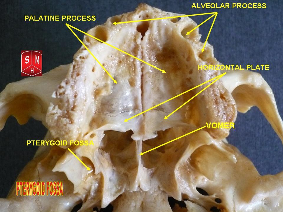
The pyramidal process of the palatine bone projects backward and lateralward from the junction of the horizontal and vertical parts, and is received into the angular interval between the lower extremities of the pterygoid plates. On its posterior surface is a smooth, grooved, triangular area, limited on either side by a rough articular furrow. The furrows articulate with the pterygoid plates, while the grooved intermediate area completes the lower part of the pterygoid fossa and gives origin to a few fibers of the Pterygoideus internus. The anterior part of the lateral surface is rough, for articulation with the tuberosity of the maxilla; its posterior part consists of a smooth triangular area which appears, in the articulated skull, between the tuberosity of the maxilla and the lower part of the lateral pterygoid plate, and completes the lower part of the infratemporal fossa. On the base of the pyramidal process, close to its union with the horizontal part, are the lesser palati ... [...More Info...] [...Related Items...] OR: [Wikipedia] [Google] [Baidu] |
Palatine Bone
In anatomy, the palatine bones () are two irregular bones of the facial skeleton in many animal species, located above the uvula in the throat. Together with the maxillae, they comprise the hard palate. (''Palate'' is derived from the Latin ''palatum''.) Structure The palatine bones are situated at the back of the nasal cavity between the maxilla and the pterygoid process of the sphenoid bone. They contribute to the walls of three cavities: the floor and lateral walls of the nasal cavity, the roof of the mouth, and the floor of the orbits. They help to form the pterygopalatine and pterygoid fossae, and the inferior orbital fissures. Each palatine bone somewhat resembles the letter L, and consists of a horizontal plate, a perpendicular plate, and three projecting processes—the pyramidal process, which is directed backward and lateral from the junction of the two parts, and the orbital and sphenoidal processes, which surmount the vertical part, and are separated by a dee ... [...More Info...] [...Related Items...] OR: [Wikipedia] [Google] [Baidu] |
Pterygoid Plate
The pterygoid processes of the sphenoid (from Greek ''pteryx'', ''pterygos'', "wing"), one on either side, descend perpendicularly from the regions where the body and the greater wings of the sphenoid bone unite. Each process consists of a medial pterygoid plate and a lateral pterygoid plate, the latter of which serve as the origins of the medial and lateral pterygoid muscles. The medial pterygoid, along with the masseter allows the jaw to move in a vertical direction as it contracts and relaxes. The lateral pterygoid allows the jaw to move in a horizontal direction during mastication (chewing). Fracture of either plate are used in clinical medicine to distinguish the Le Fort fracture classification for high impact injuries to the sphenoid and maxillary bones. The superior portion of the pterygoid processes are fused anteriorly; a vertical groove, the pterygopalatine fossa, descends on the front of the line of fusion. The plates are separated below by an angular cleft, the pt ... [...More Info...] [...Related Items...] OR: [Wikipedia] [Google] [Baidu] |
Pterygoid Fossa
The pterygoid fossa is an anatomical term for the fossa formed by the divergence of the lateral pterygoid plate and the medial pterygoid plate of the sphenoid bone. Structure The lateral and medial pterygoid plates (of the pterygoid process of the sphenoid bone) diverge behind and enclose between them a V-shaped fossa, the pterygoid fossa. This fossa faces posteriorly, and contains the medial pterygoid muscle and the tensor veli palatini muscle. See also * Pterygoid fovea * Scaphoid fossa * Pterygoid process The pterygoid processes of the sphenoid (from Greek ''pteryx'', ''pterygos'', "wing"), one on either side, descend perpendicularly from the regions where the body and the greater wings of the sphenoid bone unite. Each process consists of a me ... References Bones of the head and neck {{musculoskeletal-stub ... [...More Info...] [...Related Items...] OR: [Wikipedia] [Google] [Baidu] |
Pterygoideus Internus
The medial pterygoid muscle (or internal pterygoid muscle), is a thick, quadrilateral muscle of the face. It is supplied by the mandibular branch of the trigeminal nerve (V). It is important in mastication (chewing). Structure The medial pterygoid muscle consists of two heads. The bulk of the muscle arises as a deep head from just above the medial surface of the lateral pterygoid plate. The smaller, superficial head originates from the maxillary tuberosity and the pyramidal process of the palatine bone. Its fibers pass downward, lateral, and posterior, and are inserted, by a strong tendinous lamina, into the lower and back part of the medial surface of the ramus and angle of the mandible, as high as the mandibular foramen. The insertion joins the masseter muscle to form a common tendinous sling which allows the medial pterygoid and masseter to be powerful elevators of the jaw. Nerve supply The medial pterygoid muscle is supplied by the medial pterygoid nerve, a branch of ... [...More Info...] [...Related Items...] OR: [Wikipedia] [Google] [Baidu] |
Lesser Palatine Foramina
Behind the greater palatine foramen is the pyramidal process of the palatine bone, perforated by one or more lesser palatine foramina which carry the lesser palatine nerve, and marked by the commencement of a transverse ridge, for the attachment of the tendinous expansion of the tensor veli palatini. See also * Greater palatine foramen At either posterior angle of the hard palate is the greater palatine foramen, for the transmission of the descending palatine vessels and greater palatine nerve; and running anteriorly (forward) and medially (towards the center-line) from it is a g ... References External links * Bones of the head and neck {{musculoskeletal-stub ... [...More Info...] [...Related Items...] OR: [Wikipedia] [Google] [Baidu] |
Palatine Nerve
In neuroanatomy, the maxillary nerve (V) is one of the three branches or divisions of the trigeminal nerve, the fifth (CN V) cranial nerve. It comprises the principal functions of sensation from the maxilla, nasal cavity, sinuses, the palate and subsequently that of the mid-face, and is intermediate, both in position and size, between the ophthalmic nerve and the mandibular nerve.Illustrated Anatomy of the Head and Neck, Fehrenbach and Herring, Elsevier, 2012, page 180 Structure It begins at the middle of the trigeminal ganglion as a flattened plexiform band then it passes through the lateral wall of the cavernous sinus. It leaves the skull through the foramen rotundum, where it becomes more cylindrical in form, and firmer in texture. After leaving foramen rotundum it gives two branches to the pterygopalatine ganglion. It then crosses the pterygopalatine fossa, inclines lateralward on the back of the maxilla, and enters the orbit through the inferior orbital fissure. It then ru ... [...More Info...] [...Related Items...] OR: [Wikipedia] [Google] [Baidu] |
