|
Principal Sensory Nucleus Of Trigeminal Nerve
The principal sensory nucleus of trigeminal nerve (or chief sensory nucleus of V, main trigeminal sensory nucleus) is a group of second-order neurons which have cell bodies in the caudal pons. It receives information about discriminative sensation and light touch of the face as well as conscious proprioception of the jaw via first order neurons of CN V. * Most of the sensory information crosses the midline and travels to the contralateral ventral posteromedial nucleus (VPM) of the thalamus via the anterior trigeminothalamic tract. * However, information of the ''oral cavity'' travels to the ipsilateral VPM of the thalamus via the dorsal trigeminothalamic tract The dorsal trigeminal tract, dorsal trigeminothalamic tract, or posterior trigeminothalamic tract, is composed of second-order neuronal axons. These fibers carry sensory information about discriminative touch and conscious proprioception in the o .... {{Authority control Cranial nerve nuclei Trigeminal nerve P ... [...More Info...] [...Related Items...] OR: [Wikipedia] [Google] [Baidu] |
Second-order Neuron
In physiology, the somatosensory system is the network of neural structures in the brain and body that produce the perception of touch (haptic perception), as well as temperature (thermoception), body position (proprioception), and pain. It is a subset of the sensory nervous system, which also represents visual, auditory, olfactory, and gustatory stimuli. Somatosensation begins when mechano- and thermosensitive structures in the skin or internal organs sense physical stimuli such as pressure on the skin (see mechanotransduction, nociception). Activation of these structures, or receptors, leads to activation of peripheral sensory neurons that convey signals to the spinal cord as patterns of action potentials. Sensory information is then processed locally in the spinal cord to drive reflexes, and is also conveyed to the brain for conscious perception of touch and proprioception. Note, somatosensory information from the face and head enters the brain through peripheral senso ... [...More Info...] [...Related Items...] OR: [Wikipedia] [Google] [Baidu] |
Pons
The pons (from Latin , "bridge") is part of the brainstem that in humans and other bipeds lies inferior to the midbrain, superior to the medulla oblongata and anterior to the cerebellum. The pons is also called the pons Varolii ("bridge of Varolius"), after the Italian anatomist and surgeon Costanzo Varolio (1543–75). This region of the brainstem includes neural pathways and tracts that conduct signals from the brain down to the cerebellum and medulla, and tracts that carry the sensory signals up into the thalamus.Saladin Kenneth S.(2007) Anatomy & physiology the unity of form and function. Dubuque, IA: McGraw-Hill Structure The pons is in the brainstem situated between the midbrain and the medulla oblongata, and in front of the cerebellum. A separating groove between the pons and the medulla is the inferior pontine sulcus. The superior pontine sulcus separates the pons from the midbrain. The pons can be broadly divided into two parts: the basilar part of the pons ( ... [...More Info...] [...Related Items...] OR: [Wikipedia] [Google] [Baidu] |
Proprioception
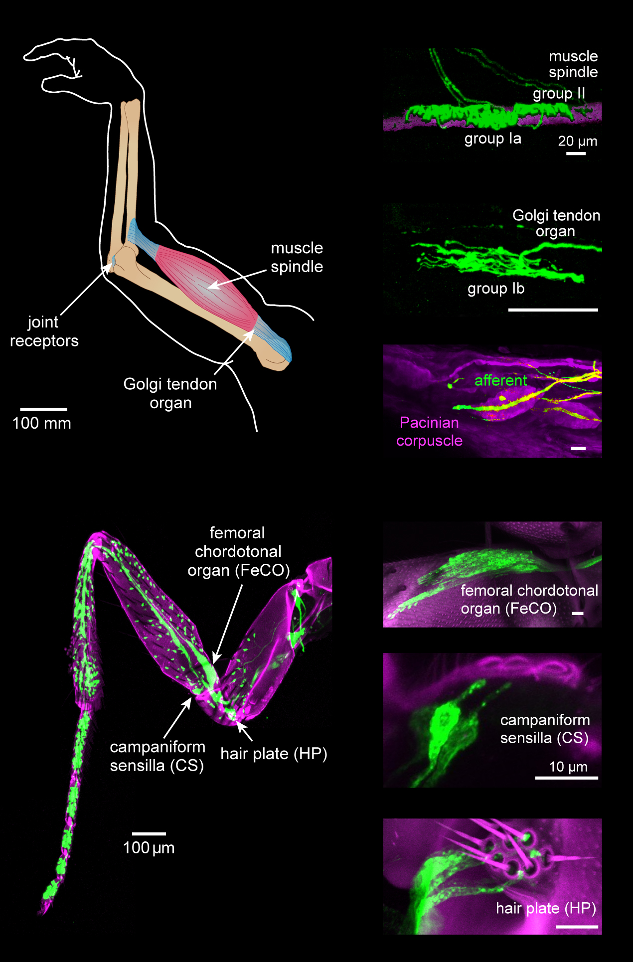
Proprioception ( ), also referred to as kinaesthesia (or kinesthesia), is the sense of self-movement, force, and body position. It is sometimes described as the "sixth sense". Proprioception is mediated by proprioceptors, mechanosensory neurons located within muscles, tendons, and joints. Most animals possess multiple subtypes of proprioceptors, which detect distinct kinematic parameters, such as joint position, movement, and load. Although all mobile animals possess proprioceptors, the structure of the sensory organs can vary across species. Proprioceptive signals are transmitted to the central nervous system, where they are integrated with information from other sensory systems, such as the visual system and the vestibular system, to create an overall representation of body position, movement, and acceleration. In many animals, sensory feedback from proprioceptors is essential for stabilizing body posture and coordinating body movement. System overview In vertebrates, limb ... [...More Info...] [...Related Items...] OR: [Wikipedia] [Google] [Baidu] |
CN V
In neuroanatomy, the trigeminal nerve ( lit. ''triplet'' nerve), also known as the fifth cranial nerve, cranial nerve V, or simply CN V, is a cranial nerve responsible for sensation in the face and motor functions such as biting and chewing; it is the most complex of the cranial nerves. Its name ("trigeminal", ) derives from each of the two nerves (one on each side of the pons) having three major branches: the ophthalmic nerve (V), the maxillary nerve (V), and the mandibular nerve (V). The ophthalmic and maxillary nerves are purely sensory, whereas the mandibular nerve supplies motor as well as sensory (or "cutaneous") functions. Adding to the complexity of this nerve is that autonomic nerve fibers as well as special sensory fibers (taste) are contained within it. The motor division of the trigeminal nerve derives from the basal plate of the embryonic pons, and the sensory division originates in the cranial neural crest. Sensory information from the face and body is proce ... [...More Info...] [...Related Items...] OR: [Wikipedia] [Google] [Baidu] |
Anatomical Terms Of Location
Standard anatomical terms of location are used to unambiguously describe the anatomy of animals, including humans. The terms, typically derived from Latin or Greek roots, describe something in its standard anatomical position. This position provides a definition of what is at the front ("anterior"), behind ("posterior") and so on. As part of defining and describing terms, the body is described through the use of anatomical planes and anatomical axes. The meaning of terms that are used can change depending on whether an organism is bipedal or quadrupedal. Additionally, for some animals such as invertebrates, some terms may not have any meaning at all; for example, an animal that is radially symmetrical will have no anterior surface, but can still have a description that a part is close to the middle ("proximal") or further from the middle ("distal"). International organisations have determined vocabularies that are often used as standard vocabularies for subdisciplines o ... [...More Info...] [...Related Items...] OR: [Wikipedia] [Google] [Baidu] |
Ventral Posteromedial Nucleus
The ventral posteromedial nucleus (VPM) is a nucleus of the thalamus. Inputs and outputs The VPM contains synapses between second and third order neurons from the anterior (ventral) trigeminothalamic tract and posterior (dorsal) trigeminothalamic tract. These neurons convey sensory information from the face and oral cavity. Third order neurons in the trigeminothalamic systems project to the postcentral gyrus. The VPM also receives taste afferent information from the solitary tract and projects to the cortical gustatory area The primary gustatory cortex is a brain structure responsible for the perception of taste. It consists of two substructures: the anterior insula on the insular lobe and the frontal operculum on the inferior frontal gyrus of the frontal lobe. B .... Subareas VPMpc The parvicellular part of the ventroposterior medial nucleus (VPMpc) is argued by some as not an actually part of the VPM, because it does not project to the somatosensory cortex as the r ... [...More Info...] [...Related Items...] OR: [Wikipedia] [Google] [Baidu] |
Anterior Trigeminothalamic Tract
The ventral trigeminal tract, ventral trigeminothalamic tract, anterior trigeminal tract, or anterior trigeminothalamic tract, is a tract composed of second order neuronal axons. These fibers carry sensory information about discriminative and crude touch, conscious proprioception, pain, and temperature from the head, face, and oral cavity. The ventral trigeminal tract connects the two major components of the brainstem trigeminal complex – the principal, or main sensory nucleus and the spinal trigeminal nucleus, to the ventral posteromedial nucleus of the thalamus. The ventral trigeminal tract is also called the anterior trigeminal lemniscus. Structure The first order neurons (from the trigeminal ganglion) enter the pons and synapse in the principal (chief sensory) nucleus or spinal trigeminal nucleus. Axons of the second order neurons cross the midline and terminate in the ventral posteromedial nucleus of the contralateral thalamus (as opposed to the ventral posterolateral nu ... [...More Info...] [...Related Items...] OR: [Wikipedia] [Google] [Baidu] |
Dorsal Trigeminothalamic Tract
The dorsal trigeminal tract, dorsal trigeminothalamic tract, or posterior trigeminothalamic tract, is composed of second-order neuronal axons. These fibers carry sensory information about discriminative touch and conscious proprioception in the oral cavity from the principal (chief sensory) nucleus of the trigeminal nerve to the ventral posteromedial (VPM) nucleus of the thalamus. The dorsal trigeminothalamic tract is also called the posterior trigeminal lemniscus. Pathway The first-order neurons (from the trigeminal ganglion) enter the pons and synapse on second-order neurons in the principal (chief sensory) nucleus. Axons of the second-order neurons then decussate to enter the trigeminal lemniscus in the midbrain and then ascend to the ventral posteromedial nucleus of the contralateral thalamus, forming the ventral trigeminothalamic tract. A subset of these fibers do not decussate and travel to the ipsilateral ventral posteromedial nucleus of the thalamus. These non ... [...More Info...] [...Related Items...] OR: [Wikipedia] [Google] [Baidu] |
Cranial Nerve Nuclei
A cranial nerve nucleus is a collection of neurons ( gray matter) in the brain stem that is associated with one or more of the cranial nerves. Axons carrying information to and from the cranial nerves form a synapse first at these nuclei. Lesions occurring at these nuclei can lead to effects resembling those seen by the severing of nerve(s) they are associated with. All the nuclei except that of the trochlear nerve (CN IV) supply nerves of the same side of the body. Structure Motor and sensory In general, motor nuclei are closer to the front ( ventral), and sensory nuclei and neurons are closer to the back ( dorsal). This arrangement mirrors the arrangement of tracts in the spinal cord. * Close to the midline are the motor efferent nuclei, such as the oculomotor nucleus, which control skeletal muscle. Just lateral to this are the autonomic (or visceral) efferent nuclei. * There is a separation, called the sulcus limitans, and lateral to this are the sensory nuclei. Near th ... [...More Info...] [...Related Items...] OR: [Wikipedia] [Google] [Baidu] |
Trigeminal Nerve
In neuroanatomy, the trigeminal nerve (literal translation, lit. ''triplet'' nerve), also known as the fifth cranial nerve, cranial nerve V, or simply CN V, is a cranial nerve responsible for Sense, sensation in the face and motor functions such as biting and chewing; it is the most complex of the cranial nerves. Its name ("trigeminal", ) derives from each of the two nerves (one on each side of the pons) having three major branches: the ophthalmic nerve (V), the maxillary nerve (V), and the mandibular nerve (V). The ophthalmic and maxillary nerves are purely sensory, whereas the mandibular nerve supplies motor as well as sensory (or "cutaneous") functions. Adding to the complexity of this nerve is that Autonomic nervous system, autonomic nerve fibers as well as special sensory fibers (taste) are contained within it. The motor division of the trigeminal nerve derives from the Basal plate (neural tube), basal plate of the embryonic pons, and the sensory division originates in t ... [...More Info...] [...Related Items...] OR: [Wikipedia] [Google] [Baidu] |


