|
Primary Mediastinal B Cell Lymphoma
Primary mediastinal B-cell lymphoma, abbreviated PMBL, is a rare type of lymphoma that forms in the mediastinum (the space in between the lungs) and predominantly affects young adults. It is a subtype of diffuse large B-cell lymphoma; however, it generally has a significantly better prognosis. Diagnosis Diagnosis requires a biopsy, so that the exact type of tissue can be determined by examination under a microscope. PMBL is generally considered a sub-type of diffuse large B-cell lymphoma, although it is also closely related to nodular sclerosing Hodgkin lymphoma (NSHL). Tumors that are even more closely related to NSHL than typical for PMBL are called gray zone lymphoma. Treatment Treatment commonly begins with months of multi-drug chemotherapy regimen. Either R-CHOP (rituximab, cyclophosphamide, doxorubicin, vincristine, prednisolone) or DA-EPOCH-R (dose-adjusted etoposide, prednisolone, vincristine, cyclophosphamide, doxorubicin, rituximab) has been typical. Other, mo ... [...More Info...] [...Related Items...] OR: [Wikipedia] [Google] [Baidu] |
Micrograph
A micrograph or photomicrograph is a photograph or digital image taken through a microscope or similar device to show a magnified image of an object. This is opposed to a macrograph or photomacrograph, an image which is also taken on a microscope but is only slightly magnified, usually less than 10 times. Micrography is the practice or art of using microscopes to make photographs. A micrograph contains extensive details of microstructure. A wealth of information can be obtained from a simple micrograph like behavior of the material under different conditions, the phases found in the system, failure analysis, grain size estimation, elemental analysis and so on. Micrographs are widely used in all fields of microscopy. Types Photomicrograph A light micrograph or photomicrograph is a micrograph prepared using an optical microscope, a process referred to as ''photomicroscopy''. At a basic level, photomicroscopy may be performed simply by connecting a camera to a microscope, th ... [...More Info...] [...Related Items...] OR: [Wikipedia] [Google] [Baidu] |
Gray Zone Lymphoma
Gray zone lymphoma, often presenting as large tumors in the mediastinum, is a type of lymphoma that is characterized by having cellular features of both classic Hodgkin's lymphomas (cHL) and large B-cell lymphomas. Sources * Traverse-Glehen A, Mediastinal gray zone lymphoma: the missing link between classic Hodgkin's lymphoma and mediastinal large B-cell lymphoma The B-cell lymphomas are types of lymphoma affecting B cells. Lymphomas are "blood cancers" in the lymph nodes. They develop more frequently in older adults and in immunocompromised individuals. B-cell lymphomas include both Hodgkin's lympho .... Am J Surg Pathol 2005 Nov; 29(11):1411-21 Hodgkin’s lymphoma and Grey-zone lymphomas Lymphoma {{oncology-stub ... [...More Info...] [...Related Items...] OR: [Wikipedia] [Google] [Baidu] |
Diffuse Large B-cell Lymphoma
Diffuse large B-cell lymphoma (DLBCL) is a cancer of B cells, a type of lymphocyte that is responsible for producing antibody, antibodies. It is the most common form of non-Hodgkin lymphoma among adults, with an annual Incidence (epidemiology), incidence of 7–8 cases per 100,000 people per year in the US and UK. This cancer occurs primarily in older individuals, with a median age of diagnosis at ~70 years, although it can occur in young adults and, in rare cases, children. DLBCL can arise in virtually any part of the body and, depending on various factors, is often a very aggressive malignancy. The first sign of this illness is typically the observation of a rapidly growing mass or tissue infiltration that is sometimes associated with systemic B symptoms, e.g. fever, Cachexia, weight loss, and night sweats. The causes of diffuse large B-cell lymphoma are not well understood. Usually DLBCL arises from normal B cells, but it can also represent a malignant transformation of other ty ... [...More Info...] [...Related Items...] OR: [Wikipedia] [Google] [Baidu] |
FDG-PET
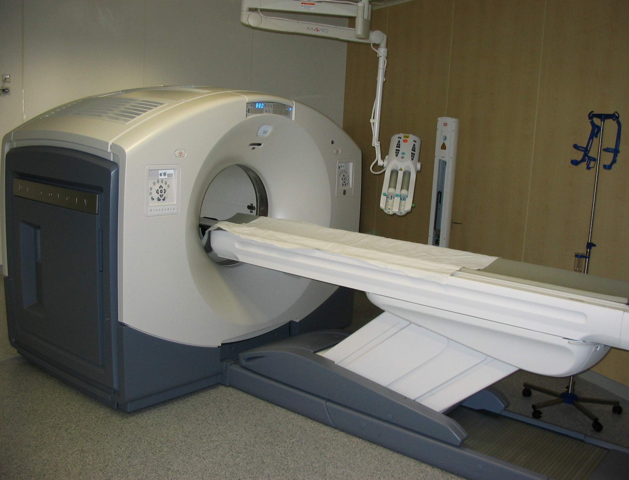
Positron emission tomography (PET) is a functional imaging technique that uses radioactive substances known as radiotracers to visualize and measure changes in metabolic processes, and in other physiological activities including blood flow, regional chemical composition, and absorption. Different tracers are used for various imaging purposes, depending on the target process within the body. For example, -FDG is commonly used to detect cancer, NaF is widely used for detecting bone formation, and oxygen-15 is sometimes used to measure blood flow. PET is a common imaging technique, a medical scintillography technique used in nuclear medicine. A radiopharmaceutical — a radioisotope attached to a drug — is injected into the body as a tracer. When the radiopharmaceutical undergoes beta plus decay, a positron is emitted, and when the positron collides with an ordinary electron, the two particles annihilate and gamma rays are emitted. These gamma rays are detected by ... [...More Info...] [...Related Items...] OR: [Wikipedia] [Google] [Baidu] |
Breast Cancer
Breast cancer is cancer that develops from breast tissue. Signs of breast cancer may include a lump in the breast, a change in breast shape, dimpling of the skin, milk rejection, fluid coming from the nipple, a newly inverted nipple, or a red or scaly patch of skin. In those with distant spread of the disease, there may be bone pain, swollen lymph nodes, shortness of breath, or yellow skin. Risk factors for developing breast cancer include obesity, a lack of physical exercise, alcoholism, hormone replacement therapy during menopause, ionizing radiation, an early age at first menstruation, having children late in life or not at all, older age, having a prior history of breast cancer, and a family history of breast cancer. About 5–10% of cases are the result of a genetic predisposition inherited from a person's parents, including BRCA1 and BRCA2 among others. Breast cancer most commonly develops in cells from the lining of milk ducts and the lobules that supply these ... [...More Info...] [...Related Items...] OR: [Wikipedia] [Google] [Baidu] |
Radiation Therapy
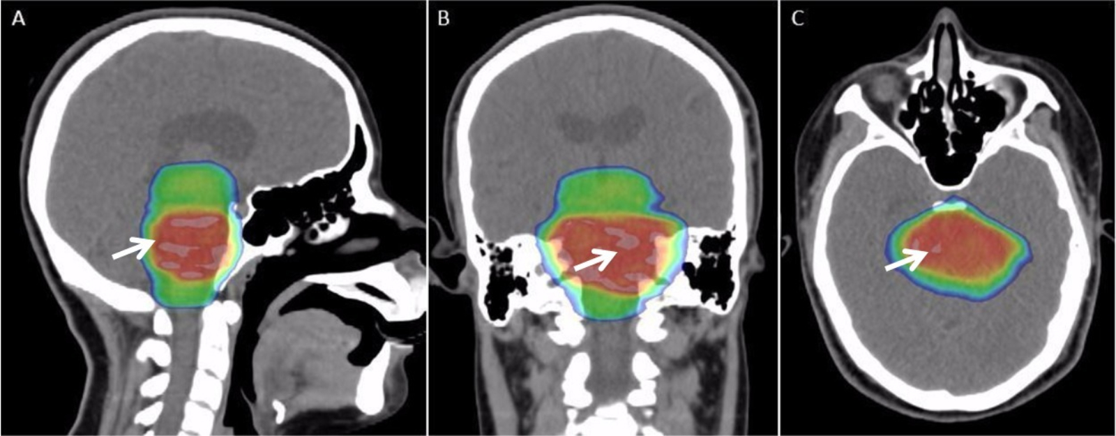
Radiation therapy or radiotherapy, often abbreviated RT, RTx, or XRT, is a therapy using ionizing radiation, generally provided as part of cancer treatment to control or kill malignant cells and normally delivered by a linear accelerator. Radiation therapy may be curative in a number of types of cancer if they are localized to one area of the body. It may also be used as part of adjuvant therapy, to prevent tumor recurrence after surgery to remove a primary malignant tumor (for example, early stages of breast cancer). Radiation therapy is synergistic with chemotherapy, and has been used before, during, and after chemotherapy in susceptible cancers. The subspecialty of oncology concerned with radiotherapy is called radiation oncology. A physician who practices in this subspecialty is a radiation oncologist. Radiation therapy is commonly applied to the cancerous tumor because of its ability to control cell growth. Ionizing radiation works by damaging the DNA of cancerous tissue ... [...More Info...] [...Related Items...] OR: [Wikipedia] [Google] [Baidu] |
DA-EPOCH-R
EPOCH is an intensive chemotherapy regimen intended for treatment of aggressive non-Hodgkin's lymphoma. It is often combined with rituximab. In this case it is called R-EPOCH or EPOCH-R.The R-EPOCH regimen consists of: # Rituximab: an anti-CD20 monoclonal antibody, which has the ability to kill B cells, be they normal or malignant; # Etoposide: a topoisomerase inhibitor from the group of epipodophyllotoxins; # Prednisolone: a glucocorticoid hormone that can cause apoptosis and lysis of both normal and malignant lymphocytes; # Oncovin, also known as vincristine: a vinca alkaloid that binds to the protein tubulin, thereby preventing the formation of microtubules and mitosis; # Cyclophosphamide: an alkylating antineoplastic agent; # Hydroxydaunorubicin, also known as doxorubicin: an anthracycline antibiotic that is able to intercalate DNA, damaging it and preventing cell division. __TOC__ Dosing regimen This regimen requires the use of prophylactic antibiotics to prevent infe ... [...More Info...] [...Related Items...] OR: [Wikipedia] [Google] [Baidu] |
Chemotherapy Regimen
A chemotherapy regimen is a regimen for chemotherapy, defining the drugs to be used, their dosage, the frequency and duration of treatments, and other considerations. In modern oncology, many regimens combine several chemotherapy drugs in combination chemotherapy. The majority of drugs used in cancer chemotherapy are cytostatic, many via cytotoxicity. A fundamental philosophy of medical oncology, including combination chemotherapy, is that different drugs work through different mechanisms, and that the results of using multiple drugs will be synergistic to some extent. Because they have different dose-limiting adverse effects, they can be given together at full doses in chemotherapy regimens. The first successful combination chemotherapy was MOPP, introduced in 1963 for lymphomas. The term " induction regimen" refers to a chemotherapy regimen used for the initial treatment of a disease. A " maintenance regimen" refers to the ongoing use of chemotherapy to reduce the chances of ... [...More Info...] [...Related Items...] OR: [Wikipedia] [Google] [Baidu] |
Nodular Sclerosing Hodgkin Lymphoma
Nodule may refer to: * Nodule (geology), a small rock or mineral cluster * Manganese nodule, a metallic concretion found on the seafloor *Nodule (medicine), a small aggregation of cells *Root nodule Root nodules are found on the roots of plants, primarily legumes, that form a symbiosis with nitrogen-fixing bacteria. Under nitrogen-limiting conditions, capable plants form a symbiotic relationship with a host-specific strain of bacteria known a ..., a growth on the roots of legumes * A feature of mollusc sculpture {{disambig ... [...More Info...] [...Related Items...] OR: [Wikipedia] [Google] [Baidu] |
H&E Stain
Hematoxylin and eosin stain ( or haematoxylin and eosin stain or hematoxylin-eosin stain; often abbreviated as H&E stain or HE stain) is one of the principal tissue stains used in histology. It is the most widely used stain in medical diagnosis and is often the gold standard. For example, when a pathologist looks at a biopsy of a suspected cancer, the histological section is likely to be stained with H&E. H&E is the combination of two histological stains: hematoxylin and eosin. The hematoxylin stains cell nuclei a purplish blue, and eosin stains the extracellular matrix and cytoplasm pink, with other structures taking on different shades, hues, and combinations of these colors. Hence a pathologist can easily differentiate between the nuclear and cytoplasmic parts of a cell, and additionally, the overall patterns of coloration from the stain show the general layout and distribution of cells and provides a general overview of a tissue sample's structure. Thus, pattern recogniti ... [...More Info...] [...Related Items...] OR: [Wikipedia] [Google] [Baidu] |
Microscope
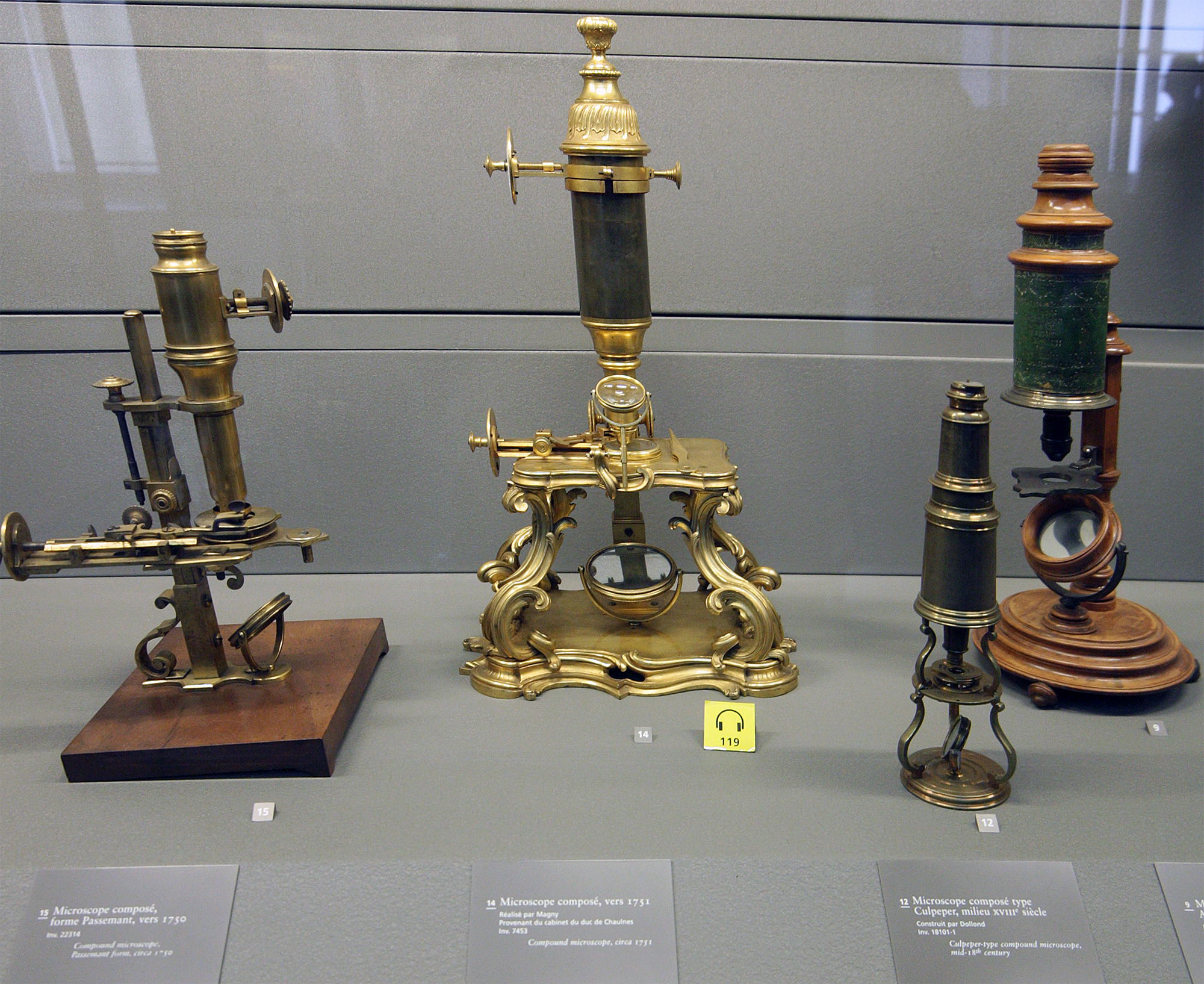
A microscope () is a laboratory instrument used to examine objects that are too small to be seen by the naked eye. Microscopy is the science of investigating small objects and structures using a microscope. Microscopic means being invisible to the eye unless aided by a microscope. There are many types of microscopes, and they may be grouped in different ways. One way is to describe the method an instrument uses to interact with a sample and produce images, either by sending a beam of light or electrons through a sample in its optical path, by detecting photon emissions from a sample, or by scanning across and a short distance from the surface of a sample using a probe. The most common microscope (and the first to be invented) is the optical microscope, which uses lenses to refract visible light that passed through a thinly sectioned sample to produce an observable image. Other major types of microscopes are the fluorescence microscope, electron microscope (both the transmi ... [...More Info...] [...Related Items...] OR: [Wikipedia] [Google] [Baidu] |


