|
Posterior Segment Of Eyeball
The posterior segment or posterior cavity is the back two-thirds of the eye that includes the anterior hyaloid membrane and all of the optical structures behind it: the vitreous humor, retina, choroid, and optic nerve.Posterior segment anatomy The portion of the posterior segment visible during (or fundoscopy) is sometimes referred to as the , or fundus. Some op ... [...More Info...] [...Related Items...] OR: [Wikipedia] [Google] [Baidu] |
Human Eye
The human eye is a sensory organ, part of the sensory nervous system, that reacts to visible light and allows humans to use visual information for various purposes including seeing things, keeping balance, and maintaining circadian rhythm. The eye can be considered as a living optical device. It is approximately spherical in shape, with its outer layers, such as the outermost, white part of the eye (the sclera) and one of its inner layers (the pigmented choroid) keeping the eye essentially light tight except on the eye's optic axis. In order, along the optic axis, the optical components consist of a first lens (the cornea—the clear part of the eye) that accomplishes most of the focussing of light from the outside world; then an aperture (the pupil) in a diaphragm (the iris—the coloured part of the eye) that controls the amount of light entering the interior of the eye; then another lens (the crystalline lens) that accomplishes the remaining focussing of light into ... [...More Info...] [...Related Items...] OR: [Wikipedia] [Google] [Baidu] |
Anterior Hyaloid Membrane
The vitreous membrane (or hyaloid membrane or vitreous cortex) is a layer of collagen separating the vitreous humour from the rest of the eye. At least two parts have been identified anatomically. The posterior hyaloid membrane separates the rear of the vitreous from the retina. It is a false anatomical membrane. The anterior hyaloid membrane separates the front of the vitreous from the lens. Andres Bernal, Jean-Marie Parel, Fabrice MannsEvidence for posterior zonular fiber attachment on the anterior hyaloid membrane "Investigative Ophthalmology and Visual Science" 2006, 47, 4708-4713. Bernal et al. describe it "as a delicate structure in the form of a thin layer that runs from the pars plana to the posterior lens, where it shares its attachment with the posterior zonule Posterior may refer to: * Posterior (anatomy), the end of an organism opposite to its head ** Buttocks, as a euphemism * Posterior horn (other) * Posterior probability, the conditional probability that is ... [...More Info...] [...Related Items...] OR: [Wikipedia] [Google] [Baidu] |
Vitreous Humor
The vitreous body (''vitreous'' meaning "glass-like"; , ) is the clear gel that fills the space between the lens and the retina of the eyeball (the vitreous chamber) in humans and other vertebrates. It is often referred to as the vitreous humor (also spelled humour, from Latin meaning liquid) or simply "the vitreous". Vitreous fluid or "liquid vitreous" is the liquid component of the vitreous gel, found after a vitreous detachment. It is not to be confused with the aqueous humor, the other fluid in the eye that is found between the cornea and lens. Structure The vitreous humor is a transparent, colorless, gelatinous mass that fills the space in the eye between the lens and the retina. It is surrounded by a layer of collagen called the vitreous membrane (or hyaloid membrane or vitreous cortex) separating it from the rest of the eye. It makes up four-fifths of the volume of the eyeball. The vitreous humour is fluid-like near the centre, and gel-like near the edges. The vitreous hu ... [...More Info...] [...Related Items...] OR: [Wikipedia] [Google] [Baidu] |
Retina
The retina (from la, rete "net") is the innermost, light-sensitive layer of tissue of the eye of most vertebrates and some molluscs. The optics of the eye create a focused two-dimensional image of the visual world on the retina, which then processes that image within the retina and sends nerve impulses along the optic nerve to the visual cortex to create visual perception. The retina serves a function which is in many ways analogous to that of the film or image sensor in a camera. The neural retina consists of several layers of neurons interconnected by synapses and is supported by an outer layer of pigmented epithelial cells. The primary light-sensing cells in the retina are the photoreceptor cells, which are of two types: rods and cones. Rods function mainly in dim light and provide monochromatic vision. Cones function in well-lit conditions and are responsible for the perception of colour through the use of a range of opsins, as well as high-acuity vision used for task ... [...More Info...] [...Related Items...] OR: [Wikipedia] [Google] [Baidu] |
Choroid
The choroid, also known as the choroidea or choroid coat, is a part of the uvea, the vascular layer of the eye, and contains connective tissues, and lies between the retina and the sclera. The human choroid is thickest at the far extreme rear of the eye (at 0.2 mm), while in the outlying areas it narrows to 0.1 mm. The choroid provides oxygen and nourishment to the outer layers of the retina. Along with the ciliary body and iris, the choroid forms the uveal tract. The structure of the choroid is generally divided into four layers (classified in order of furthest away from the retina to closest): *Haller's layer - outermost layer of the choroid consisting of larger diameter blood vessels; *Sattler's layer - layer of medium diameter blood vessels; * Choriocapillaris - layer of capillaries; and *Bruch's membrane (synonyms: Lamina basalis, Complexus basalis, Lamina vitra) - innermost layer of the choroid. Blood supply There are two circulations of the eye: the retin ... [...More Info...] [...Related Items...] OR: [Wikipedia] [Google] [Baidu] |
Optic Nerve
In neuroanatomy, the optic nerve, also known as the second cranial nerve, cranial nerve II, or simply CN II, is a paired cranial nerve that transmits visual system, visual information from the retina to the brain. In humans, the optic nerve is derived from optic stalks during the seventh week of development and is composed of retinal ganglion cell axons and glial cells; it extends from the optic disc to the optic chiasma and continues as the optic tract to the lateral geniculate nucleus, Pretectal area, pretectal nuclei, and superior colliculus. Structure The optic nerve has been classified as the second of twelve paired cranial nerves, but it is technically part of the central nervous system, rather than the peripheral nervous system because it is derived from an out-pouching of the diencephalon (optic stalks) during embryonic development. As a consequence, the fibers of the optic nerve are covered with myelin produced by oligodendrocytes, rather than Schwann cells of the per ... [...More Info...] [...Related Items...] OR: [Wikipedia] [Google] [Baidu] |
Ophthalmoscopy
Ophthalmoscopy, also called funduscopy, is a test that allows a health professional to see inside the fundus of the eye and other structures using an ophthalmoscope (or funduscope). It is done as part of an eye examination and may be done as part of a routine physical examination. It is crucial in determining the health of the retina, optic disc, and vitreous humor. The pupil is a hole through which the eye's interior will be viewed. Opening the pupil wider (dilating it) is a simple and effective way to better see the structures behind it. Therefore, dilation of the pupil ( mydriasis) is often accomplished with medicated eye drops before funduscopy. However, although dilated fundus examination is ideal, undilated examination is more convenient and is also helpful (albeit not as comprehensive), and it is the most common type in primary care. An alternative or complement to ophthalmoscopy is to perform a fundus photography, where the image can be analysed later by a professional. ... [...More Info...] [...Related Items...] OR: [Wikipedia] [Google] [Baidu] |
Posterior Pole
In ophthalmology, the posterior pole is the back of the eye, usually referring to the retina between the optic disc and the macula.Cassin, B. and Solomon, S. ''Dictionary of Eye Terminology''. Gainesville, Florida: Triad Publishing Company, 1990. See also * Fundus (eye) The fundus of the eye is the interior surface of the eye opposite the lens and includes the retina, optic disc, macula, fovea, and posterior pole.Cassin, B. and Solomon, S. ''Dictionary of Eye Terminology''. Gainesville, Florida: Triad Publish ... References Human eye anatomy {{eye-stub ... [...More Info...] [...Related Items...] OR: [Wikipedia] [Google] [Baidu] |
Fundus (eye)
The fundus of the eye is the interior surface of the eye opposite the lens and includes the retina, optic disc, macula, fovea, and posterior pole.Cassin, B. and Solomon, S. ''Dictionary of Eye Terminology''. Gainesville, Florida: Triad Publishing Company, 1990. The fundus can be examined by ophthalmoscopy and/or fundus photography. Variation The color of the fundus varies both between and within species. In one study of primates the retina is blue, green, yellow, orange, and red; only the human fundus (from a lightly pigmented blond person) is red. The major differences noted among the "higher" primate species were size and regularity of the border of macular area, size and shape of the optic disc, apparent 'texturing' of retina, and pigmentation of retina. Clinical significance Medical signs that can be detected from observation of eye fundus (generally by funduscopy) include hemorrhages, exudates, cotton wool spots, blood vessel abnormalities (tortuosity, pulsation and n ... [...More Info...] [...Related Items...] OR: [Wikipedia] [Google] [Baidu] |
Ophthalmologist
Ophthalmology ( ) is a surgery, surgical subspecialty within medicine that deals with the diagnosis and treatment of eye disorders. An ophthalmologist is a physician who undergoes subspecialty training in medical and surgical eye care. Following a medical degree, a doctor specialising in ophthalmology must pursue additional postgraduate residency (medicine), residency training specific to that field. This may include a one-year integrated internship that involves more general medical training in other fields such as internal medicine or general surgery. Following residency, additional specialty training (or fellowship) may be sought in a particular aspect of eye pathology. Ophthalmologists prescribe medications to treat eye diseases, implement laser therapy, and perform surgery when needed. Ophthalmologists provide both primary and specialty eye care - medical and surgical. Most ophthalmologists participate in academic research on eye diseases at some point in their training an ... [...More Info...] [...Related Items...] OR: [Wikipedia] [Google] [Baidu] |
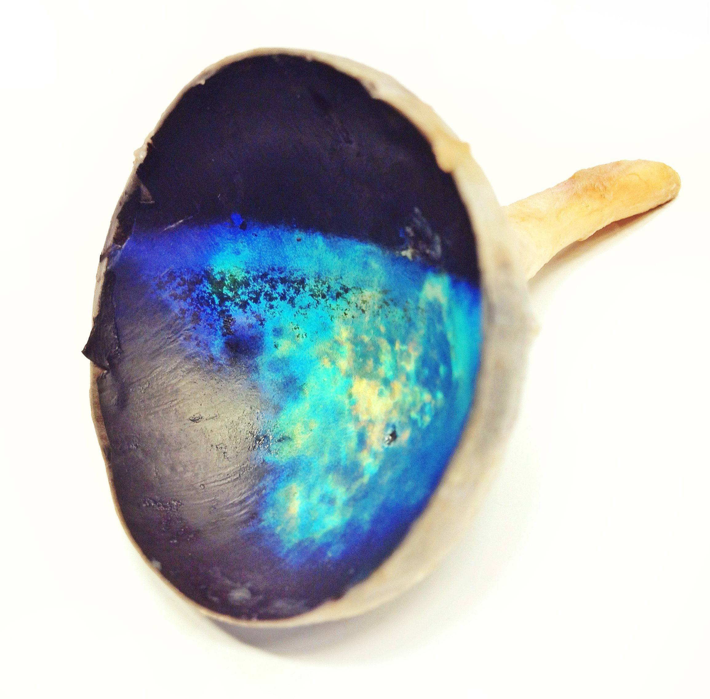
Tapetum Lucidum
The ''tapetum lucidum'' ( ; ; ) is a layer of tissue in the eye of many vertebrates and some other animals. Lying immediately behind the retina, it is a retroreflector. It reflects visible light back through the retina, increasing the light available to the photoreceptors (although slightly blurring the image). The tapetum lucidum contributes to the superior night vision of some animals. Many of these animals are nocturnal, especially carnivores, while others are deep sea animals. Similar adaptations occur in some species of spiders. Haplorhine primates, including humans, are diurnal and lack a ''tapetum lucidum''. Function and mechanism Presence of a ''tapetum lucidum'' enables animals to see in dimmer light than would otherwise be possible. The ''tapetum lucidum'', which is iridescent, reflects light roughly on the interference principles of thin-film optics, as seen in other iridescent tissues. However, the ''tapetum lucidum'' cells are leucophores, not iridophores. ... [...More Info...] [...Related Items...] OR: [Wikipedia] [Google] [Baidu] |
Anterior Segment
The anterior segment or anterior cavity is the front third of the eye that includes the structures in front of the vitreous humour: the cornea, iris, ciliary body, and lens.Cassin, B. and Solomon, S. ''Dictionary of Eye Terminology''. Gainesville, Florida: Triad Publishing Company, 1990."Departments. Anterior segment." Cantabrian Institute of Ophthalmology. Within the anterior segment are two fluid-filled spaces: * the between the posterior surface of the cornea (i.e. the ) and the iris. * the [...More Info...] [...Related Items...] OR: [Wikipedia] [Google] [Baidu] |


