|
Ophthalmologist
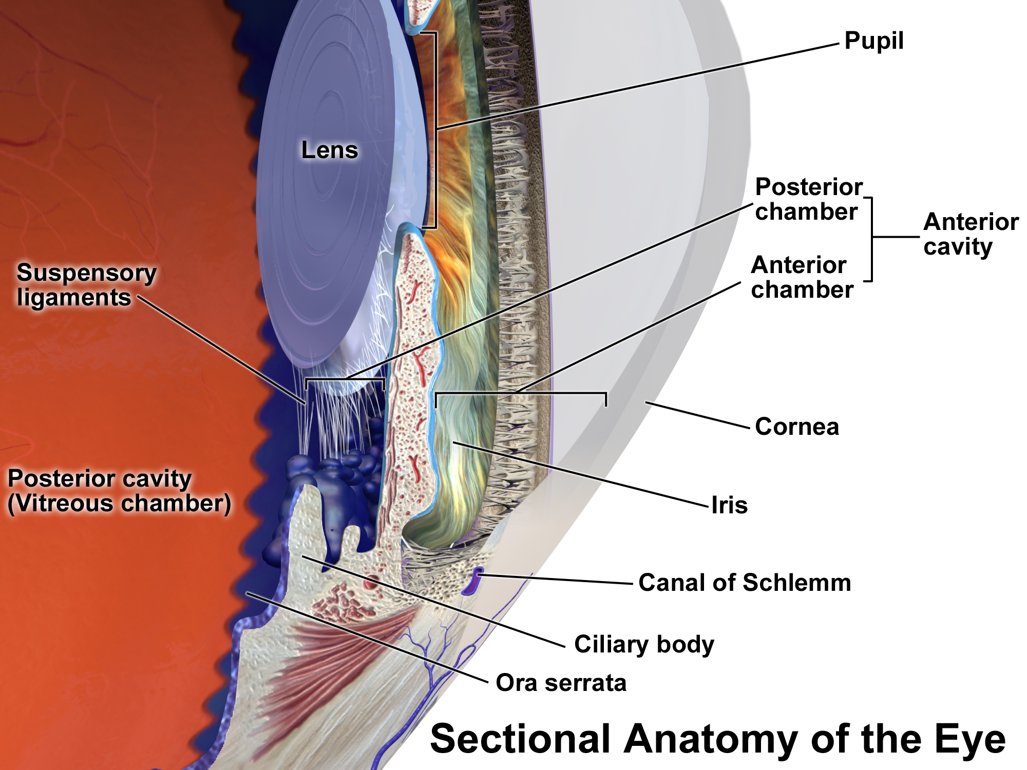
Ophthalmology ( ) is a surgery, surgical subspecialty within medicine that deals with the diagnosis and treatment of eye disorders. An ophthalmologist is a physician who undergoes subspecialty training in medical and surgical eye care. Following a medical degree, a doctor specialising in ophthalmology must pursue additional postgraduate residency (medicine), residency training specific to that field. This may include a one-year integrated internship that involves more general medical training in other fields such as internal medicine or general surgery. Following residency, additional specialty training (or fellowship) may be sought in a particular aspect of eye pathology. Ophthalmologists prescribe medications to treat eye diseases, implement laser therapy, and perform surgery when needed. Ophthalmologists provide both primary and specialty eye care - medical and surgical. Most ophthalmologists participate in academic research on eye diseases at some point in their training an ... [...More Info...] [...Related Items...] OR: [Wikipedia] [Google] [Baidu] |
Neuro-ophthalmology
Neuro-ophthalmology is an academically-oriented subspecialty that merges the fields of neurology and ophthalmology, often dealing with complex systemic diseases that have manifestations in the visual system. Neuro-ophthalmologists initially complete a residency in either neurology or ophthalmology, then do a fellowship in the complementary field. Since diagnostic studies can be normal in patients with significant neuro-ophthalmic disease, a detailed medical history and physical exam is essential, and neuro-ophthalmologists often spend a significant amount of time with their patients. Common pathology referred to a neuro-ophthalmologist includes afferent visual system disorders (e.g. optic neuritis, optic neuropathy, papilledema, brain tumors or strokes) and efferent visual system disorders (e.g. anisocoria, diplopia, ophthalmoplegia, ptosis, nystagmus, blepharospasm, seizures of the eye or eye muscles, and hemifacial spasm). The largest international society of neuro-ophthalmologis ... [...More Info...] [...Related Items...] OR: [Wikipedia] [Google] [Baidu] |
Graves' Ophthalmopathy
Graves’ ophthalmopathy, also known as thyroid eye disease (TED), is an autoimmune inflammatory disorder of the orbit and periorbital tissues, characterized by upper eyelid retraction, lid lag, swelling, redness ( erythema), conjunctivitis, and bulging eyes (exophthalmos). It occurs most commonly in individuals with Graves' disease, and less commonly in individuals with Hashimoto's thyroiditis, or in those who are euthyroid. It is part of a systemic process with variable expression in the eyes, thyroid, and skin, caused by autoantibodies that bind to tissues in those organs. The autoantibodies target the fibroblasts in the eye muscles, and those fibroblasts can differentiate into fat cells ( adipocytes). Fat cells and muscles expand and become inflamed. Veins become compressed and are unable to drain fluid, causing edema. Annual incidence is 16/100,000 in women, 3/100,000 in men. About 3–5% have severe disease with intense pain, and sight-threatening corneal ulceration ... [...More Info...] [...Related Items...] OR: [Wikipedia] [Google] [Baidu] |
American Academy Of Ophthalmology
The American Academy of Ophthalmology (Academy) is a professional medical association of ophthalmologists. It is headquartered in San Francisco, California. Its membership of 32,000 medical doctors includes more than 90 percent of practicing ophthalmologists in the United States as well as over 7,000 members abroad. The Academy's stated mission is "to protect sight and empower lives by serving as an advocate for patients and the public, leading ophthalmic education, and advancing the profession of ophthalmology." History The academy has its origins in the American Academy of Ophthalmology and Otolaryngology (AAOO), founded in 1896 as a medical association of both ophthalmologists and otolaryngologists. The Academy was founded when the AAOO split in 1979 and divided into separate academies for each specialty. Like most medical associations, the Academy collects dues, provides continuing education and seminars for its members, including its four-day annual meeting. Outside the ... [...More Info...] [...Related Items...] OR: [Wikipedia] [Google] [Baidu] |
Ptosis (eyelid)
Ptosis, also known as blepharoptosis, is a drooping or falling of the upper eyelid. The drooping may be worse after being awake longer when the individual's muscles are tired. This condition is sometimes called "lazy eye", but that term normally refers to the condition amblyopia. If severe enough and left untreated, the drooping eyelid can cause other conditions, such as amblyopia or astigmatism. This is why it is especially important for this disorder to be treated in children at a young age, before it can interfere with vision development. The term is from Greek 'fall, falling'. Signs and symptoms Signs and symptoms typically seen in this condition include: * The eyelid(s) may appear to droop. * Droopy eyelids can give the face a false appearance of being fatigued, disinterested, or even sinister. * The eyelid may not protect the eye as effectively, allowing it to dry out. * Sagging upper eyelids can partially block the person's field of view. * Obstructed vision may ca ... [...More Info...] [...Related Items...] OR: [Wikipedia] [Google] [Baidu] |
Ophthalmoscopy
Ophthalmoscopy, also called funduscopy, is a test that allows a health professional to see inside the fundus of the eye and other structures using an ophthalmoscope (or funduscope). It is done as part of an eye examination and may be done as part of a routine physical examination. It is crucial in determining the health of the retina, optic disc, and vitreous humor. The pupil is a hole through which the eye's interior will be viewed. Opening the pupil wider (dilating it) is a simple and effective way to better see the structures behind it. Therefore, dilation of the pupil (mydriasis) is often accomplished with medicated eye drops before funduscopy. However, although dilated fundus examination is ideal, undilated examination is more convenient and is also helpful (albeit not as comprehensive), and it is the most common type in primary care. An alternative or complement to ophthalmoscopy is to perform a fundus photography, where the image can be analysed later by a profess ... [...More Info...] [...Related Items...] OR: [Wikipedia] [Google] [Baidu] |
Glaucoma
Glaucoma is a group of eye diseases that result in damage to the optic nerve (or retina) and cause vision loss. The most common type is open-angle (wide angle, chronic simple) glaucoma, in which the drainage angle for fluid within the eye remains open, with less common types including closed-angle (narrow angle, acute congestive) glaucoma and normal-tension glaucoma. Open-angle glaucoma develops slowly over time and there is no pain. Peripheral vision may begin to decrease, followed by central vision, resulting in blindness if not treated. Closed-angle glaucoma can present gradually or suddenly. The sudden presentation may involve severe eye pain, blurred vision, mid-dilated pupil, redness of the eye, and nausea. Vision loss from glaucoma, once it has occurred, is permanent. Eyes affected by glaucoma are referred to as being glaucomatous. Risk factors for glaucoma include increasing age, high pressure in the eye, a family history of glaucoma, and use of steroid medicatio ... [...More Info...] [...Related Items...] OR: [Wikipedia] [Google] [Baidu] |
Eye Neoplasm
Eye neoplasms can affect all parts of the eye, and can be a benign tumor or a malignant tumor (cancer). Eye cancers can be primary (starts within the eye) or metastatic cancer (spread to the eye from another organ). The two most common cancers that spread to the eye from another organ are breast cancer and lung cancer. Other less common sites of origin include the prostate, kidney, thyroid, skin, colon and blood or bone marrow. Types Tumors in the eye and orbit can be benign like dermoid cysts, or malignant like rhabdomyosarcoma and retinoblastoma. Malignant The most common eyelid tumor is called basal cell carcinoma. This tumor can grow around the eye but rarely spreads to other parts of the body. Other types of common eyelid cancers include squamous carcinoma, sebaceous carcinoma and malignant melanoma. The most common orbital malignancy is '' orbital lymphoma''. This tumor can be diagnosed by biopsy with histopathologic and immunohistochemical analysis. Most patients ... [...More Info...] [...Related Items...] OR: [Wikipedia] [Google] [Baidu] |
Uveitis
Uveitis () is inflammation of the uvea, the pigmented layer of the eye between the inner retina and the outer fibrous layer composed of the sclera and cornea. The uvea consists of the middle layer of pigmented vascular structures of the eye and includes the iris, ciliary body, and choroid. Uveitis is described anatomically, by the part of the eye affected, as anterior, intermediate or posterior, or panuveitic if all parts are involved. Anterior uveitis ( iridocyclytis) is the most common, with the incidence of uveitis overall affecting approximately 1:4500, most commonly those between the ages of 20-60. Symptoms include eye pain, eye redness, floaters and blurred vision, and ophthalmic examination may show dilated ciliary blood vessels and the presence of cells in the anterior chamber. Uveitis may arise spontaneously, have a genetic component, or be associated with an autoimmune disease or infection. While the eye is a relatively protected environment, its immune mechan ... [...More Info...] [...Related Items...] OR: [Wikipedia] [Google] [Baidu] |
Cataract
A cataract is a cloudy area in the lens of the eye that leads to a decrease in vision. Cataracts often develop slowly and can affect one or both eyes. Symptoms may include faded colors, blurry or double vision, halos around light, trouble with bright lights, and trouble seeing at night. This may result in trouble driving, reading, or recognizing faces. Poor vision caused by cataracts may also result in an increased risk of falling and depression. Cataracts cause 51% of all cases of blindness and 33% of visual impairment worldwide. Cataracts are most commonly due to aging but may also occur due to trauma or radiation exposure, be present from birth, or occur following eye surgery for other problems. Risk factors include diabetes, longstanding use of corticosteroid medication, smoking tobacco, prolonged exposure to sunlight, and alcohol. The underlying mechanism involves accumulation of clumps of protein or yellow-brown pigment in the lens that reduces transmission of ... [...More Info...] [...Related Items...] OR: [Wikipedia] [Google] [Baidu] |
Optical Coherence Tomography
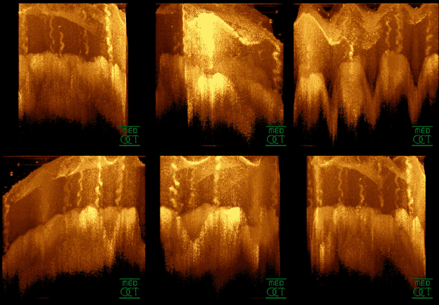
Optical coherence tomography (OCT) is an imaging technique that uses low-coherence light to capture micrometer-resolution, two- and three-dimensional images from within optical scattering media (e.g., biological tissue). It is used for medical imaging and industrial nondestructive testing (NDT). Optical coherence tomography is based on low-coherence interferometry, typically employing near-infrared light. The use of relatively long wavelength light allows it to penetrate into the scattering medium. Confocal microscopy, another optical technique, typically penetrates less deeply into the sample but with higher resolution. Depending on the properties of the light source (superluminescent diodes, ultrashort pulsed lasers, and supercontinuum lasers have been employed), optical coherence tomography has achieved sub-micrometer resolution (with very wide-spectrum sources emitting over a ~100 nm wavelength range). Optical coherence tomography is one of a class of optical tom ... [...More Info...] [...Related Items...] OR: [Wikipedia] [Google] [Baidu] |
Glossary Of Medicine
This glossary of medical terms is a list of definitions about medicine, its sub-disciplines, and related fields. A * Aarskog–Scott syndrome – (AAS) A rare, inherited (X-linked) disease characterized by short stature, facial abnormalities, skeletal and genital anomalies. *Abdomen – The part of the body between the chest and pelvis, which contains most of the tubelike organs of the digestive tract, as well as several solid organs. * Abdominal external oblique muscle – The largest, and outermost, of the three flat muscles of the lateral anterior abdominal wall. * Abdominal internal oblique muscle – A muscle of the abdominal wall, which lies below the external oblique and just above the transverse abdominal muscles. * Abductor pollicis brevis muscle – A muscle in the hand that abducts (straightens) the thumb. * Abductor pollicis longus muscle – One of the extrinsic muscles of the hand. Its major function is to abduct the thumb at the wrist. * Abscess – A collect ... [...More Info...] [...Related Items...] OR: [Wikipedia] [Google] [Baidu] |
Diabetic Retinopathy
Diabetic retinopathy (also known as diabetic eye disease), is a medical condition in which damage occurs to the retina due to diabetes mellitus. It is a leading cause of blindness in developed countries. Diabetic retinopathy affects up to 80 percent of those who have had both type 1 and type 2 diabetes for 20 years or more. In at least 90% of new cases, progression to more aggressive forms of sight threatening retinopathy and maculopathy could be reduced with proper treatment and monitoring of the eyes The longer a person has diabetes, the higher his or her chances of developing diabetic retinopathy. Each year in the United States, diabetic retinopathy accounts for 12% of all new cases of blindness. It is also the leading cause of blindness in people aged 20 to 64. Signs and symptoms Nearly all people with diabetes develop some degree of retina damage ("retinopathy") over several decades with the disease. For many, that damage can only be detected by a retinal exam, and h ... [...More Info...] [...Related Items...] OR: [Wikipedia] [Google] [Baidu] |


_PHIL_4284_lores.jpg)
