|
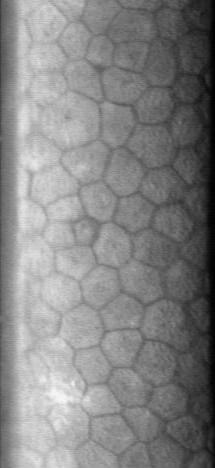
Posterior Polymorphous Corneal Dystrophy 3
Posterior polymorphous corneal dystrophy (PPCD; sometimes also ''Schlichting dystrophy'') is a type of corneal dystrophy, characterised by changes in Descemet's membrane and endothelial layer. Symptoms mainly consist of decreased vision due to corneal edema. In some cases they are present from birth, other patients are asymptomatic. Histopathological analysis shows that the cells of endothelium have some characteristics of epithelial cells and have become multilayered. The disease was first described in 1916 by Koeppe as ''keratitis bullosa interna''. Genetics PPCD type 2 is linked to the mutations in COL8A2, and PPCD type 3 mutations in ZEB1 gene, but the underlying genetic disturbance in PPCD type 1 is unknown. Pathophysiology Vacuoles are demonstrated in the posterior parts of the cornea. The vesicles are located on the endothelial surface. The corneal endothelium is normally a single layer of cells that lose their mitotic potential after development is complete. In posterior ... [...More Info...] [...Related Items...] OR: [Wikipedia] [Google] [Baidu] |
Descemet's Membrane
Descemet's membrane ( or the Descemet membrane) is the basement membrane that lies between the corneal proper substance, also called stroma, and the endothelial layer of the cornea. It is composed of different kinds of collagen (Type IV and VIII) than the stroma. The endothelial layer is located at the posterior of the cornea. Descemet's membrane, as the basement membrane for the endothelial layer, is secreted by the single layer of squamous epithelial cells that compose the endothelial layer of the cornea. Structure Its thickness ranges from 3 μm at birth to 8–10 μm in adults.Johnson DH, Bourne WM, Campbell RJ: The ultrastructure of Descemet's membrane. I. Changes with age in normal cornea. Arch Ophthalmol 100:1942, 1982 The corneal endothelium is a single layer of squamous cells covering the surface of the cornea that faces the anterior chamber. Clinical significance Significant damage to the membrane may require a corneal transplant. Damage caused by the hereditary co ... [...More Info...] [...Related Items...] OR: [Wikipedia] [Google] [Baidu] |
Corneal Endothelium
The corneal endothelium is a single layer of endothelial cells on the inner surface of the cornea. It faces the chamber formed between the cornea and the iris. The corneal endothelium are specialized, flattened, mitochondria-rich cells that line the posterior surface of the cornea and face the anterior chamber of the eye. The corneal endothelium governs fluid and solute transport across the posterior surface of the cornea and maintains the cornea in the slightly dehydrated state that is required for optical transparency. Embryology and anatomy The corneal endothelium is embryologically derived from the neural crest. The postnatal total endothelial cellularity of the cornea (approximately 300,000 cells per cornea) is achieved as early as the second trimester of gestation. Thereafter the endothelial cell density (but not the absolute number of cells) rapidly declines, as the fetal cornea grows in surface area, achieving a final adult density of approximately 2400 - 3200 cells ... [...More Info...] [...Related Items...] OR: [Wikipedia] [Google] [Baidu] |
COL8A2
Collagen alpha-2(VIII) chain is a protein that in humans is encoded by the ''COL8A2'' gene In biology, the word gene (from , ; "... Wilhelm Johannsen coined the word gene to describe the Mendelian units of heredity..." meaning ''generation'' or ''birth'' or ''gender'') can have several different meanings. The Mendelian gene is a b .... Mutations of the gene are linked to posterior polymorphous dystrophy type 2. References Further reading * * * * * * * * * * * * * * * * Collagens {{gene-1-stub ... [...More Info...] [...Related Items...] OR: [Wikipedia] [Google] [Baidu] |
ZEB1
Zinc finger E-box-binding homeobox 1 is a protein that in humans is encoded by the ''ZEB1'' gene. ZEB1 (previously known as TCF8) encodes a zinc finger and homeodomain transcription factor that represses T-lymphocyte-specific IL2 gene expression by binding to a negative regulatory domain 100 nucleotides 5-prime of the IL2 transcription start site. ZEB1 and its mammalian paralog ZEB2 belongs to the Zeb family within the ZF (zinc finger) class of homeodomain transcription factors. ZEB1 protein has 7 zinc fingers and 1 homeodomain. The structure of the homeodomain is shown on the right. Clinical significance Mutations of the gene are linked to posterior polymorphous corneal dystrophy 3. ZEB1 downregulates E-cadherin and induces epithelial to mesenchymal transition in breast and other carcinomas. A recent study suggested its contributing role in lung cancer Lung cancer, also known as lung carcinoma (since about 98–99% of all lung cancers are carcinomas), is a malignant lun ... [...More Info...] [...Related Items...] OR: [Wikipedia] [Google] [Baidu] |
Cornea
The cornea is the transparent front part of the eye that covers the iris, pupil, and anterior chamber. Along with the anterior chamber and lens, the cornea refracts light, accounting for approximately two-thirds of the eye's total optical power. In humans, the refractive power of the cornea is approximately 43 dioptres. The cornea can be reshaped by surgical procedures such as LASIK. While the cornea contributes most of the eye's focusing power, its focus is fixed. Accommodation (the refocusing of light to better view near objects) is accomplished by changing the geometry of the lens. Medical terms related to the cornea often start with the prefix "'' kerat-''" from the Greek word κέρας, ''horn''. Structure The cornea has unmyelinated nerve endings sensitive to touch, temperature and chemicals; a touch of the cornea causes an involuntary reflex to close the eyelid. Because transparency is of prime importance, the healthy cornea does not have or need blood vessels with ... [...More Info...] [...Related Items...] OR: [Wikipedia] [Google] [Baidu] |
Glaucoma
Glaucoma is a group of eye diseases that result in damage to the optic nerve (or retina) and cause vision loss. The most common type is open-angle (wide angle, chronic simple) glaucoma, in which the drainage angle for fluid within the eye remains open, with less common types including closed-angle (narrow angle, acute congestive) glaucoma and normal-tension glaucoma. Open-angle glaucoma develops slowly over time and there is no pain. Peripheral vision may begin to decrease, followed by central vision, resulting in blindness if not treated. Closed-angle glaucoma can present gradually or suddenly. The sudden presentation may involve severe eye pain, blurred vision, mid-dilated pupil, redness of the eye, and nausea. Vision loss from glaucoma, once it has occurred, is permanent. Eyes affected by glaucoma are referred to as being glaucomatous. Risk factors for glaucoma include increasing age, high pressure in the eye, a family history of glaucoma, and use of steroid medication. F ... [...More Info...] [...Related Items...] OR: [Wikipedia] [Google] [Baidu] |
Corneal Dystrophy
Corneal dystrophy is a group of rare hereditary disorders characterised by bilateral abnormal deposition of substances in the transparent front part of the eye called the cornea. Signs and symptoms Corneal dystrophy may not significantly affect vision in the early stages. However, it does require proper evaluation and treatment for restoration of optimal vision. Corneal dystrophies usually manifest themselves during the first or second decade but sometimes later. It appears as grayish white lines, circles, or clouding of the cornea. Corneal dystrophy can also have a crystalline appearance. There are over 20 corneal dystrophies that affect all parts of the cornea. These diseases share many traits: * They are usually inherited. * They affect the right and left eyes equally. * They are not caused by outside factors, such as injury or diet. * Most progress gradually. * Most usually begin in one of the five corneal layers and may later spread to nearby layers. * Most do not affect oth ... [...More Info...] [...Related Items...] OR: [Wikipedia] [Google] [Baidu] |


