|
Polychromasia
Polychromasia is a disorder where there is an abnormally high number of immature Red blood cell, red blood cells found in the bloodstream as a result of being prematurely released from the bone marrow during blood formation (''poly''- refers to ''many'', and ''-chromasia'' means ''color''.) These cells are often shades of grayish-blue. Polychromasia is usually a sign of bone marrow stress as well as immature red blood cells. 3 types are recognized, with types 1 and 2 being referred to as 'young red blood cells' and type 3 as 'old red blood cells'. Giemsa stain is used to distinguish all three types of blood smears. The young cells will generally stain gray or blue in the cytoplasm. These young red blood cells are commonly called reticulocytes. All polychromatophilic cells are reticulocytes, however, not all reticulocytes are polychromatophilic. In the old blood cells, the cytoplasm either stains a light orange or does not stain at all. Causes Red blood cells can be released prema ... [...More Info...] [...Related Items...] OR: [Wikipedia] [Google] [Baidu] |
Reticulocyte Index
The reticulocyte production index (RPI), also called a corrected reticulocyte count (CRC), is a calculated value used in the diagnosis of anemia. This calculation is necessary because the raw reticulocyte count is misleading in anemic patients. The problem arises because the reticulocyte count is not really a ''count'' but rather a ''percentage'': it reports the number of reticulocytes as a percentage of the reference ranges for blood tests#Red blood cells, number of red blood cells. In anemia, the patient's red blood cells are depleted, creating an erroneously elevated reticulocyte count. Physiology Reticulocytes are newly produced red blood cells. They are slightly larger than totally mature red blood cells, and have some residual ribosomal RNA. The presence of RNA allows a visible blue stain to bind or, in the case of fluorescent dye, result in a different brightness. This allows them to be detected and counted as a distinct population.Adamson JW, Longo DL. Anemia and polyc ... [...More Info...] [...Related Items...] OR: [Wikipedia] [Google] [Baidu] |
Anemia
Anemia or anaemia (British English) is a blood disorder in which the blood has a reduced ability to carry oxygen due to a lower than normal number of red blood cells, or a reduction in the amount of hemoglobin. When anemia comes on slowly, the symptoms are often vague, such as tiredness, weakness, shortness of breath, headaches, and a reduced ability to exercise. When anemia is acute, symptoms may include confusion, feeling like one is going to pass out, loss of consciousness, and increased thirst. Anemia must be significant before a person becomes noticeably pale. Symptoms of anemia depend on how quickly hemoglobin decreases. Additional symptoms may occur depending on the underlying cause. Preoperative anemia can increase the risk of needing a blood transfusion following surgery. Anemia can be temporary or long term and can range from mild to severe. Anemia can be caused by blood loss, decreased red blood cell production, and increased red blood cell breakdown. Causes o ... [...More Info...] [...Related Items...] OR: [Wikipedia] [Google] [Baidu] |
Normocytic Anemia
Normocytic anemia is a type of anemia and is a common issue that occurs for men and women typically over 85 years old. Its prevalence increases with age, reaching 44 percent in men older than 85 years. The most common type of normocytic anemia is anemia of chronic disease. Classification A normocytic anemia is when the red blood cells (RBCs) are of normal size. Normocytic anemia is defined when the mean corpuscular volume (MCV) is between 80 and 100 femtolitres (fL), which is within the normal and expected range. However, the hematocrit and hemoglobin are decreased. In contrast, microcytic anemias are defined as an anemia with a mean corpuscular volume (MCV) less than 80 fL and macrocytic anemias have a mean corpuscular volume over 100 fL. Diagnosis To aid with determining the underlying cause of the normocytic anemia, a lab test is done on reticulocyte count. A reticulocyte count that is high, normal or low will aid with the classification process. A high reticulocyte count sig ... [...More Info...] [...Related Items...] OR: [Wikipedia] [Google] [Baidu] |
Red Blood Cell
Red blood cells (RBCs), also referred to as red cells, red blood corpuscles (in humans or other animals not having nucleus in red blood cells), haematids, erythroid cells or erythrocytes (from Greek ''erythros'' for "red" and ''kytos'' for "hollow vessel", with ''-cyte'' translated as "cell" in modern usage), are the most common type of blood cell and the vertebrate's principal means of delivering oxygen (O2) to the body tissues—via blood flow through the circulatory system. RBCs take up oxygen in the lungs, or in fish the gills, and release it into tissues while squeezing through the body's capillaries. The cytoplasm of a red blood cell is rich in hemoglobin, an iron-containing biomolecule that can bind oxygen and is responsible for the red color of the cells and the blood. Each human red blood cell contains approximately 270 million hemoglobin molecules. The cell membrane is composed of proteins and lipids, and this structure provides properties essential for physiolo ... [...More Info...] [...Related Items...] OR: [Wikipedia] [Google] [Baidu] |
Fibrosis
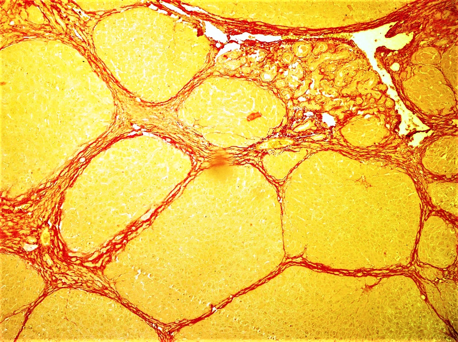
Fibrosis, also known as fibrotic scarring, is a pathological wound healing in which connective tissue replaces normal parenchymal tissue to the extent that it goes unchecked, leading to considerable tissue remodelling and the formation of permanent scar tissue. Repeated injuries, chronic inflammation and repair are susceptible to fibrosis where an accidental excessive accumulation of extracellular matrix components, such as the collagen is produced by fibroblasts, leading to the formation of a permanent fibrotic scar. In response to injury, this is called scarring, and if fibrosis arises from a single cell line, this is called a fibroma. Physiologically, fibrosis acts to deposit connective tissue, which can interfere with or totally inhibit the normal architecture and function of the underlying organ or tissue. Fibrosis can be used to describe the pathological state of excess deposition of fibrous tissue, as well as the process of connective tissue deposition in healing. Define ... [...More Info...] [...Related Items...] OR: [Wikipedia] [Google] [Baidu] |
Max Askanazy
Max Askanazy (24 February 1865, Stallupönen, East Prussia – 23 October 1940, Geneva, Switzerland) was a German-Swiss pathologist. In 1890 he received his medical doctorate from the University of Königsberg, where he worked for several years in its pathological institute. In 1903 he obtained the title of professor. In 1905 he succeeded Friedrich Wilhelm Zahn (1845-1904), as professor of general pathology at the University of Geneva, a position he maintained until 1939. Askanazy made contributions in the fields of hematology and parasitology, also conducting important research of bone pathology and the formation of tumors in humans. In 1898 he was the first scientist to describe Hürthle cells, and in 1904 he was the first to link osteitis fibrosa cystica with parathyroid tumors. In 1921, he provided an early description of Schaumann bodies (''kalkdrusen''), and two years later, he was the first to describe a gastric carcinoid tumor. In 1928, he founded the ''Société interna ... [...More Info...] [...Related Items...] OR: [Wikipedia] [Google] [Baidu] |
William Henry Howell
William Henry Howell (February 20, 1860 – February 6, 1945) was an American physiologist. He pioneered the use of heparin as a blood anti-coagulant. Early life William Henry Howell was born on February 20, 1860, in Baltimore, Maryland. He graduated from the Baltimore City College high school in 1878. He was educated at Johns Hopkins University, from which he graduated in 1881 with a Bachelor of Arts. He taught at the University of Michigan and at Harvard before becoming professor at Johns Hopkins in 1893. He received a Doctor of Medicine from the University of Michigan in 1890. He graduated with a PhD from Johns Hopkins in 1894. He also studied at Trinity College and the University of Edinburgh. Career Howell served as associate professor of physiology at Johns Hopkins in 1888 and 1889. He served as a full professor at the University of Michigan from 1889 to 1892. He then served as associate professor of physiology at Harvard Medical School from 1892 to 1893. He then moved ba ... [...More Info...] [...Related Items...] OR: [Wikipedia] [Google] [Baidu] |
Hematocrit
The hematocrit () (Ht or HCT), also known by several other names, is the volume percentage (vol%) of red blood cells (RBCs) in blood, measured as part of a blood test. The measurement depends on the number and size of red blood cells. It is normally 40.7–50.3% for males and 36.1–44.3% for females. It is a part of a person's complete blood count results, along with hemoglobin concentration, white blood cell count and platelet count. Because the purpose of red blood cells is to transfer oxygen from the lungs to body tissues, a blood sample's hematocrit—the red blood cell volume percentage—can become a point of reference of its capability of delivering oxygen. Hematocrit levels that are too high or too low can indicate a blood disorder, dehydration, or other medical conditions. An abnormally low hematocrit may suggest anemia, a decrease in the total amount of red blood cells, while an abnormally high hematocrit is called polycythemia. Both are potentially life-threatening di ... [...More Info...] [...Related Items...] OR: [Wikipedia] [Google] [Baidu] |
Yolk Sac
The yolk sac is a membranous sac attached to an embryo, formed by cells of the hypoblast layer of the bilaminar embryonic disc. This is alternatively called the umbilical vesicle by the Terminologia Embryologica (TE), though ''yolk sac'' is far more widely used. In humans, the yolk sac is important in early embryonic blood supply, and much of it is incorporated into the primordial gut during the fourth week of embryonic development. In humans The yolk sac is the first element seen within the gestational sac during pregnancy, usually at 3 days gestation. The yolk sac is situated on the front (ventral) part of the embryo; it is lined by extra-embryonic endoderm, outside of which is a layer of extra-embryonic mesenchyme, derived from the epiblast. Blood is conveyed to the wall of the yolk sac by the primitive aorta and after circulating through a wide-meshed capillary plexus, is returned by the vitelline veins to the tubular heart of the embryo. This constitutes the vitell ... [...More Info...] [...Related Items...] OR: [Wikipedia] [Google] [Baidu] |
Hematopoiesis
Haematopoiesis (, from Greek , 'blood' and 'to make'; also hematopoiesis in American English; sometimes also h(a)emopoiesis) is the formation of blood cellular components. All cellular blood components are derived from haematopoietic stem cells. In a healthy adult person, approximately – new blood cells are produced daily in order to maintain steady state levels in the peripheral circulation.Semester 4 medical lectures at Uppsala University 2008 by Leif Jansson Process Haematopoietic stem cells (HSCs) Haematopoietic stem cells (HSCs) reside in the medulla of the bone (bone marrow) and have the unique ability to give rise to all of the different mature blood cell types and tissues. HSCs are self-renewing cells: when they differentiate, at least some of their daughter cells remain as HSCs so the pool of stem cells is not depleted. This phenomenon is called asymmetric division. The other daughters of HSCs ( myeloid and lymphoid progenitor cells) can follow any of the other ... [...More Info...] [...Related Items...] OR: [Wikipedia] [Google] [Baidu] |
Hemolysis
Hemolysis or haemolysis (), also known by several other names, is the rupturing (lysis) of red blood cells (erythrocytes) and the release of their contents (cytoplasm) into surrounding fluid (e.g. blood plasma). Hemolysis may occur in vivo or in vitro. One cause of hemolysis is the action of hemolysins, toxins that are produced by certain pathogenic bacteria or fungi. Another cause is intense physical exercise. Hemolysins damage the red blood cell's cytoplasmic membrane, causing lysis and eventually cell death. Etymology From hemo- + -lysis, from , "blood") + , "loosening"). Inside the body Hemolysis inside the body can be caused by a large number of medical conditions, including some parasites (''e.g.'', ''Plasmodium''), some autoimmune disorders (''e.g.'', autoimmune haemolytic anaemia, drug-induced hemolytic anemia, atypical hemolytic uremic syndrome (aHUS)), some genetic disorders (''e.g.'', Sickle-cell disease or G6PD deficiency), or blood with too low a solute conc ... [...More Info...] [...Related Items...] OR: [Wikipedia] [Google] [Baidu] |
Coombs Test
A Coombs test, also known as antiglobulin test (AGT), is either of two blood tests used in immunohematology. They are the direct and indirect Coombs tests. The direct Coombs test detects antibodies that are stuck to the surface of the red blood cells. Since these antibodies sometimes destroy red blood cells, a person can be anemic and this test can help clarify the condition. The indirect Coombs detects antibodies that are floating freely in the blood. These antibodies could act against certain red blood cells and the test can be done to diagnose reactions to a blood transfusion. The direct Coombs test is used to test for autoimmune hemolytic anemia—that is, a condition where the immune system breaks down red blood cells, leading to anemia. The direct Coombs test is used to detect antibodies or complement proteins attached to the surface of red blood cells. To perform the test, a blood sample is taken and the red blood cells are washed (removing the patient's own plasma and unbou ... [...More Info...] [...Related Items...] OR: [Wikipedia] [Google] [Baidu] |


