|
Pituicytoma
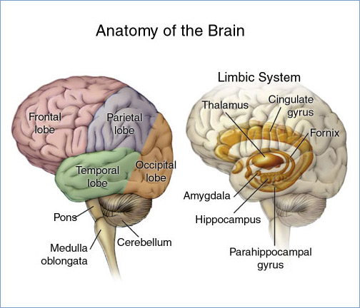
Pituicytoma is a rare brain tumor. It grows at the base of the brain from the pituitary gland. This tumor is thought to be derived from the parenchymal cells of the posterior lobe of the pituitary gland, called pituicytes. Some researchers believe that they arise from the folliculostellate cells in the anterior lobe of the pituitary. As such, it is a low-grade glioma. It occurs in adults and symptoms include visual disturbance and endocrine dysfunction. They are often mistaken for pituitary adenoma Pituitary adenomas are tumors that occur in the pituitary gland. Most pituitary tumors are benign, approximately 35% are invasive and just 0.1% to 0.2% are carcinomas. References Fu ...
|
Pituicytes
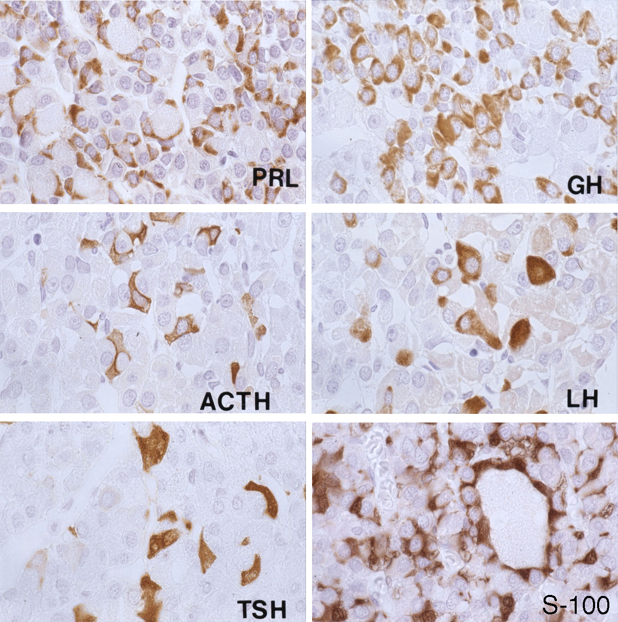
Pituicytes are glial cells of the posterior pituitary. Their main role is to assist in the storage and release of neurohypophysial hormones. Structure Pituicytes are located in the pars nervosa of the posterior pituitary and interspersed with unmyelinated axons and Herring bodies. They generally stain dark purple with an H&E stain and are among the easiest structures to identify in the region. Pituicytes have an irregular and branched shape which resembles that of another type of glial cell: the astrocyte. Like astrocytes, their cytoplasm presents specific intermediate filaments made up of glial fibrillary acidic protein (GFAP). Function Pituicytes are similar to astrocytes, another type of glial cell. Their main role is to assist in the storage and release of hormones of the posterior pituitary. Pituicytes surround axonal endings and regulate hormone secretion by releasing their processes from these endings. Clinical significance Pituicytomas are rare tumors that arise from p ... [...More Info...] [...Related Items...] OR: [Wikipedia] [Google] [Baidu] |
H&E Stain
Hematoxylin and eosin stain ( or haematoxylin and eosin stain or hematoxylin-eosin stain; often abbreviated as H&E stain or HE stain) is one of the principal tissue stains used in histology. It is the most widely used stain in medical diagnosis and is often the gold standard. For example, when a pathologist looks at a biopsy of a suspected cancer, the histological section is likely to be stained with H&E. H&E is the combination of two histological stains: hematoxylin and eosin. The hematoxylin stains cell nuclei a purplish blue, and eosin stains the extracellular matrix and cytoplasm pink, with other structures taking on different shades, hues, and combinations of these colors. Hence a pathologist can easily differentiate between the nuclear and cytoplasmic parts of a cell, and additionally, the overall patterns of coloration from the stain show the general layout and distribution of cells and provides a general overview of a tissue sample's structure. Thus, pattern recogniti ... [...More Info...] [...Related Items...] OR: [Wikipedia] [Google] [Baidu] |
Brain Tumor
A brain tumor occurs when abnormal cells form within the brain. There are two main types of tumors: malignant tumors and benign (non-cancerous) tumors. These can be further classified as primary tumors, which start within the brain, and secondary tumors, which most commonly have spread from tumors located outside the brain, known as brain metastasis tumors. All types of brain tumors may produce symptoms that vary depending on the size of the tumor and the part of the brain that is involved. Where symptoms exist, they may include headaches, seizures, problems with vision, vomiting and mental changes. Other symptoms may include difficulty walking, speaking, with sensations, or unconsciousness. The cause of most brain tumors is unknown. Uncommon risk factors include exposure to vinyl chloride, Epstein–Barr virus, ionizing radiation, and inherited syndromes such as neurofibromatosis, tuberous sclerosis, and von Hippel-Lindau Disease. Studies on mobile phone exposure hav ... [...More Info...] [...Related Items...] OR: [Wikipedia] [Google] [Baidu] |
Pituitary Gland
In vertebrate anatomy, the pituitary gland, or hypophysis, is an endocrine gland, about the size of a chickpea and weighing, on average, in humans. It is a protrusion off the bottom of the hypothalamus at the base of the brain. The hypophysis rests upon the hypophyseal fossa of the sphenoid bone in the center of the middle cranial fossa and is surrounded by a small bony cavity (sella turcica) covered by a dural fold (diaphragma sellae). The anterior pituitary (or adenohypophysis) is a lobe of the gland that regulates several physiological processes including stress, growth, reproduction, and lactation. The intermediate lobe synthesizes and secretes melanocyte-stimulating hormone. The posterior pituitary (or neurohypophysis) is a lobe of the gland that is functionally connected to the hypothalamus by the median eminence via a small tube called the pituitary stalk (also called the infundibular stalk or the infundibulum). Hormones secreted from the pituitary gland ... [...More Info...] [...Related Items...] OR: [Wikipedia] [Google] [Baidu] |
Parenchymal Cells
Parenchyma () is the bulk of functional substance in an animal organ or structure such as a tumour. In zoology it is the name for the tissue that fills the interior of flatworms. Etymology The term ''parenchyma'' is New Latin from the word παρέγχυμα ''parenchyma'' meaning 'visceral flesh', and from παρεγχεῖν ''parenchyma'' meaning 'to pour in' from παρα- ''para-'' 'beside' + ἐν ''en-'' 'in' + χεῖν ''chyma'' 'to pour'. Originally, Erasistratus and other anatomists used it to refer to certain human tissues. Later, it was also applied to plant tissues by Nehemiah Grew. Structure The parenchyma is the ''functional'' parts of an organ, or of a structure such as a tumour in the body. This is in contrast to the stroma, which refers to the ''structural'' tissue of organs or of structures, namely, the connective tissues. Brain The brain parenchyma refers to the functional tissue in the brain that is made up of the two types of brain cell, neurons a ... [...More Info...] [...Related Items...] OR: [Wikipedia] [Google] [Baidu] |
Posterior Lobe
The cerebellum (Latin for "little brain") is a major feature of the hindbrain of all vertebrates. Although usually smaller than the cerebrum, in some animals such as the mormyrid fishes it may be as large as or even larger. In humans, the cerebellum plays an important role in motor control. It may also be involved in some cognitive functions such as attention and language as well as emotional control such as regulating fear and pleasure responses, but its movement-related functions are the most solidly established. The human cerebellum does not initiate movement, but contributes to coordination, precision, and accurate timing: it receives input from sensory systems of the spinal cord and from other parts of the brain, and integrates these inputs to fine-tune motor activity. Cerebellar damage produces disorders in fine movement, equilibrium, posture, and motor learning in humans. Anatomically, the human cerebellum has the appearance of a separate structure attached to the bot ... [...More Info...] [...Related Items...] OR: [Wikipedia] [Google] [Baidu] |
Folliculostellate Cell
A Folliculostellate (FS) cell is a type of non- endocrine cell found in the anterior lobe of the pituitary gland. Histology and ultrastructure Rinehart and Farquhar first discovered FS cells through electron microscopy of the anterior pituitary gland. Vila-Porcile named these non-endocrine cells "folliculo-stellate" cells in 1972 due to their stellate (star) shape, and their location lining the lumen of small follicules in the anterior pituitary. Unlike the majority of cells in the anterior pituitary, they are non-endocrine and agranular. They have long cytoplasmic processes which interlock to form a mesh, within which the endocrine cells reside. They typically have a large number of microvilli on their apical side, and contain lysosomes, suggesting phagocytotic activity. Gap junctions can be seen between the FS cells and the adjacent endocrine cells when viewed under an electron microscope. Cell properties Using pituitary slices, studies have been conducted that have illu ... [...More Info...] [...Related Items...] OR: [Wikipedia] [Google] [Baidu] |
Anterior Lobe
The anterior lobe of cerebellum is the portion of the cerebellum responsible for mediating unconscious proprioception. Inputs into the anterior lobe of the cerebellum are mainly from the spinal cord. It is sometimes equated to the "paleocerebellum". Clinical significance Anterior lobe syndrome When a person gets most of their calories from alcohol (chronic alcoholism) the anterior lobe can deteriorate due to malnutrition. This is known as anterior lobe syndrome, and it causes unsteady gait. Additional images File:Anterior lobe of cerebellum -- animation.gif, Animation. Anterior lobe shown in red. File:Anterior lobe of cerebellum --- animation.gif, Close up animation. Anterior lobe shown in red. File:Cerebellar lobes by Sanjoy Sanyal.webm, Dissection video (1 min 20 s). Demonstrating the three cerebellar lobes. References External links * NIF Search - Anterior Lobe of the Cerebellumvia the Neuroscience Information Framework The Neuroscience Information Framework is a ... [...More Info...] [...Related Items...] OR: [Wikipedia] [Google] [Baidu] |
Glioma
A glioma is a type of tumor that starts in the glial cells of the brain or the spine. Gliomas comprise about 30 percent of all brain tumors and central nervous system tumours, and 80 percent of all malignant brain tumours. Signs and symptoms Symptoms of gliomas depend on which part of the central nervous system is affected. A brain glioma can cause headaches, vomiting, seizures, and cranial nerve disorders as a result of increased intracranial pressure. A glioma of the optic nerve can cause visual loss. Spinal cord gliomas can cause pain, weakness, or numbness in the extremities. Gliomas do not usually metastasize by the bloodstream, but they can spread via the cerebrospinal fluid and cause "drop metastases" to the spinal cord. Complex visual hallucinations have been described as a symptom of low-grade glioma. A child who has a subacute disorder of the central nervous system that produces cranial nerve abnormalities (especially of cranial nerve VII and the lower bulbar nerv ... [...More Info...] [...Related Items...] OR: [Wikipedia] [Google] [Baidu] |
Visual Disturbance
A vision disorder is an impairment of the sense of vision. Vision disorder is not the same as an eye disease. Although many vision disorders do have their immediate cause in the eye, there are many other causes that may occur at other locations in the optic pathway. Causes There are many eye conditions that can lead to vision disorder. Some of which are as follows: *Age-Related Macular Degeneration (ARMD): ARMD is a retinal degeneration disease specifically associated with macula blood vessels, which can result in central vision impairment. It is strongly linked to advancing age, as well as European ancestry. * Bulging eyes: where the eye (one or both) protrudes or distends out of its orbit. Left untreated, bulging eyes may lead to eye dryness, pain and vision loss * Cytomegalovirus (CMV) Retinitis: This is an inflammation of the retina caused by infection, which can result in blindness. It occurs in people experiencing suppressed immune systems, most commonly by Acquired Immu ... [...More Info...] [...Related Items...] OR: [Wikipedia] [Google] [Baidu] |
Endocrine Dysfunction
Endocrine diseases are disorders of the endocrine system. The branch of medicine associated with endocrine disorders is known as endocrinology. Types of disease Broadly speaking, endocrine disorders may be subdivided into three groups: # Endocrine gland hypofunction/hyposecretion (leading to hormone deficiency) # Endocrine gland hyperfunction/hypersecretion (leading to hormone excess) # Tumours (benign or malignant) of endocrine glands Endocrine disorders are often quite complex, involving a mixed picture of hyposecretion and hypersecretion because of the feedback mechanisms involved in the endocrine system. For example, most forms of hyperthyroidism are associated with an excess of thyroid hormone and a low level of thyroid stimulating hormone. List of diseases Glucose homeostasis disorders * Diabetes ** Type 1 Diabetes ** Type 2 Diabetes ** Gestational Diabetes ** Mature Onset Diabetes of the Young * Hypoglycemia ** Idiopathic hypoglycemia ** Insulinoma * Glucagonoma Thyro ... [...More Info...] [...Related Items...] OR: [Wikipedia] [Google] [Baidu] |
Pituitary Adenoma
Pituitary adenomas are tumors that occur in the pituitary gland. Most pituitary tumors are benign, approximately 35% are invasive and just 0.1% to 0.2% are carcinomas.Pituitary Tumors Treatment (PDQ®)–Health Professional Version NIH National Cancer Institute Pituitary adenomas represent from 10% to 25% of all intracranial and the estimated prevalence rate in the general population is approximately 17%. Non-invasive and non-secreting pituitary adenomas are considered to be |


