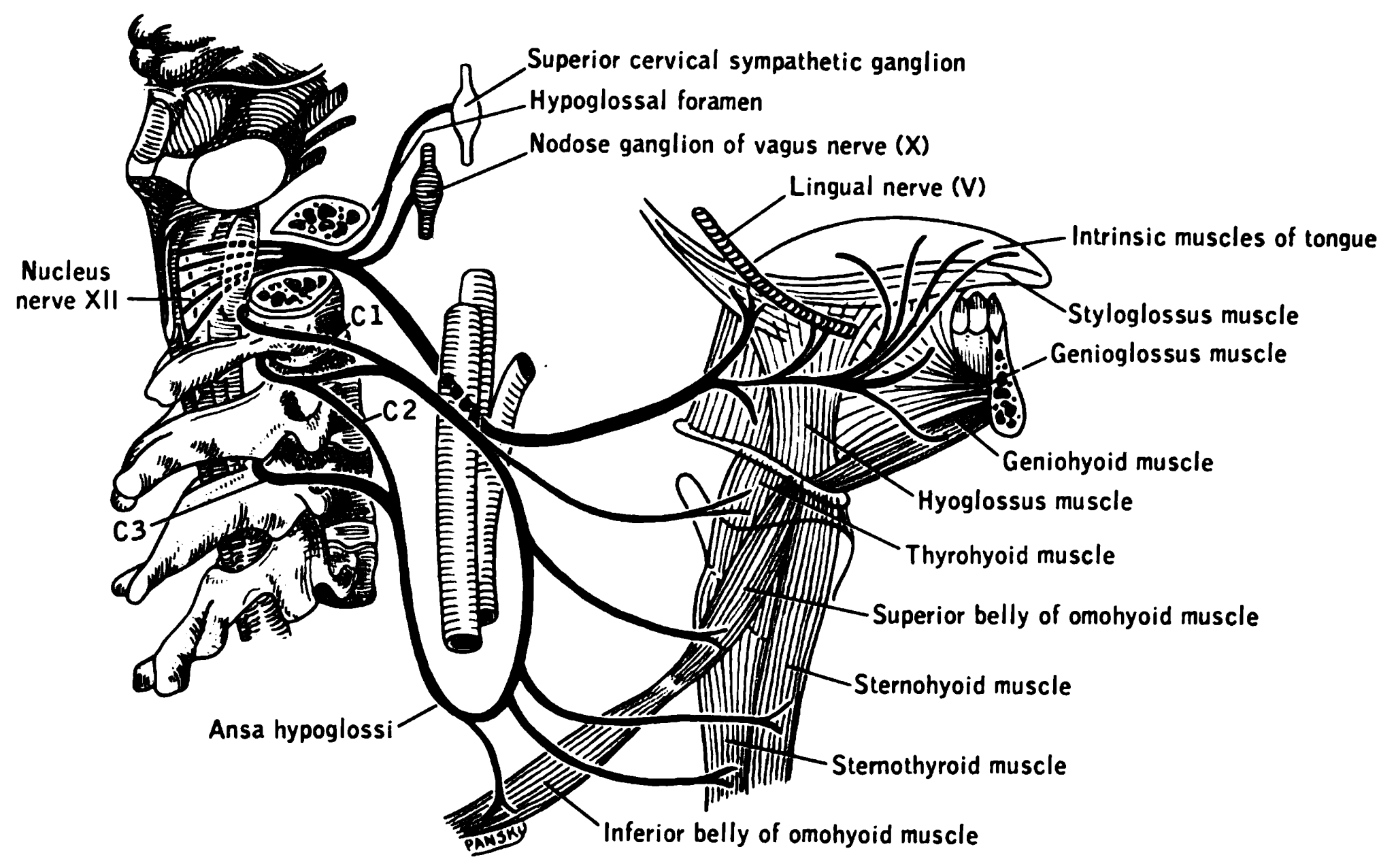
|
Pharyngeal Branch Of The Vagus Nerve
The pharyngeal branch of the vagus nerve, the principal motor nerve of the pharynx, arises from the upper part of the ganglion nodosum, and consists principally of filaments from the cranial portion of the accessory nerve. It passes across the internal carotid artery to the upper border of the Constrictor pharyngis medius, where it divides into numerous filaments, which join with branches from the glossopharyngeal, sympathetic, and external laryngeal to form the pharyngeal plexus. From the plexus, branches are distributed to the muscles and mucous membrane of the pharynx (except the stylopharyngeus, which is innervated by the glossopharyngeal nerve (CN IX)) and the muscles of the soft palate, except the Tensor veli palatini, which is supplied by the nerve to tensor veli palatini, a branch of the nerve to medial pterygoid (which itself is a branch of the mandibular nerve - CNV/3). A minute filament descends and joins the hypoglossal nerve as it winds around the occipital artery ... [...More Info...] [...Related Items...] OR: [Wikipedia] [Google] [Baidu] |
Glossopharyngeal
The glossopharyngeal nerve (), also known as the ninth cranial nerve, cranial nerve IX, or simply CN IX, is a cranial nerve that exits the brainstem from the sides of the upper Medulla oblongata, medulla, just anterior (closer to the nose) to the vagus nerve. Being a mixed nerve (sensorimotor), it carries afferent sensory and efferent motor information. The motor division of the glossopharyngeal nerve is derived from the Basal plate (neural tube), basal plate of the embryonic medulla oblongata, whereas the sensory division originates from the cranial neural crest. Structure From the anterior portion of the medulla oblongata, the glossopharyngeal nerve passes laterally across or below the Flocculus (cerebellar), flocculus, and leaves the skull through the central part of the jugular foramen. From the superior and inferior ganglia in jugular foramen, it has its own sheath of dura mater. The inferior ganglion on the inferior surface of petrous part of temporal is related with a tri ... [...More Info...] [...Related Items...] OR: [Wikipedia] [Google] [Baidu] |
Mucous Membrane
A mucous membrane or mucosa is a membrane that lines various cavities in the body of an organism and covers the surface of internal organs. It consists of one or more layers of epithelial cells overlying a layer of loose connective tissue. It is mostly of endodermal origin and is continuous with the skin at body openings such as the eyes, eyelids, ears, inside the nose, inside the mouth, lips, the genital areas, the urethral opening and the anus. Some mucous membranes secrete mucus, a thick protective fluid. The function of the membrane is to stop pathogens and dirt from entering the body and to prevent bodily tissues from becoming dehydrated. Structure The mucosa is composed of one or more layers of epithelial cells that secrete mucus, and an underlying lamina propria of loose connective tissue. The type of cells and type of mucus secreted vary from organ to organ and each can differ along a given tract. Mucous membranes line the digestive, respiratory and reproductive trac ... [...More Info...] [...Related Items...] OR: [Wikipedia] [Google] [Baidu] |
Vagus Nerve
The vagus nerve, also known as the tenth cranial nerve, cranial nerve X, or simply CN X, is a cranial nerve that interfaces with the parasympathetic control of the heart, lungs, and digestive tract. It comprises two nerves—the left and right vagus nerves—but they are typically referred to collectively as a single subsystem. The vagus is the longest nerve of the autonomic nervous system in the human body and comprises both sensory and motor fibers. The sensory fibers originate from neurons of the nodose ganglion, whereas the motor fibers come from neurons of the dorsal motor nucleus of the vagus and the nucleus ambiguus. The vagus was also historically called the pneumogastric nerve. Structure Upon leaving the medulla oblongata between the olive and the inferior cerebellar peduncle, the vagus nerve extends through the jugular foramen, then passes into the carotid sheath between the internal carotid artery and the internal jugular vein down to the neck, chest, and abdom ... [...More Info...] [...Related Items...] OR: [Wikipedia] [Google] [Baidu] |
Yale School Of Medicine
The Yale School of Medicine is the graduate medical school at Yale University, a private research university in New Haven, Connecticut. It was founded in 1810 as the Medical Institution of Yale College and formally opened in 1813. The primary teaching hospital for the school is Yale New Haven Hospital. The school is home to the Harvey Cushing/John Hay Whitney Medical Library, one of the largest modern medical libraries which is known for its historical collections. The faculty includes 70 National Academy of Sciences members, 47 National Academy of Medicine members, and 13 Howard Hughes Medical Institute investigators. '' U.S. News & World Report'' currently ranks the Yale School of Medicine 10th in the country for research and 59th in primary care. The MD program is highly selective; for the class of 2022, the school received 4,968 applications to fill 104 seats. The median GPA for the class was 3.89, and the median MCAT was 521. Education The School of Medicine offers the Doc ... [...More Info...] [...Related Items...] OR: [Wikipedia] [Google] [Baidu] |
Pharyngeal Nerve
The pharyngeal nerve is a small branch of the maxillary nerve, arising from the posterior part of the pterygopalatine ganglion. It passes through the palatovaginal canal with the pharyngeal branch of the maxillary artery, and is distributed to the mucous membrane of the nasal part of the pharynx, behind the auditory tube. See also * Pharyngeal branch of vagus nerve The pharyngeal branch of the vagus nerve, the principal motor nerve of the pharynx, arises from the upper part of the ganglion nodosum, and consists principally of filaments from the cranial portion of the accessory nerve. It passes across the i ... References External links Trigeminal nerve {{Neuroanatomy-stub ... [...More Info...] [...Related Items...] OR: [Wikipedia] [Google] [Baidu] |
Occipital Artery
The occipital artery arises from the external carotid artery opposite the facial artery. Its path is below the posterior belly of digastric to the occipital region. This artery supplies blood to the back of the scalp and sternocleidomastoid muscles, and deep muscles in the back and neck. Structure At its origin, it is covered by the posterior belly of the digastricus and the stylohyoideus, and the hypoglossal nerve winds around it from behind forward; higher up, it crosses the internal carotid artery, the internal jugular vein, and the vagus and accessory nerves. It next ascends to the interval between the transverse process of the atlas and the mastoid process of the temporal bone, and passes horizontally backward, grooving the surface of the latter bone, being covered by the sternocleidomastoideus, splenius capitis, longissimus capitis, and digastricus, and resting upon the rectus capitis lateralis, the obliquus superior, and semispinalis capitis. It then changes its course and ... [...More Info...] [...Related Items...] OR: [Wikipedia] [Google] [Baidu] |
Hypoglossal Nerve
The hypoglossal nerve, also known as the twelfth cranial nerve, cranial nerve XII, or simply CN XII, is a cranial nerve that innervates all the extrinsic and intrinsic muscles of the tongue except for the palatoglossus, which is innervated by the vagus nerve. CN XII is a nerve with a solely motor function. The nerve arises from the hypoglossal nucleus in the medulla as a number of small rootlets, passes through the hypoglossal canal and down through the neck, and eventually passes up again over the tongue muscles it supplies into the tongue. The nerve is involved in controlling tongue movements required for speech and swallowing, including sticking out the tongue and moving it from side to side. Damage to the nerve or the neural pathways which control it can affect the ability of the tongue to move and its appearance, with the most common sources of damage being injury from trauma or surgery, and motor neuron disease. The first recorded description of the nerve is by Herophil ... [...More Info...] [...Related Items...] OR: [Wikipedia] [Google] [Baidu] |
Tensor Veli Palatini
The tensor veli palatini muscle (tensor palati or tensor muscle of the velum palatinum) is a broad, thin, ribbon-like muscle in the head that tenses the soft palate. Structure The tensor veli palatini is found anterior-lateral to the levator veli palatini muscle. It arises by a flat lamella from the scaphoid fossa at the base of the medial pterygoid plate, from the spina angularis of the sphenoid and from the lateral wall of the cartilage of the auditory tube. Descending vertically between the medial pterygoid plate and the medial pterygoid muscle, it ends in a tendon which winds around the pterygoid hamulus, being retained in this situation by some of the fibers of origin of the medial pterygoid muscle. Between the tendon and the hamulus is a small bursa. The tendon then passes medially and is inserted into the palatine aponeurosis and into the surface behind the transverse ridge on the horizontal part of the palatine bone. Nerve supply The tensor veli palatini muscle is ... [...More Info...] [...Related Items...] OR: [Wikipedia] [Google] [Baidu] |
Soft Palate
The soft palate (also known as the velum, palatal velum, or muscular palate) is, in mammals, the soft tissue constituting the back of the roof of the mouth. The soft palate is part of the palate of the mouth; the other part is the hard palate. The soft palate is distinguished from the hard palate at the front of the mouth in that it does not contain bone. Structure Muscles The five muscles of the soft palate play important roles in swallowing and breathing. The muscles are: # Tensor veli palatini, which is involved in swallowing # Palatoglossus, involved in swallowing # Palatopharyngeus, involved in breathing # Levator veli palatini, involved in swallowing # Musculus uvulae, which moves the uvula These muscles are innervated by the pharyngeal plexus via the vagus nerve, with the exception of the tensor veli palatini. The tensor veli palatini is innervated by the mandibular division of the trigeminal nerve (V3). Function The soft palate is moveable, consisting of muscle f ... [...More Info...] [...Related Items...] OR: [Wikipedia] [Google] [Baidu] |
Stylopharyngeus
The stylopharyngeus is a muscle in the head that stretches between the temporal styloid process and the pharynx. Structure The stylopharyngeus is a long, slender muscle, cylindrical above, flattened below. It arises from the medial side of the base of the temporal styloid process, passes downward along the side of the pharynx between the superior pharyngeal constrictor and the middle pharyngeal constrictor, and spreads out beneath the mucous membrane. Some of its fibers are lost in the constrictor muscles while others, joining the palatopharyngeus muscle, are inserted into the posterior border of the thyroid cartilage. The glossopharyngeal nerve runs on the lateral side of this muscle, and crosses over it to reach the tongue. Nerve supply The stylopharyngeus is the only muscle in the pharynx innervated by the glossopharyngeal nerve (CN IX) via branchial motor neurons with their cell bodies in the rostral part of the nucleus ambiguus. Development Embryological origin is the ... [...More Info...] [...Related Items...] OR: [Wikipedia] [Google] [Baidu] |
External Laryngeal
The superior laryngeal nerve is a branch of the vagus nerve. It arises from the middle of the inferior ganglion of vagus nerve and in its course receives a branch from the superior cervical ganglion of the sympathetic nervous system. The superior laryngeal nerve consists of two branches: the internal laryngeal nerve (sensory), which supplies sensory fibers to the laryngeal mucosa, and the external laryngeal nerve (motor), which innervates the cricothyroid muscle. The superior laryngeal nerve innervates the two cricothyroid muscles. The recurrent laryngeal nerve gets its name from the fact that it loops below the aorta on its way to the intrinsic muscles of the larynx. The left recurrent laryngeal nerve passes under and around the aorta on its way to the larynx, whereas the right recurrent laryngeal nerve passes under and around the subclavian artery. Because the aorta is inferior to the subclavian artery, the left recurrent laryngeal nerve is a bit longer than the right recurren ... [...More Info...] [...Related Items...] OR: [Wikipedia] [Google] [Baidu] |
Vagus
The vagus nerve, also known as the tenth cranial nerve, cranial nerve X, or simply CN X, is a cranial nerve that interfaces with the parasympathetic control of the heart, lungs, and digestive tract. It comprises two nerves—the left and right vagus nerves—but they are typically referred to collectively as a single subsystem. The vagus is the longest nerve of the autonomic nervous system in the human body and comprises both sensory and motor fibers. The sensory fibers originate from neurons of the nodose ganglion, whereas the motor fibers come from neurons of the dorsal motor nucleus of the vagus and the nucleus ambiguus. The vagus was also historically called the pneumogastric nerve. Structure Upon leaving the medulla oblongata between the olive and the inferior cerebellar peduncle, the vagus nerve extends through the jugular foramen, then passes into the carotid sheath between the internal carotid artery and the internal jugular vein down to the neck, chest, and abdomen, wh ... [...More Info...] [...Related Items...] OR: [Wikipedia] [Google] [Baidu] |


