|
Perinuclear Vacuolization
Vacuolization is the formation of vacuoles or vacuole-like structures, within or adjacent to cells. Perinuclear vacuolization of epidermal keratinocytes is most likely inconsequential when not observed in combination with other pathologic findings. In dermatopathology "vacuolization" often refers specifically to vacuoles in the basal cell-basement membrane zone area, where it is an unspecific sign of disease.Kumar, Vinay; Fausto, Nelso; Abbas, Abul (2004) ''Robbins & Cotran Pathologic Basis of Disease'' (7th ed.). Saunders. Page 1230. . It may be a sign of for example vacuolar interface dermatitis, which in turn has many causes. It is one of the components of koilocytosis, which may be present in potentially pre- cancerous cervical, oral and anal lesions. See also * Skin lesion * Skin disease * List of skin diseases Many skin conditions affect the human integumentary system—the organ system covering the entire surface of the body and composed of skin, hair, nails, a ... [...More Info...] [...Related Items...] OR: [Wikipedia] [Google] [Baidu] |
Micrograph Of Perinuclear Vacuolization, Annotated
A micrograph or photomicrograph is a photograph or digital image taken through a microscope or similar device to show a magnified image of an object. This is opposed to a macrograph or photomacrograph, an image which is also taken on a microscope but is only slightly magnified, usually less than 10 times. Micrography is the practice or art of using microscopes to make photographs. A micrograph contains extensive details of microstructure. A wealth of information can be obtained from a simple micrograph like behavior of the material under different conditions, the phases found in the system, failure analysis, grain size estimation, elemental analysis and so on. Micrographs are widely used in all fields of microscopy. Types Photomicrograph A light micrograph or photomicrograph is a micrograph prepared using an optical microscope, a process referred to as ''photomicroscopy''. At a basic level, photomicroscopy may be performed simply by connecting a camera to a microscope, t ... [...More Info...] [...Related Items...] OR: [Wikipedia] [Google] [Baidu] |
Ameloblastoma - High Mag
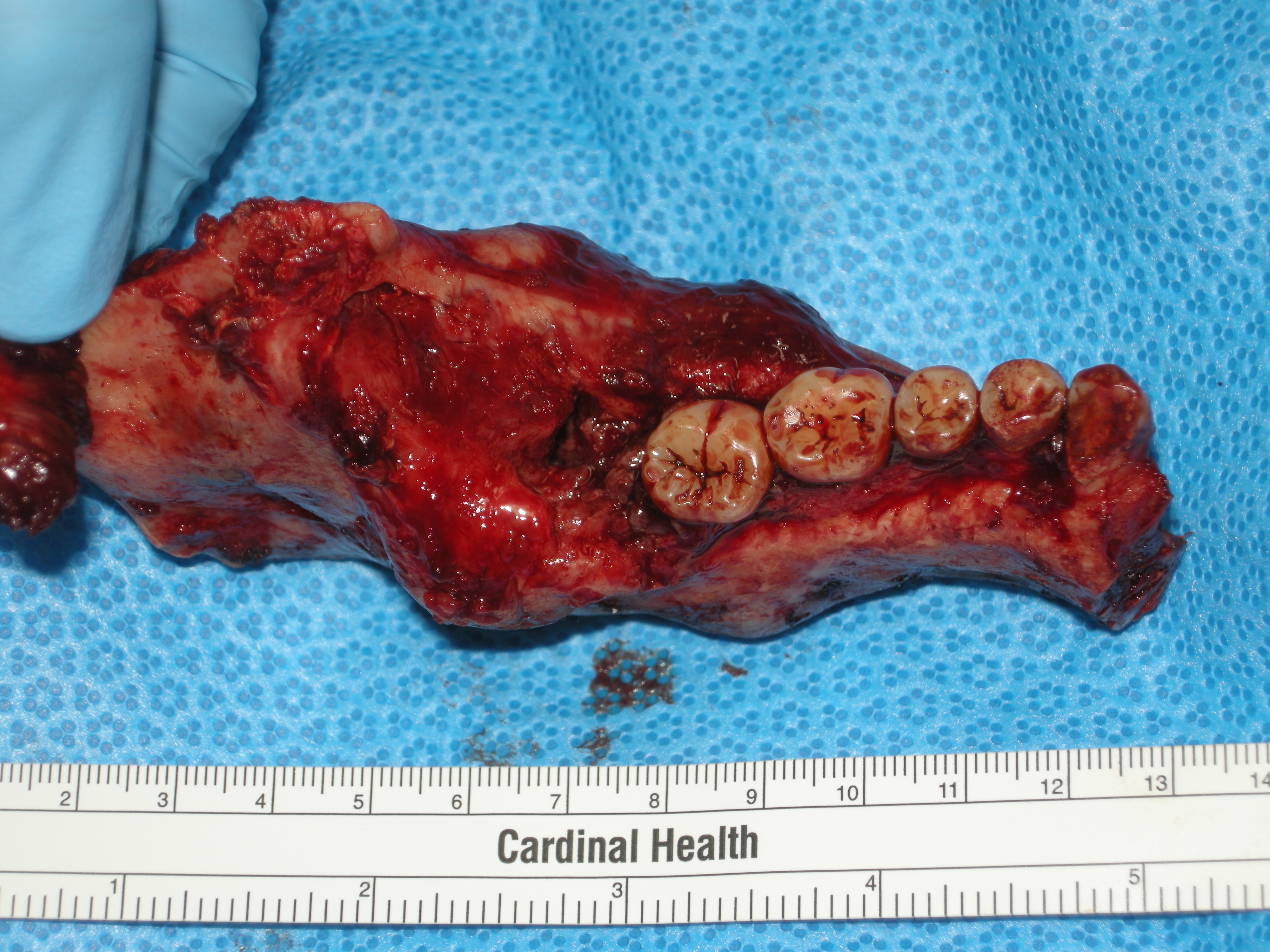
Ameloblastoma is a rare, benign or cancerous tumor of odontogenic epithelium (ameloblasts, or outside portion, of the Tooth development, teeth during development) much more commonly appearing in the human mandible, lower jaw than the maxilla, upper jaw. It was recognized in 1827 by Cusack. This type of odontogenic neoplasm was designated as an ''adamantinoma'' in 1885 by the French physician Louis-Charles Malassez. It was finally renamed to the modern name ''ameloblastoma'' in 1930 by Ivey and Churchill. While these tumors are rarely malignant or metastatic (that is, they rarely spread to other parts of the body), and progress slowly, the resulting lesions can cause severe abnormalities of the face and jaw leading to severe disfiguration. Additionally, as abnormal cell growth easily infiltrates and destroys surrounding bony tissues, wide surgical excision is required to treat this disorder. If an aggressive tumor is left untreated, it can obstruct the nasal and oral airways making ... [...More Info...] [...Related Items...] OR: [Wikipedia] [Google] [Baidu] |
Vacuoles
A vacuole () is a membrane-bound organelle which is present in plant and fungal cells and some protist, animal, and bacterial cells. Vacuoles are essentially enclosed compartments which are filled with water containing inorganic and organic molecules including enzymes in solution, though in certain cases they may contain solids which have been engulfed. Vacuoles are formed by the fusion of multiple membrane vesicles and are effectively just larger forms of these. The organelle has no basic shape or size; its structure varies according to the requirements of the cell. Discovery Contractile vacuoles ("stars") were first observed by Spallanzani (1776) in protozoa, although mistaken for respiratory organs. Dujardin (1841) named these "stars" as ''vacuoles''. In 1842, Schleiden applied the term for plant cells, to distinguish the structure with cell sap from the rest of the protoplasm. In 1885, de Vries named the vacuole membrane as tonoplast. Function The function and significa ... [...More Info...] [...Related Items...] OR: [Wikipedia] [Google] [Baidu] |
Nuclear Envelope
The nuclear envelope, also known as the nuclear membrane, is made up of two lipid bilayer membranes that in eukaryotic cells surround the nucleus, which encloses the genetic material. The nuclear envelope consists of two lipid bilayer membranes: an inner nuclear membrane and an outer nuclear membrane. The space between the membranes is called the perinuclear space. It is usually about 10–50 nm wide. The outer nuclear membrane is continuous with the endoplasmic reticulum membrane. The nuclear envelope has many nuclear pores that allow materials to move between the cytosol and the nucleus. Intermediate filament proteins called lamins form a structure called the nuclear lamina on the inner aspect of the inner nuclear membrane and give structural support to the nucleus. Structure The nuclear envelope is made up of two lipid bilayer membranes, an inner nuclear membrane and an outer nuclear membrane. These membranes are connected to each other by nuclear pores. Two sets of in ... [...More Info...] [...Related Items...] OR: [Wikipedia] [Google] [Baidu] |
Epidermis
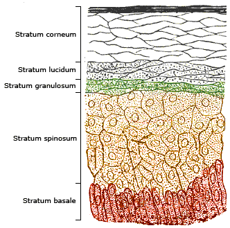
The epidermis is the outermost of the three layers that comprise the skin, the inner layers being the dermis and hypodermis. The epidermis layer provides a barrier to infection from environmental pathogens and regulates the amount of water released from the body into the atmosphere through transepidermal water loss. The epidermis is composed of multiple layers of flattened cells that overlie a base layer (stratum basale) composed of columnar cells arranged perpendicularly. The layers of cells develop from stem cells in the basal layer. The human epidermis is a familiar example of epithelium, particularly a stratified squamous epithelium. The word epidermis is derived through Latin , itself and . Something related to or part of the epidermis is termed epidermal. Structure Cellular components The epidermis primarily consists of keratinocytes ( proliferating basal and differentiated suprabasal), which comprise 90% of its cells, but also contains melanocytes, Langerhans ... [...More Info...] [...Related Items...] OR: [Wikipedia] [Google] [Baidu] |
Keratinocyte
Keratinocytes are the primary type of Cell (biology), cell found in the epidermis (skin), epidermis, the outermost layer of the skin. In humans, they constitute 90% of epidermal skin cells. Basal cells in the stratum basale, basal layer (''stratum basale'') of the skin are sometimes referred to as basal keratinocytes. Keratinocytes form a barrier against environmental damage by heat, UV radiation, Dehydration, water loss, pathogenic bacteria, fungi, parasites, and viruses. A number of structural proteins, enzymes, lipids, and antimicrobial peptides contribute to maintain the important barrier function of the skin. Keratinocytes differentiate from epidermal stem cells in the lower part of the epidermis and migrate towards the surface, finally becoming corneocytes and eventually be shed off, which happens every 40 to 56 days in humans. Function The primary function of keratinocytes is the formation of a barrier against environmental damage by heat, UV radiation, Dehydration, wat ... [...More Info...] [...Related Items...] OR: [Wikipedia] [Google] [Baidu] |
Dermatopathology
Dermatopathology (from Greek , ''derma'' 'skin' + , ''pathos'' 'fate, harm' + , '' -logia'' 'study of') is a joint subspecialty of dermatology and pathology or surgical pathology that focuses on the study of cutaneous diseases at a microscopic and molecular level. It also encompasses analyses of the potential causes of skin diseases at a basic level. Dermatopathologists work in close association with clinical dermatologists, with many possessing further clinical training in dermatology. Dermatologists are able to recognize most skin diseases based on their appearances, anatomic distributions, and behavior. Sometimes, however, those criteria do not allow a conclusive diagnosis to be made, and a skin biopsy is taken to be examined under the microscope or are subject to other molecular tests. That process reveals the histology of the disease and results in a specific diagnostic interpretation. In some cases, additional specialized testing needs to be performed on biopsies, including i ... [...More Info...] [...Related Items...] OR: [Wikipedia] [Google] [Baidu] |
Basement Membrane
The basement membrane is a thin, pliable sheet-like type of extracellular matrix that provides cell and tissue support and acts as a platform for complex signalling. The basement membrane sits between Epithelium, epithelial tissues including mesothelium and endothelium, and the underlying connective tissue. Structure As seen with the electron microscope, the basement membrane is composed of two layers, the basal lamina and the reticular lamina. The underlying connective tissue attaches to the basal lamina with collagen VII anchoring fibrils and fibrillin microfibrils. The basal lamina layer can further be subdivided into two layers based on their visual appearance in electron microscopy. The lighter-colored layer closer to the epithelium is called the lamina lucida, while the denser-colored layer closer to the connective tissue is called the lamina densa. The Electron microscope, electron-dense lamina densa layer is about 30–70 nanometers thick and consists of an underlying ... [...More Info...] [...Related Items...] OR: [Wikipedia] [Google] [Baidu] |
Vacuolar Interface Dermatitis
Vacuolar interface dermatitis (VAC, also known as liquefaction degeneration, vacuolar alteration or hydropic degeneration) is a dermatitis with vacuolization at the dermoepidermal junction, with lymphocytic inflammation at the epidermis and dermis. Causes An interface dermatitis with vacuolar alteration, not otherwise specified, may be caused by viral exanthems, phototoxic dermatitis, acute radiation dermatitis, erythema dyschromicum perstans, lupus erythematosus and dermatomyositis Dermatomyositis (DM) is a long-term inflammatory disorder which affects skin and the muscles. Its symptoms are generally a skin rash and worsening muscle weakness over time. These may occur suddenly or develop over months. Other symptoms may inc .... References {{reflist Dermatologic terminology ... [...More Info...] [...Related Items...] OR: [Wikipedia] [Google] [Baidu] |
Koilocytosis
A koilocyte is a squamous epithelial cell that has undergone a number of structural changes, which occur as a result of infection of the cell by human papillomavirus (HPV). Identification of these cells by pathologists can be useful in diagnosing various HPV-associated lesions. Koilocytosis Koilocytosis or koilocytic atypia or koilocytotic atypia are terms used in histology and cytology to describe the presence of koilocytes in a specimen. Koilocytes may have the following cellular changes: * Nuclear enlargement (two to three times normal size). * Irregularity of the nuclear membrane contour, creating a wrinkled or raisinoid appearance. * A darker than normal staining pattern in the nucleus, known as hyperchromasia. * A clear area around the nucleus, known as a perinuclear halo or perinuclear cytoplasmic vacuolization. Collectively, these types of changes are called a cytopathic effect; various types of cytopathic effect can be seen in many different cell types infected by many ... [...More Info...] [...Related Items...] OR: [Wikipedia] [Google] [Baidu] |
Cervical Intraepithelial Neoplasia
Cervical intraepithelial neoplasia (CIN), also known as cervical dysplasia, is the abnormal growth of cells on the surface of the cervix that could potentially lead to cervical cancer. More specifically, CIN refers to the potentially precancerous transformation of cells of the cervix. CIN most commonly occurs at the squamocolumnar junction of the cervix, a transitional area between the squamous epithelium of the vagina and the columnar epithelium of the endocervix. It can also occur in vaginal walls and vulvar epithelium. CIN is graded on a 1–3 scale, with 3 being the most abnormal (see classification section below). Human papillomavirus (HPV) infection is necessary for the development of CIN, but not all with this infection develop cervical cancer. Many women with HPV infection never develop CIN or cervical cancer. Typically, HPV resolves on its own. However, those with an HPV infection that lasts more than one or two years have a higher risk of developing a higher grade of CIN. ... [...More Info...] [...Related Items...] OR: [Wikipedia] [Google] [Baidu] |
HPV-positive Oropharyngeal Cancer
Human papillomavirus-positive oropharyngeal cancer (HPV-positive OPC or HPV+OPC), is a cancer (squamous cell carcinoma) of the throat caused by the human papillomavirus type 16 virus (HPV16). In the past, cancer of the oropharynx (throat) was associated with the use of alcohol or tobacco or both, but the majority of cases are now associated with the HPV virus, acquired by having oral contact with the genitals (oral-genital sex) of a person who has a genital HPV infection. Risk factors include having a large number of sexual partners, a history of oral-genital sex or anal–oral sex, having a female partner with a history of either an abnormal Pap smear or cervical dysplasia, having chronic periodontitis, and, among men, younger age at first intercourse and a history of genital warts. HPV-positive OPC is considered a separate disease from HPV-negative oropharyngeal cancer (also called HPV negative-OPC and HPV-OPC). HPV-positive OPC presents in one of four ways: as an asymptoma ... [...More Info...] [...Related Items...] OR: [Wikipedia] [Google] [Baidu] |


.jpg)
_EM_(new_version).jpg)