|
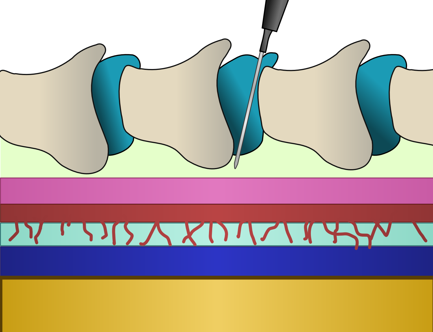
Peridural
Epidural administration (from Ancient Greek ἐπί, , upon" + ''dura mater'') is a method of medication administration in which a medicine is injected into the epidural space around the spinal cord. The epidural route is used by physicians and nurse anesthetists to administer local anesthetic agents, analgesics, diagnostic medicines such as radiocontrast agents, and other medicines such as glucocorticoids. Epidural administration involves the placement of a catheter into the epidural space, which may remain in place for the duration of the treatment. The technique of intentional epidural administration of medication was first described in 1921 by Spanish military surgeon Fidel Pagés. In the United States, over 50% of childbirths involve the use of epidural anesthesia. Epidural anaesthesia causes a loss of sensation, including pain, by blocking the transmission of signals through nerve fibres in or near the spinal cord. For this reason, epidurals are commonly used for pain cont ... [...More Info...] [...Related Items...] OR: [Wikipedia] [Google] [Baidu] |
Route Of Administration
A route of administration in pharmacology and toxicology is the way by which a medication, drug, fluid, poison, or other substance is taken into the body. Routes of administration are generally classified by the location at which the substance is applied. Common examples include oral administration, oral and intravenous therapy, intravenous administration. Routes can also be classified based on where the target of action is. Action may be topical medication, topical (local), enteral administration, enteral (system-wide effect, but delivered through the gastrointestinal tract), or #Parenteral, parenteral (systemic action, but delivered by routes other than the GI tract). Route of administration and dosage form are aspects of drug delivery. Classification Routes of administration are usually classified by application location (or exposition). The route or course the active substance takes from application location to the location where it has its target effect is usually rather ... [...More Info...] [...Related Items...] OR: [Wikipedia] [Google] [Baidu] |
Epidural Space
In anatomy, the epidural space is the potential space between the dura mater and vertebrae ( spine). The anatomy term "epidural space" has its origin in the Ancient Greek language; , "on, upon" + dura mater also known as "epidural cavity", "extradural space" or "peridural space". In humans the epidural space contains lymphatics, spinal nerve roots, loose connective tissue, adipose tissue, small arteries, dural venous sinuses and a network of internal vertebral venous plexuses. Cranial epidural space In the skull, the periosteal layer of the dura mater adheres to the inner surface of the skull bones while the meningeal layer lays over the arachnoid mater. Between them is the epidural space. The two layers of the dura mater separate at several places, with the meningeal layer projecting deeper into the brain parenchyma forming fibrous septa that compartmentalize the brain tissue. At these sites, the epidural space is wide enough to house the epidural venous sinuses. There are fo ... [...More Info...] [...Related Items...] OR: [Wikipedia] [Google] [Baidu] |
Lumbar
In tetrapod anatomy, lumbar is an adjective that means ''of or pertaining to the abdominal segment of the torso, between the diaphragm and the sacrum.'' The lumbar region is sometimes referred to as the lower spine, or as an area of the back in its proximity. In human anatomy the five lumbar vertebrae (vertebrae in the lumbar region of the back) are the largest and strongest in the movable part of the spinal column, and can be distinguished by the absence of a foramen in the transverse process, and by the absence of facets on the sides of the body. In most mammals, the lumbar region of the spine curves outward. The actual spinal cord terminates between vertebrae one and two of this series, called L1 and L2. The nervous tissue that extends below this point are individual strands that collectively form the cauda equina. In between each lumbar vertebra a nerve root exits, and these nerve roots come together again to form the largest single nerve in the human body, the sciatic n ... [...More Info...] [...Related Items...] OR: [Wikipedia] [Google] [Baidu] |
Nerve Fibres
An axon (from Greek ἄξων ''áxōn'', axis), or nerve fiber (or nerve fibre: see spelling differences), is a long, slender projection of a nerve cell, or neuron, in vertebrates, that typically conducts electrical impulses known as action potentials away from the nerve cell body. The function of the axon is to transmit information to different neurons, muscles, and glands. In certain sensory neurons (pseudounipolar neurons), such as those for touch and warmth, the axons are called afferent nerve fibers and the electrical impulse travels along these from the periphery to the cell body and from the cell body to the spinal cord along another branch of the same axon. Axon dysfunction can be the cause of many inherited and acquired neurological disorders that affect both the peripheral and central neurons. Nerve fibers are classed into three typesgroup A nerve fibers, group B nerve fibers, and group C nerve fibers. Groups A and B are myelinated, and group C are unmyelinated. The ... [...More Info...] [...Related Items...] OR: [Wikipedia] [Google] [Baidu] |
Fluoroscopy
Fluoroscopy () is an imaging technique that uses X-rays to obtain real-time moving images of the interior of an object. In its primary application of medical imaging, a fluoroscope () allows a physician to see the internal structure and function of a patient, so that the pumping action of the heart or the motion of swallowing, for example, can be watched. This is useful for both diagnosis and therapy and occurs in general radiology, interventional radiology, and image-guided surgery. In its simplest form, a fluoroscope consists of an X-ray source and a fluorescent screen, between which a patient is placed. However, since the 1950s most fluoroscopes have included X-ray image intensifiers and cameras as well, to improve the image's visibility and make it available on a remote display screen. For many decades, fluoroscopy tended to produce live pictures that were not recorded, but since the 1960s, as technology improved, recording and playback became the norm. Fluoroscopy is s ... [...More Info...] [...Related Items...] OR: [Wikipedia] [Google] [Baidu] |
Portable Ultrasound
Portable ultrasound is a modality of medical ultrasonography that utilizes small and light devices, compared to the console-style ultrasound machines that preceded them. In most cases these mobile ultrasound systems could be carried by hand and in some cases even operated for a time on battery power alone. The first portable ultrasound machines arrived in the early 1980s but battery powered systems that could be easily carried did not arrive until the late 1990s. History The ADR 2130, designed by Marty Wilcox was the first portable ultrasound unit commercially available in the United States, being released in 1975. This unit weighed about 25 lbs, had 3 linear probes to choose from and used an oscilloscope for a display instead of a TV monitor. ADR was later purchased by ATL which later spun off its portable technology into a company that became Sonosite. ATL was later purchased by Philips while Sonosite came out with its first battery powered portable offering, the Sonosite 180 ... [...More Info...] [...Related Items...] OR: [Wikipedia] [Google] [Baidu] |
Nerve
A nerve is an enclosed, cable-like bundle of nerve fibers (called axons) in the peripheral nervous system. A nerve transmits electrical impulses. It is the basic unit of the peripheral nervous system. A nerve provides a common pathway for the electrochemical nerve impulses called action potentials that are transmitted along each of the axons to peripheral organs or, in the case of sensory nerves, from the periphery back to the central nervous system. Each axon, within the nerve, is an extension of an individual neuron, along with other supportive cells such as some Schwann cells that coat the axons in myelin. Within a nerve, each axon is surrounded by a layer of connective tissue called the endoneurium. The axons are bundled together into groups called fascicles, and each fascicle is wrapped in a layer of connective tissue called the perineurium. Finally, the entire nerve is wrapped in a layer of connective tissue called the epineurium. Nerve cells (often called neurons) are f ... [...More Info...] [...Related Items...] OR: [Wikipedia] [Google] [Baidu] |
Lumbar Spine
The lumbar vertebrae are, in human anatomy, the five vertebrae between the rib cage and the pelvis. They are the largest segments of the vertebral column and are characterized by the absence of the foramen transversarium within the transverse process (since it is only found in the cervical region) and by the absence of facets on the sides of the body (as found only in the thoracic region). They are designated L1 to L5, starting at the top. The lumbar vertebrae help support the weight of the body, and permit movement. Human anatomy General characteristics The adjacent figure depicts the general characteristics of the first through fourth lumbar vertebrae. The fifth vertebra contains certain peculiarities, which are detailed below. As with other vertebrae, each lumbar vertebra consists of a ''vertebral body'' and a ''vertebral arch''. The vertebral arch, consisting of a pair of ''pedicles'' and a pair of ''laminae'', encloses the ''vertebral foramen'' (opening) and sup ... [...More Info...] [...Related Items...] OR: [Wikipedia] [Google] [Baidu] |
Respiratory Depression
Hypoventilation (also known as respiratory depression) occurs when ventilation is inadequate (''hypo'' meaning "below") to perform needed respiratory gas exchange. By definition it causes an increased concentration of carbon dioxide (hypercapnia) and respiratory acidosis. Hypoventilation is not synonymous with respiratory arrest, in which breathing ceases entirely and death occurs within minutes due to hypoxia and leads rapidly into complete anoxia, although both are medical emergencies. Hypoventilation can be considered a precursor to hypoxia and its lethality is attributed to hypoxia with carbon dioxide toxicity. Causes Hypoventilation may be caused by: *A medical condition such as stroke affecting the brainstem *Voluntary breath-holding or underbreathing, for example, hypoventilation training or the Buteyko method. *Medication or drugs, typically when taken in accidental or intentional overdose. Opioids and benzodiazepines in particular are known to cause respiratory depress ... [...More Info...] [...Related Items...] OR: [Wikipedia] [Google] [Baidu] |
Headache
Headache is the symptom of pain in the face, head, or neck. It can occur as a migraine, tension-type headache, or cluster headache. There is an increased risk of depression in those with severe headaches. Headaches can occur as a result of many conditions. There are a number of different classification systems for headaches. The most well-recognized is that of the International Headache Society, which classifies it into more than 150 types of primary and secondary headaches. Causes of headaches may include dehydration; fatigue; sleep deprivation; stress; the effects of medications (overuse) and recreational drugs, including withdrawal; viral infections; loud noises; head injury; rapid ingestion of a very cold food or beverage; and dental or sinus issues (such as sinusitis). Treatment of a headache depends on the underlying cause, but commonly involves pain medication (especially in case of migraine or cluster headache). A headache is one of the most commonly experienced ... [...More Info...] [...Related Items...] OR: [Wikipedia] [Google] [Baidu] |
Inflammation
Inflammation (from la, wikt:en:inflammatio#Latin, inflammatio) is part of the complex biological response of body tissues to harmful stimuli, such as pathogens, damaged cells, or Irritation, irritants, and is a protective response involving immune cells, blood vessels, and molecular mediators. The function of inflammation is to eliminate the initial cause of cell injury, clear out necrotic cells and tissues damaged from the original insult and the inflammatory process, and initiate tissue repair. The five cardinal signs are heat, pain, redness, swelling, and Functio laesa, loss of function (Latin ''calor'', ''dolor'', ''rubor'', ''tumor'', and ''functio laesa''). Inflammation is a generic response, and therefore it is considered as a mechanism of innate immune system, innate immunity, as compared to adaptive immune system, adaptive immunity, which is specific for each pathogen. Too little inflammation could lead to progressive tissue destruction by the harmful stimulus (e.g. b ... [...More Info...] [...Related Items...] OR: [Wikipedia] [Google] [Baidu] |
Steroids
A steroid is a biologically active organic compound with four rings arranged in a specific molecular configuration. Steroids have two principal biological functions: as important components of cell membranes that alter membrane fluidity; and as signaling molecules. Hundreds of steroids are found in plants, animals and fungi. All steroids are manufactured in cells from the sterols lanosterol (opisthokonts) or cycloartenol (plants). Lanosterol and cycloartenol are derived from the cyclization of the triterpene squalene. The steroid core structure is typically composed of seventeen carbon atoms, bonded in four " fused" rings: three six-member cyclohexane rings (rings A, B and C in the first illustration) and one five-member cyclopentane ring (the D ring). Steroids vary by the functional groups attached to this four-ring core and by the oxidation state of the rings. Sterols are forms of steroids with a hydroxy group at position three and a skeleton derived from cholestane. '' ... [...More Info...] [...Related Items...] OR: [Wikipedia] [Google] [Baidu] |

.jpg)


