|
Palmar Cutaneous Branch
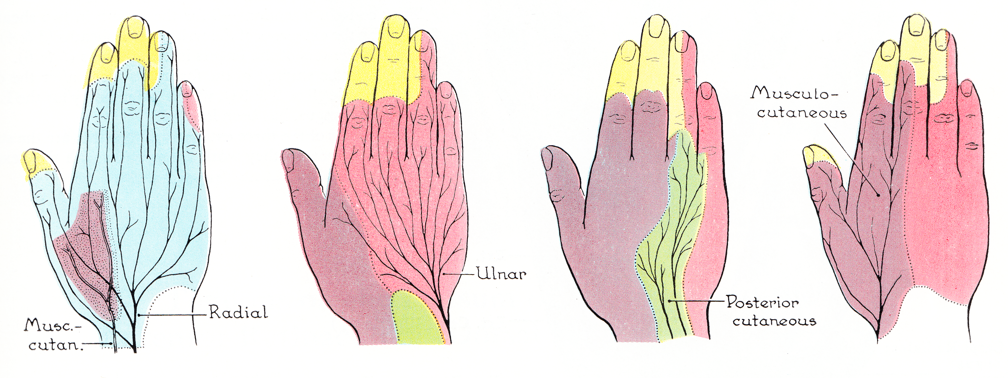
The palmar branch of the median nerve is a branch of the median nerve which arises at the distal part of the forearm. Branches It pierces the palmar carpal ligament, and divides into a lateral and a medial branch; * The ''lateral branch'' supplies the skin over the ball of the thumb, and communicates with the volar branch of the lateral antebrachial cutaneous nerve. * The ''medial branch'' supplies the skin of the Hand, palm and communicates with the palmar cutaneous branch of the ulnar. Clinical significance Unlike most of the median nerve innervation of the hand, the palmar branch travels superficial to the Flexor retinaculum of the hand. Therefore, this portion of the median nerve usually remains functioning during carpal tunnel syndrome. Additional images File:Gray812and814.PNG, Diagram of segmental distribution of the cutaneous nerves of the right upper extremity. References * http://nervesurgery.wustl.edu/NerveImages/Anatomy%20and%20Physiology/AP-Median-Nerve---I ... [...More Info...] [...Related Items...] OR: [Wikipedia] [Google] [Baidu] |
Median Nerve
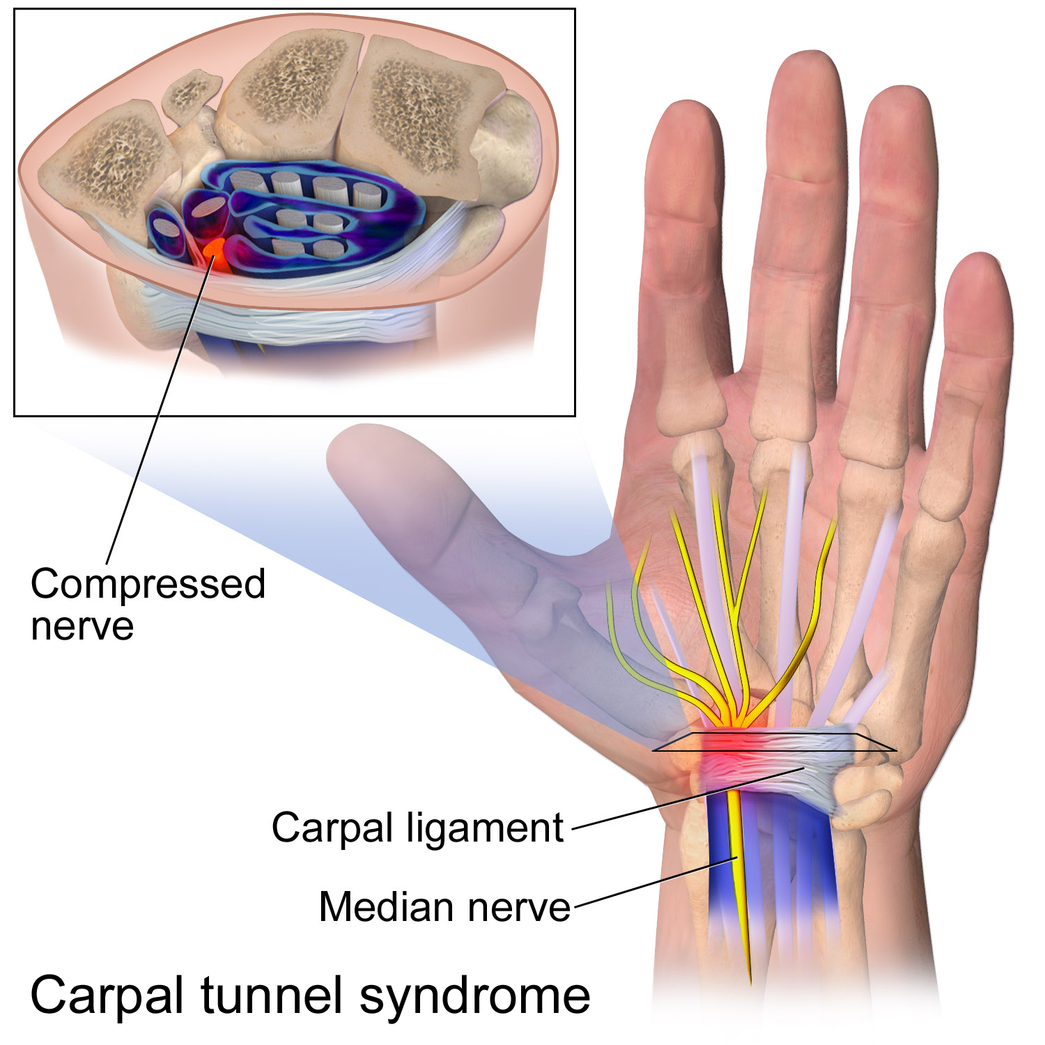
The median nerve is a nerve in humans and other animals in the upper limb. It is one of the five main nerves originating from the brachial plexus. The median nerve originates from the lateral and medial cords of the brachial plexus, and has contributions from ventral roots of C5-C7 (lateral cord) and C8 and T1 (medial cord). The median nerve is the only nerve that passes through the carpal tunnel. Carpal tunnel syndrome is the disability that results from the median nerve being pressed in the carpal tunnel. Structure The median nerve arises from the branches from lateral and medial cords of the brachial plexus, courses through the anterior part of arm, forearm, and hand, and terminates by supplying the muscles of the hand. Arm After receiving inputs from both the lateral and medial cords of the brachial plexus, the median nerve enters the arm from the axilla at the inferior margin of the teres major muscle. It then passes vertically down and courses lateral to the brachial ar ... [...More Info...] [...Related Items...] OR: [Wikipedia] [Google] [Baidu] |
Distal
Standard anatomical terms of location are used to unambiguously describe the anatomy of animals, including humans. The terms, typically derived from Latin or Greek roots, describe something in its standard anatomical position. This position provides a definition of what is at the front ("anterior"), behind ("posterior") and so on. As part of defining and describing terms, the body is described through the use of anatomical planes and anatomical axes. The meaning of terms that are used can change depending on whether an organism is bipedal or quadrupedal. Additionally, for some animals such as invertebrates, some terms may not have any meaning at all; for example, an animal that is radially symmetrical will have no anterior surface, but can still have a description that a part is close to the middle ("proximal") or further from the middle ("distal"). International organisations have determined vocabularies that are often used as standard vocabularies for subdisciplines of anatom ... [...More Info...] [...Related Items...] OR: [Wikipedia] [Google] [Baidu] |
Forearm
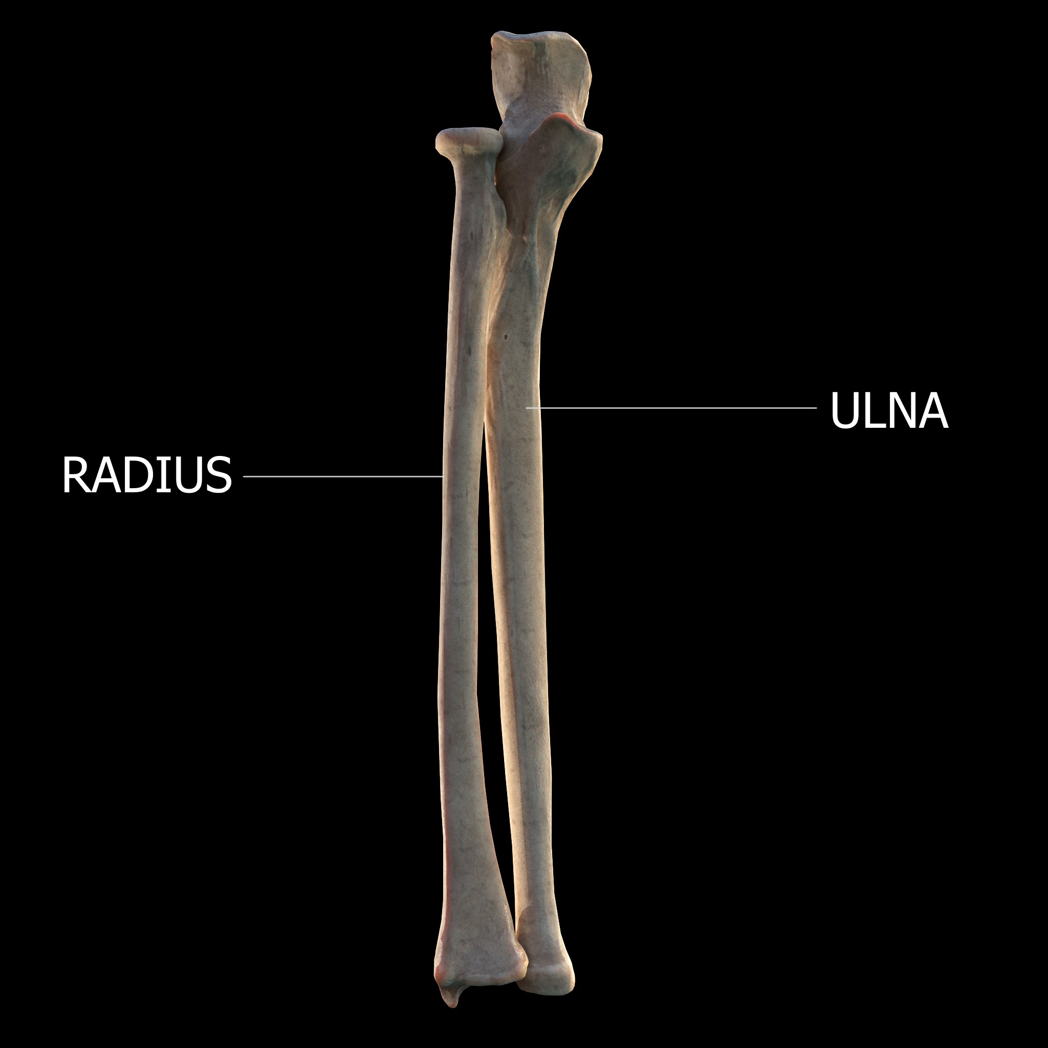
The forearm is the region of the upper limb between the elbow and the wrist. The term forearm is used in anatomy to distinguish it from the arm, a word which is most often used to describe the entire appendage of the upper limb, but which in anatomy, technically, means only the region of the upper arm, whereas the lower "arm" is called the forearm. It is homologous to the region of the leg that lies between the knee and the ankle joints, the crus. The forearm contains two long bones, the radius and the ulna, forming the two radioulnar joints. The interosseous membrane connects these bones. Ultimately, the forearm is covered by skin, the anterior surface usually being less hairy than the posterior surface. The forearm contains many muscles, including the flexors and extensors of the wrist, flexors and extensors of the digits, a flexor of the elbow (brachioradialis), and pronators and supinators that turn the hand to face down or upwards, respectively. In cross-section, the for ... [...More Info...] [...Related Items...] OR: [Wikipedia] [Google] [Baidu] |
Palmar Carpal Ligament
The palmar carpal ligament (also volar carpal ligament or ''Guyon's Tunnel'') is the thickened portion of antebrachial fascia on the anterior of the wrist. It is officially unnamed.Moore, Keith L., Arthur F. Dalley II: ''Clinically Oriented Anatomy'', 4th ed. Lippincott, Williams & Wilkins,1999. The palmar carpal ligament is a different structure than the flexor retinaculum of the hand, but the two are frequently confused. The palmar carpal ligament lies superficial and proximal to the flexor retinaculum. The ulnar nerve and the ulnar artery run through the ulnar canal, which is deep to the palmar carpal ligament and superficial to the flexor retinaculum. The palmar carpal ligament is continuous with the extensor retinaculum of the hand, which is located on the posterior side of the wrist. References See also * Flexor retinaculum of the hand * Extensor retinaculum of the hand * Antebrachial fascia The antebrachial fascia (antibrachial fascia or deep fascia of forearm) conti ... [...More Info...] [...Related Items...] OR: [Wikipedia] [Google] [Baidu] |
Thumb
The thumb is the first digit of the hand, next to the index finger. When a person is standing in the medical anatomical position (where the palm is facing to the front), the thumb is the outermost digit. The Medical Latin English noun for thumb is ''pollex'' (compare ''hallux'' for big toe), and the corresponding adjective for thumb is ''pollical''. Definition Thumb and fingers The English word ''finger'' has two senses, even in the context of appendages of a single typical human hand: # Any of the five terminal members of the hand. # Any of the four terminal members of the hand, other than the thumb Linguistically, it appears that the original sense was the first of these two: (also rendered as ) was, in the inferred Proto-Indo-European language, a suffixed form of (or ), which has given rise to many Indo-European-family words (tens of them defined in English dictionaries) that involve, or stem from, concepts of fiveness. The thumb shares the following with each of the o ... [...More Info...] [...Related Items...] OR: [Wikipedia] [Google] [Baidu] |
Lateral Antebrachial Cutaneous Nerve
The lateral antebrachial cutaneous nerve (or lateral cutaneous nerve of forearm) (branch of musculocutaneous nerve, also sometimes spelled "antebrachial") passes behind the cephalic vein, and divides, opposite the elbow-joint, into a volar and a dorsal branch. Volar branch The volar branch (ramus volaris; anterior branch) descends along the radial border of the forearm to the wrist, and supplies the skin over the lateral half of its volar surface. At the wrist-joint it is placed in front of the radial artery, and some filaments, piercing the deep fascia, accompany that vessel to the dorsal surface of the carpus. The nerve then passes downward to the ball of the thumb, where it ends in cutaneous filaments. It communicates with the superficial branch of the radial nerve, and with the palmar cutaneous branch of the median nerve. Dorsal branch The dorsal branch (ramus dorsalis; posterior branch) descends, along the dorsal surface of the radial side of the forearm to the wrist. ... [...More Info...] [...Related Items...] OR: [Wikipedia] [Google] [Baidu] |
Hand
A hand is a prehensile, multi-fingered appendage located at the end of the forearm or forelimb of primates such as humans, chimpanzees, monkeys, and lemurs. A few other vertebrates such as the koala (which has two opposable thumbs on each "hand" and fingerprints extremely similar to human fingerprints) are often described as having "hands" instead of paws on their front limbs. The raccoon is usually described as having "hands" though opposable thumbs are lacking. Some evolutionary anatomists use the term ''hand'' to refer to the appendage of digits on the forelimb more generally—for example, in the context of whether the three digits of the bird hand involved the same homologous loss of two digits as in the dinosaur hand. The human hand usually has five digits: four fingers plus one thumb; these are often referred to collectively as five fingers, however, whereby the thumb is included as one of the fingers. It has 27 bones, not including the sesamoid bone, the number o ... [...More Info...] [...Related Items...] OR: [Wikipedia] [Google] [Baidu] |
Palmar Cutaneous Branch Of The Ulnar
The palmar branch of the ulnar nerve arises about five cm proximal to the wrist from where the ulnar nerve splits into palmar and dorsal branches. It supplies sensory innervation to a small area in the palmar surface of the wrist. The palmar branch represents the continuation of the ulnar nerve as it crosses the flexor retinaculum of the hand on the lateral side of the pisiform bone, medial to and a little behind the ulnar artery. Some sources state that it ends by dividing into a superficial and a deep branch. (Other sources state that the [...More Info...] [...Related Items...] OR: [Wikipedia] [Google] [Baidu] |
Flexor Retinaculum Of The Hand
The flexor retinaculum (transverse carpal ligament, or anterior annular ligament) is a fibrous band on the palmar side of the hand near the wrist. It arches over the carpal bones of the hands, covering them and forming the carpal tunnel. Structure The flexor retinaculum is a strong, fibrous band that covers the carpal bones on the palmar side of the hand near the wrist. It attaches to the bones near the radius and ulna. On the ulnar side, the flexor retinaculum attaches to the pisiform bone and the hook of the hamate bone. On the radial side, it attaches to the tubercle of the scaphoid bone, and to the medial part of the palmar surface and the ridge of the trapezium bone. The flexor retinaculum is continuous with the palmar carpal ligament, and deeper with the palmar aponeurosis. The ulnar artery and ulnar nerve, and the cutaneous branches of the median and ulnar nerves, pass on top of the flexor retinaculum. On the radial side of the retinaculum is the tendon of the flexor c ... [...More Info...] [...Related Items...] OR: [Wikipedia] [Google] [Baidu] |
Carpal Tunnel Syndrome
Carpal tunnel syndrome (CTS) is the collection of symptoms and signs associated with median neuropathy at the carpal tunnel. Most CTS is related to idiopathic compression of the median nerve as it travels through the wrist at the carpal tunnel (IMNCT). Idiopathic means that there is no other disease process contributing to pressure on the nerve. As with most structural issues, it occurs in both hands, and the strongest risk factor is genetics. Other conditions can cause CTS such as wrist fracture or rheumatoid arthritis. After fracture, swelling, bleeding, and deformity compress the median nerve. With rheumatoid arthritis, the enlarged synovial lining of the tendons causes compression. The main symptoms are numbness and tingling in the thumb, index finger, middle finger and the thumb side of the ring finger. People often report pain, but pain without tingling is not characteristic of IMNCT. Rather, the numbness can be so intense that it is described as painful. Symptoms are ... [...More Info...] [...Related Items...] OR: [Wikipedia] [Google] [Baidu] |


