|
Median Nerve
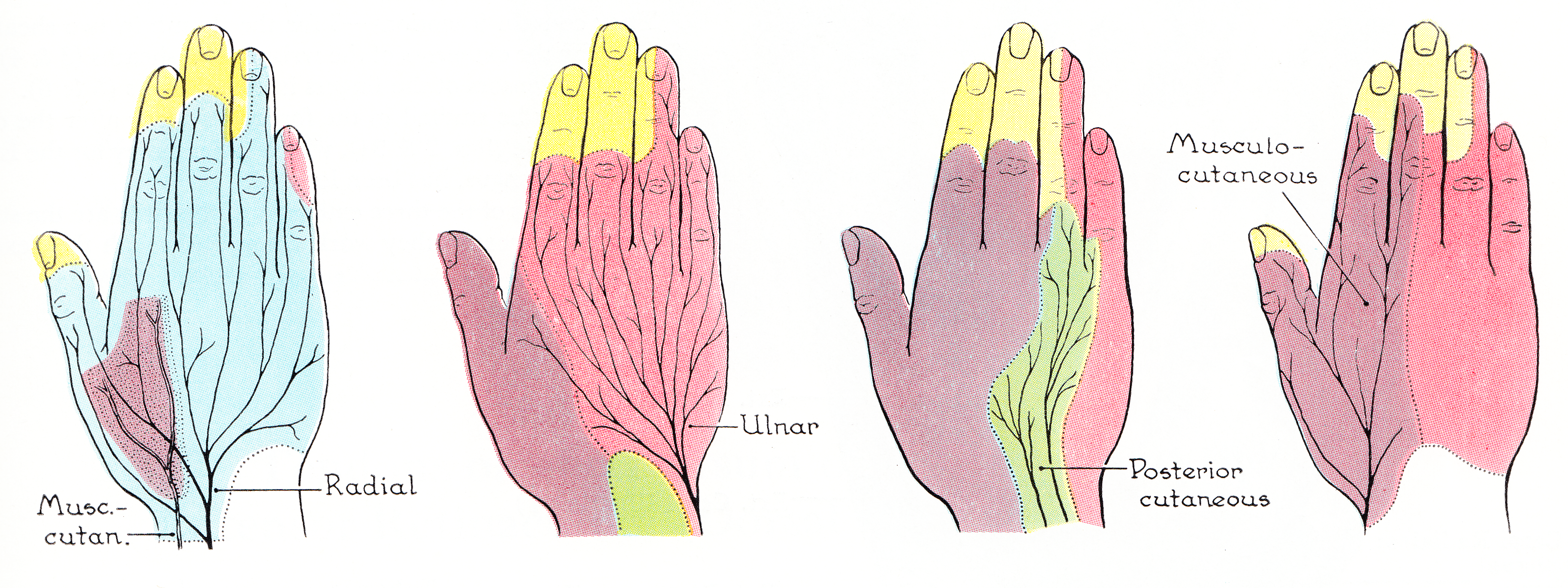
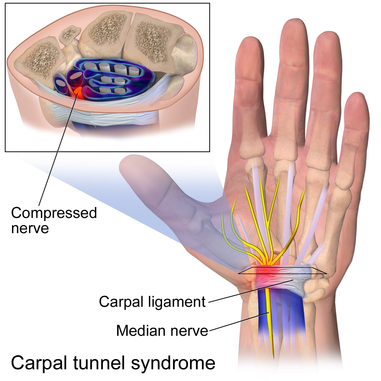
The median nerve is a nerve in humans and other animals in the upper limb. It is one of the five main nerves originating from the brachial plexus. The median nerve originates from the lateral and medial cords of the brachial plexus, and has contributions from ventral roots of C6-C7 (lateral cord) and C8 and T1 (medial cord). The median nerve is the only nerve that passes through the carpal tunnel. Carpal tunnel syndrome is the disability that results from the median nerve being pressed in the carpal tunnel. Structure The median nerve arises from the branches from lateral and medial cords of the brachial plexus, courses through the anterior part of arm, forearm, and hand, and terminates by supplying the muscles of the hand. Arm After receiving inputs from both the lateral and medial cords of the brachial plexus, the median nerve enters the arm from the axilla at the inferior margin of the teres major muscle. It then passes vertically down and courses lateral to the brac ... [...More Info...] [...Related Items...] OR: [Wikipedia] [Google] [Baidu] |
Gray's Anatomy
''Gray's Anatomy'' is a reference book of human anatomy written by Henry Gray, illustrated by Henry Vandyke Carter and first published in London in 1858. It has had multiple revised editions, and the current edition, the 42nd (October 2020), remains a standard reference, often considered "the doctors' bible". Earlier editions were called ''Anatomy: Descriptive and Surgical'', ''Anatomy of the Human Body'' and ''Gray's Anatomy: Descriptive and Applied'', but the book's name is commonly shortened to, and later editions are titled, ''Gray's Anatomy''. The book is widely regarded as an extremely influential work on the subject. Publication history Origins The English anatomist Henry Gray was born in 1827. He studied the development of the endocrine glands and spleen and in 1853 was appointed Lecturer on Anatomy at St George's Hospital Medical School in London. In 1855, he approached his colleague Henry Vandyke Carter with his idea to produce an inexpensive and access ... [...More Info...] [...Related Items...] OR: [Wikipedia] [Google] [Baidu] |
Biceps Brachii
The biceps or biceps brachii (, "two-headed muscle of the arm") is a large muscle that lies on the front of the upper arm between the shoulder and the elbow. Both heads of the muscle arise on the scapula and join to form a single muscle belly which is attached to the upper forearm. While the long head of the biceps crosses both the shoulder and elbow joints, its main function is at the elbow where it flexes and supinates the forearm. Both these movements are used when opening a bottle with a corkscrew: first biceps screws in the cork (supination), then it pulls the cork out (flexion). Structure The biceps is one of three muscles in the anterior compartment of the upper arm, along with the brachialis muscle and the coracobrachialis muscle, with which the biceps shares a nerve supply. The biceps muscle has two heads, the short head and the long head, distinguished according to their origin at the coracoid process and supraglenoid tubercle of the scapula, respectively. ... [...More Info...] [...Related Items...] OR: [Wikipedia] [Google] [Baidu] |
Flexor Carpi Radialis
In anatomy, flexor carpi radialis is a muscle of the human forearm that acts to flex and (radially) abduct the hand. The Latin ''carpus'' means wrist; hence flexor carpi is a flexor of the wrist. Origin and insertion The flexor carpi radialis is one of four muscles in the superficial layer of the anterior compartment of the forearm. This muscle originates from the medial epicondyle of the humerus as part of the common flexor tendon. It runs just laterally of flexor digitorum superficialis and inserts on the anterior aspect of the base of the second metacarpal, and has small slips to both the third metacarpal and trapezium tuberosity. The tendon of the flexor carpi radialis is visible on the anterior surface of the forearm, just proximal to the wrist, when the wrist is flexed. It is the tendon seen most lateral, closest to the thumb. Nerve and artery Like most flexors of the anterior compartment of the forearm, FCR is innervated by the median nerve, specifically by axons fro ... [...More Info...] [...Related Items...] OR: [Wikipedia] [Google] [Baidu] |
Flexor Retinaculum Of The Hand
The flexor retinaculum (transverse carpal ligament or anterior annular ligament) is a fibrous band on the palmar side of the hand near the wrist. It arches over the carpal bones of the hands, covering them and forming the carpal tunnel. Structure The flexor retinaculum is a strong, fibrous band that covers the carpal bones on the palmar side of the hand near the wrist. It attaches to the bones near the radius and ulna. On the ulnar side, the flexor retinaculum attaches to the pisiform bone and the hook of the hamate bone. On the radial side, it attaches to the tubercle of the scaphoid bone, and to the medial part of the palmar surface and the ridge of the trapezium bone. The flexor retinaculum is continuous with the palmar carpal ligament, and deeper with the palmar aponeurosis. The ulnar artery and ulnar nerve, and the cutaneous branches of the median and ulnar nerves, pass on top of the flexor retinaculum. On the radial side of the retinaculum is the tendon of the flexor car ... [...More Info...] [...Related Items...] OR: [Wikipedia] [Google] [Baidu] |
Anterior Interosseous Artery
The anterior interosseous artery (volar interosseous artery) is an artery in the forearm. It is a branch of the common interosseous artery. Course It passes down the forearm on the palmar surface of the interosseous membrane. It is accompanied by the palmar interosseous branch of the median nerve, and overlapped by the contiguous margins of the flexor digitorum profundus and flexor pollicis longus muscles, giving off in this situation muscular branches, and the nutrient arteries of the radius and ulna. At the upper border of the pronator quadratus muscle it pierces the interosseous membrane and reaches the back of the forearm, where it anastomoses with the dorsal interosseous artery. It then descends, in company with the terminal portion of the dorsal interosseous nerve, to the back of the wrist to join the dorsal carpal network. The anterior interosseous artery may give off a slender branch, the median artery, which accompanies the median nerve, and gives offsets to its ... [...More Info...] [...Related Items...] OR: [Wikipedia] [Google] [Baidu] |
Median Artery
The median artery is an artery that is occasionally found in humans and other animals. The prevalence was around 10% in people born in the mid-1880s compared to 30% in those born in the late 20th century, and 35% of people born as of 2020; a significant increase in a fairly short period of time, when it comes to evolution. When the median artery prevalence reaches 50% or more, it should not be considered as a variant, but as a ‘normal’ human structure. "This increase could have resulted from mutations of genes involved in median artery development or health problems in mothers during pregnancy, or both. If this trend continues, a majority of people will have median artery of the forearm by 2100." When present, it is found in the forearm, between the radial artery and ulnar artery. It runs with the median nerve The median nerve is a nerve in humans and other animals in the upper limb. It is one of the five main nerves originating from the brachial plexus. The median nerve ... [...More Info...] [...Related Items...] OR: [Wikipedia] [Google] [Baidu] |
Flexor Digitorum Profundus
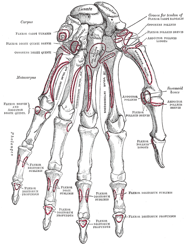
The flexor digitorum profundus or flexor digitorum communis profundus is a muscle in the forearm of humans that flexes the fingers (also known as digits). It is considered an Muscles of the hand#Extrinsic, extrinsic hand muscle because it acts on the hand while its muscle belly is located in the forearm. Together the Flexor pollicis longus muscle, flexor pollicis longus, Pronator quadratus muscle, pronator quadratus, and flexor digitorum profundus form the deep layer of ventral forearm muscles.Platzer 2004, p 162 The muscle is named . Structure Flexor digitorum profundus originates in the upper 3/4 of the anterior and medial surfaces of the ulna, interosseous membrane and deep fascia of the forearm. The muscle fans out into four tendons (one to each of the second to fifth fingers) to the palmar base of the distal phalanges, distal phalanx. Along with the flexor digitorum superficialis, it has long tendons that run down the arm and through the carpal tunnel and attach to the p ... [...More Info...] [...Related Items...] OR: [Wikipedia] [Google] [Baidu] |
Flexor Digitorum Superficialis
Flexor digitorum superficialis (''flexor digitorum sublimis'') or flexor digitorum communis sublimis is an extrinsic flexor muscle of the fingers at the proximal interphalangeal joints. It is in the anterior compartment of the forearm. It is sometimes considered to be the deepest part of the superficial layer of this compartment, and sometimes considered to be a distinct, "intermediate layer" of this compartment. It is relatively common for the Flexor digitorum superficialis to be missing from the little finger, bilaterally and unilaterally, which can cause problems when diagnosing a little finger injury. Structure The muscle has two classically described heads – the humeroulnar and radial – and it is between these heads that the median nerve and ulnar artery pass. The ulnar collateral ligament of elbow joint gives its origin to part of this muscle. Four long tendons come off this muscle near the wrist and travel through the carpal tunnel formed by the flexor retinacu ... [...More Info...] [...Related Items...] OR: [Wikipedia] [Google] [Baidu] |
Ulnar Artery
The ulnar artery is the main blood vessel, with oxygenated blood, of the Human Anatomical Terms#Anatomical directions, medial aspects of the forearm. It arises from the brachial artery and terminates in the superficial palmar arch, which joins with the superficial branch of the radial artery. It is palpable on the anterior and medial aspect of the wrist. Along its course, it is accompanied by a similarly named vein or veins, the ulnar vein or ulnar veins. The ulnar artery, the larger of the two terminal branches of the brachial, begins a little below the bend of the Elbow-joint, elbow in the cubital fossa, and, passing obliquely downward, reaches the ulnar side of the forearm at a point about midway between the elbow and the wrist. It then runs along the ulnar border to the wrist, crosses the transverse carpal ligament on the radial side of the pisiform bone, and immediately beyond this bone divides into two branches, which enter into the formation of the Superficial palmar a ... [...More Info...] [...Related Items...] OR: [Wikipedia] [Google] [Baidu] |
Aponeurosis
An aponeurosis (; : aponeuroses) is a flattened tendon by which muscle attaches to bone or fascia. Aponeuroses exhibit an ordered arrangement of collagen fibres, thus attaining high tensile strength in a particular direction while being vulnerable to tensional or shear forces in other directions. They have a shiny, whitish-silvery color, are histologically similar to tendons, and are very sparingly supplied with blood vessels and nerves. When dissected, aponeuroses are papery and peel off by sections. The primary regions with thick aponeuroses are in the ventral abdominal region, the dorsal lumbar region, the ventriculus in birds, and the palmar (palms) and plantar (soles) regions. Anatomy Anterior abdominal aponeuroses The anterior abdominal aponeuroses are located just superficial to the rectus abdominis muscle. It has for its borders the external oblique, pectoralis muscles, and the latissimus dorsi. Posterior lumbar aponeuroses The posterior lumbar aponeuroses are sit ... [...More Info...] [...Related Items...] OR: [Wikipedia] [Google] [Baidu] |
Bicipital Aponeurosis
The bicipital aponeurosis (also known as lacertus fibrosus) is a broad aponeurosis of the biceps brachii, which is located in the cubital fossa of the elbow. It separates superficial from deep structures in much of the fossa. Structure The bicipital aponeurosis originates from the distal insertion of the biceps brachii, and inserts into the deep fascia of the forearm. The biceps tendon inserts on the radial tuberosity, and the bicipital aponeurosis lies medially to it. It reinforces the cubital fossa, helping to protect the brachial artery and the median nerve running underneath. Variations Some individuals (about 3% of the population) have a ''superficial ulnar artery'' that runs superficially to the bicipital aponeurosis instead of underneath it. These individuals are at risk for accidental injury to the ulnar artery during venipuncture. Clinical significance The bicipital aponeurosis is superficial to the brachial artery and the median nerve, but deep to the median cu ... [...More Info...] [...Related Items...] OR: [Wikipedia] [Google] [Baidu] |
Pronator Teres
The pronator teres is a muscle (located mainly in the forearm) that, along with the pronator quadratus, serves to pronate the forearm (turning it so that the palm faces posteriorly when from the anatomical position). It has two origins, at the medial humeral supracondylar ridge and the medial side of the coronoid process of the ulna and inserts near the middle of the radius. Structure The pronator teres has two heads—humeral and ulnar. * The humeral head, the larger and more superficial, arises from the medial supracondylar ridge immediately superior to the medial epicondyle of the humerus, and from the common flexor tendon (which arises from the medial epicondyle). * The ulnar head (or ulnar tuberosity) is a thin fasciculus, which arises from the medial side of the coronoid process of the ulna, and joins the preceding at an acute angle. The median nerve enters the forearm between the two heads of the muscle, and is separated from the ulnar artery by the ulnar head. T ... [...More Info...] [...Related Items...] OR: [Wikipedia] [Google] [Baidu] |


