|
P19 Cell
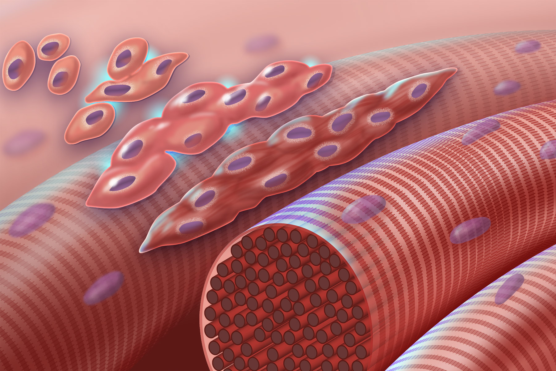
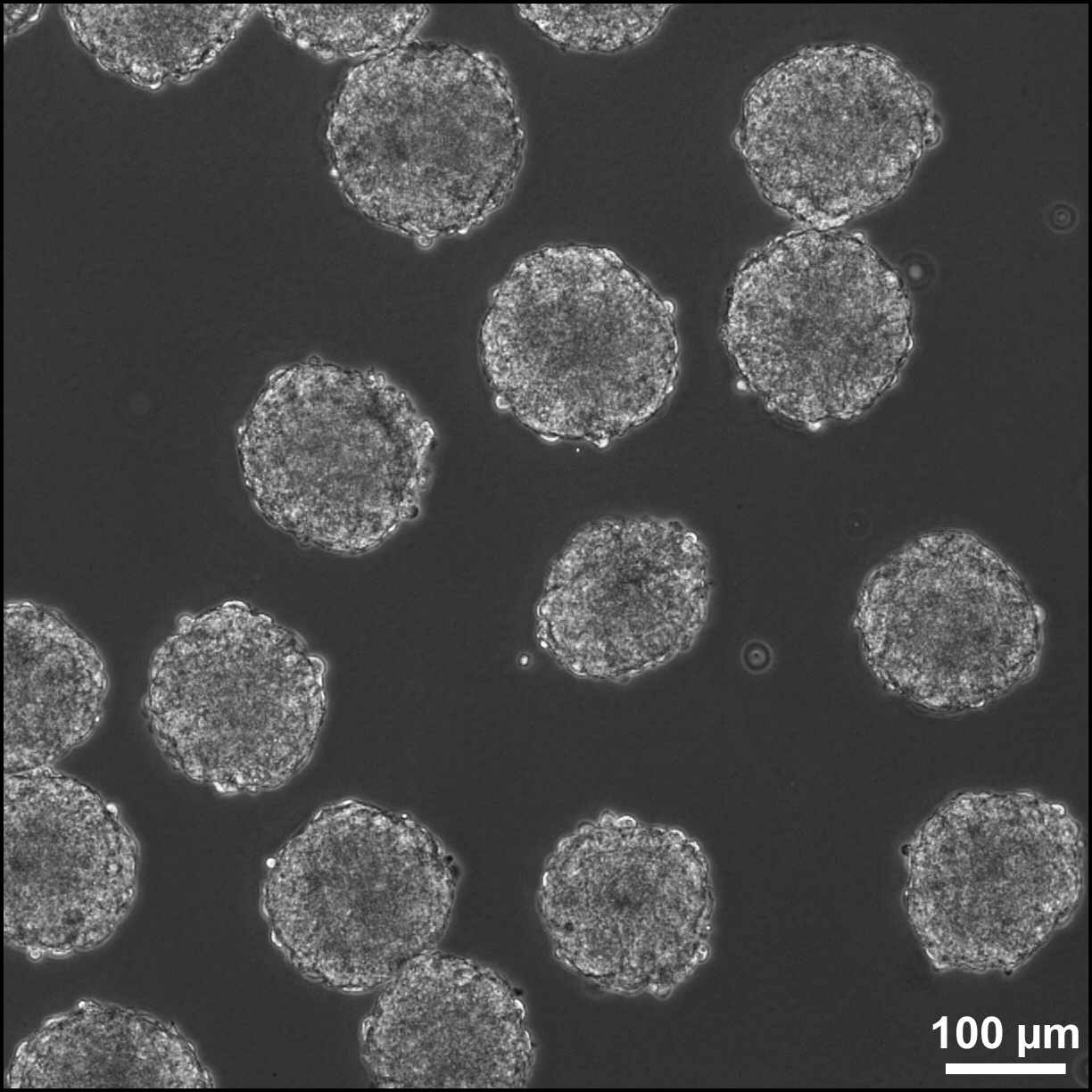
P19 cells is an embryonic carcinoma cell line derived from an embryo-derived teratocarcinoma in mice. The cell line is pluripotent and can differentiate into cell types of all three germ layers. Also, it is the most characterized embryonic carcinoma (EC) cell line that can be induced into cardiac muscle cells and neuronal cells by different specific treatments. Indeed, exposing aggregated P19 cells to dimethyl sulfoxide (DMSO) induces differentiation into cardiac and skeletal muscle. Also, exposing P19 cells to retinoic acid (RA) can differentiate them into neuronal cells. Origin of the P19 cell line Cancer cells in humans may result in the patient's death if the aggressive cancer cell grows and metastasizes. However, researchers utilize these cells to study the development of cancer cells in order to find more specific treatments. For developmental biologists, embryonal carcinoma, which is derived from teratocarcinoma, is a good object for developmental study. In 1982, McBurney an ... [...More Info...] [...Related Items...] OR: [Wikipedia] [Google] [Baidu] |
Beta-catenin
Catenin beta-1, also known as beta-catenin (β-catenin), is a protein that in humans is encoded by the ''CTNNB1'' gene. Beta-catenin is a dual function protein, involved in regulation and coordination of cell–cell adhesion and gene transcription. In humans, the CTNNB1 protein is encoded by the ''CTNNB1'' gene. In ''Drosophila'', the homologous protein is called ''armadillo''. β-catenin is a subunit of the cadherin protein complex and acts as an intracellular signal transducer in the Wnt signaling pathway. It is a member of the catenin protein family and homologous to γ-catenin, also known as plakoglobin. Beta-catenin is widely expressed in many tissues. In cardiac muscle, beta-catenin localizes to adherens junctions in intercalated disc structures, which are critical for electrical and mechanical coupling between adjacent cardiomyocytes. Mutations and overexpression of β-catenin are associated with many cancers, including hepatocellular carcinoma, colorectal carcinoma, lun ... [...More Info...] [...Related Items...] OR: [Wikipedia] [Google] [Baidu] |
Blastocyst
The blastocyst is a structure formed in the early embryonic development of mammals. It possesses an inner cell mass (ICM) also known as the ''embryoblast'' which subsequently forms the embryo, and an outer layer of trophoblast cells called the trophectoderm. This layer surrounds the inner cell mass and a fluid-filled cavity known as the blastocoel. In the late blastocyst the trophectoderm is known as the trophoblast. The trophoblast gives rise to the chorion and amnion, the two fetal membranes that surround the embryo. The placenta derives from the embryonic chorion (the portion of the chorion that develops villi) and the underlying uterine tissue of the mother. The name "blastocyst" arises from the Greek ' ("a sprout") and ' ("bladder, capsule"). In other animals this is a structure consisting of an undifferentiated ball of cells and is called a blastula. In humans, blastocyst formation begins about five days after fertilization when a fluid-filled cavity opens up in the mor ... [...More Info...] [...Related Items...] OR: [Wikipedia] [Google] [Baidu] |
Myogenesis
Myogenesis is the formation of skeletal muscular tissue, particularly during embryonic development. Muscle fibers generally form through the fusion of precursor myoblasts into multinucleated fibers called ''myotubes''. In the early development of an embryo, myoblasts can either proliferate, or differentiate into a myotube. What controls this choice in vivo is generally unclear. If placed in cell culture, most myoblasts will proliferate if enough fibroblast growth factor (FGF) or another growth factor is present in the medium surrounding the cells. When the growth factor runs out, the myoblasts cease division and undergo terminal differentiation into myotubes. Myoblast differentiation proceeds in stages. The first stage, involves cell cycle exit and the commencement of expression of certain genes. The second stage of differentiation involves the alignment of the myoblasts with one another. Studies have shown that even rat and chick myoblasts can recognise and align with one a ... [...More Info...] [...Related Items...] OR: [Wikipedia] [Google] [Baidu] |
Neurogenesis
Neurogenesis is the process by which nervous system cells, the neurons, are produced by neural stem cells (NSCs). It occurs in all species of animals except the porifera (sponges) and placozoans. Types of NSCs include neuroepithelial cells (NECs), radial glial cells (RGCs), basal progenitors (BPs), intermediate neuronal precursors (INPs), subventricular zone astrocytes, and subgranular zone radial astrocytes, among others. Neurogenesis is most active during embryonic development and is responsible for producing all the various types of neurons of the organism, but it continues throughout adult life in a variety of organisms. Once born, neurons do not divide (see mitosis), and many will live the lifespan of the animal. Neurogenesis in mammals Developmental neurogenesis During embryonic development, the mammalian central nervous system (CNS; brain and spinal cord) is derived from the neural tube, which contains NSCs that will later generate neurons. However, neurogenesis does ... [...More Info...] [...Related Items...] OR: [Wikipedia] [Google] [Baidu] |
Choline Acetyltransferase
Choline acetyltransferase (commonly abbreviated as ChAT, but sometimes CAT) is a transferase enzyme responsible for the synthesis of the neurotransmitter acetylcholine. ChAT catalyzes the transfer of an acetyl group from the coenzyme acetyl-CoA to choline, yielding acetylcholine (ACh). ChAT is found in high concentration in cholinergic neurons, both in the central nervous system (CNS) and peripheral nervous system (PNS). As with most nerve terminal proteins, ChAT is produced in the body of the neuron and is transported to the nerve terminal, where its concentration is highest. Presence of ChAT in a nerve cell classifies this cell as a "cholinergic" neuron. In humans, the choline acetyltransferase enzyme is encoded by the ''CHAT'' gene. History Choline acetyltransferase was first described by David Nachmansohn and A. L. Machado in 1943. A German biochemist, Nachmansohn had been studying the process of nerve impulse conduction and utilization of energy-yielding chemical reacti ... [...More Info...] [...Related Items...] OR: [Wikipedia] [Google] [Baidu] |
Fibroblast
A fibroblast is a type of cell (biology), biological cell that synthesizes the extracellular matrix and collagen, produces the structural framework (Stroma (tissue), stroma) for animal Tissue (biology), tissues, and plays a critical role in wound healing. Fibroblasts are the most common cells of connective tissue in animals. Structure Fibroblasts have a branched cytoplasm surrounding an elliptical, speckled cell nucleus, nucleus having two or more nucleoli. Active fibroblasts can be recognized by their abundant Endoplasmic reticulum#Rough endoplasmic reticulum, rough endoplasmic reticulum. Inactive fibroblasts (called fibrocytes) are smaller, spindle-shaped, and have a reduced amount of rough endoplasmic reticulum. Although disjointed and scattered when they have to cover a large space, fibroblasts, when crowded, often locally align in parallel clusters. Unlike the epithelial cells lining the body structures, fibroblasts do not form flat monolayers and are not restricted by a ... [...More Info...] [...Related Items...] OR: [Wikipedia] [Google] [Baidu] |
Astroglia
Astrocytes (from Ancient Greek , , "star" + , , "cavity", "cell"), also known collectively as astroglia, are characteristic star-shaped glial cells in the brain and spinal cord. They perform many functions, including biochemical control of endothelial cells that form the blood–brain barrier, provision of nutrients to the nervous tissue, maintenance of extracellular ion balance, regulation of cerebral blood flow, and a role in the repair and scarring process of the brain and spinal cord following infection and traumatic injuries. The proportion of astrocytes in the brain is not well defined; depending on the counting technique used, studies have found that the astrocyte proportion varies by region and ranges from 20% to 40% of all glia. Another study reports that astrocytes are the most numerous cell type in the brain. Astrocytes are the major source of cholesterol in the central nervous system. Apolipoprotein E transports cholesterol from astrocytes to neurons and other glial ce ... [...More Info...] [...Related Items...] OR: [Wikipedia] [Google] [Baidu] |
Glial Cell
Glia, also called glial cells (gliocytes) or neuroglia, are non-neuronal cells in the central nervous system (brain and spinal cord) and the peripheral nervous system that do not produce electrical impulses. They maintain homeostasis, form myelin in the peripheral nervous system, and provide support and protection for neurons. In the central nervous system, glial cells include oligodendrocytes, astrocytes, ependymal cells, and microglia, and in the peripheral nervous system they include Schwann cells and satellite cells. Function They have four main functions: *to surround neurons and hold them in place *to supply nutrients and oxygen to neurons *to insulate one neuron from another *to destroy pathogens and remove dead neurons. They also play a role in neurotransmission and synaptic connections, and in physiological processes such as breathing. While glia were thought to outnumber neurons by a ratio of 10:1, recent studies using newer methods and reappraisal of historical quant ... [...More Info...] [...Related Items...] OR: [Wikipedia] [Google] [Baidu] |
Embryoid Body
Embryoid bodies (EBs) are three-dimensional aggregates of pluripotent stem cells. EBs are differentiation of human embryonic stem cells into embryoid bodies comprising the three embryonic germ layers. Background The pluripotent cell types that comprise embryoid bodies include embryonic stem cells (ESCs) derived from the blastocyst stage of embryos from mouse (mESC), primate, and human (hESC) sources. Additionally, EBs can be formed from embryonic stem cells derived through alternative techniques, including somatic cell nuclear transfer or the reprogramming of somatic cells to yield induced pluripotent stem cells (iPS). Similar to ESCs cell culture, cultured in monolayer formats, ESCs within embryoid bodies undergo differentiation and cell specification along the three germ layer, germ lineages – endoderm, ectoderm, and mesoderm – which comprise all somatic (biology), somatic cell types. In contrast to monolayer cultures, however, the spheroid structures that are ... [...More Info...] [...Related Items...] OR: [Wikipedia] [Google] [Baidu] |
Cell Differentiate
Cell most often refers to: * Cell (biology), the functional basic unit of life Cell may also refer to: Locations * Monastic cell, a small room, hut, or cave in which a religious recluse lives, alternatively the small precursor of a monastery with only a few monks or nuns * Prison cell, a room used to hold people in prisons Groups of people * Cell, a group of people in a cell group, a form of Christian church organization * Cell, a unit of a clandestine cell system, a penetration-resistant form of a secret or outlawed organization * Cellular organizational structure, such as in business management Science, mathematics, and technology Computing and telecommunications * Cell (EDA), a term used in an electronic circuit design schematics * Cell (microprocessor), a microprocessor architecture developed by Sony, Toshiba, and IBM * Memory cell (computing), the basic unit of (volatile or non-volatile) computer memory * Cell, a unit in a database table or spreadsheet, formed by the ... [...More Info...] [...Related Items...] OR: [Wikipedia] [Google] [Baidu] |
Exponential Growth
Exponential growth is a process that increases quantity over time. It occurs when the instantaneous rate of change (that is, the derivative) of a quantity with respect to time is proportional to the quantity itself. Described as a function, a quantity undergoing exponential growth is an exponential function of time, that is, the variable representing time is the exponent (in contrast to other types of growth, such as quadratic growth). If the constant of proportionality is negative, then the quantity decreases over time, and is said to be undergoing exponential decay instead. In the case of a discrete domain of definition with equal intervals, it is also called geometric growth or geometric decay since the function values form a geometric progression. The formula for exponential growth of a variable at the growth rate , as time goes on in discrete intervals (that is, at integer times 0, 1, 2, 3, ...), is x_t = x_0(1+r)^t where is the value of at ... [...More Info...] [...Related Items...] OR: [Wikipedia] [Google] [Baidu] |
Endoderm
Endoderm is the innermost of the three primary germ layers in the very early embryo. The other two layers are the ectoderm (outside layer) and mesoderm (middle layer). Cells migrating inward along the archenteron form the inner layer of the gastrula, which develops into the endoderm. The endoderm consists at first of flattened cells, which subsequently become columnar. It forms the epithelial lining of multiple systems. In plant biology, endoderm corresponds to the innermost part of the cortex ( bark) in young shoots and young roots often consisting of a single cell layer. As the plant becomes older, more endoderm will lignify. Production The following chart shows the tissues produced by the endoderm. The embryonic endoderm develops into the interior linings of two tubes in the body, the digestive and respiratory tube. Liver and pancreas cells are believed to derive from a common precursor. In humans, the endoderm can differentiate into distinguishable organs after 5 week ... [...More Info...] [...Related Items...] OR: [Wikipedia] [Google] [Baidu] |


.jpg)


