|
Osteochondroma
Osteochondromas are the most common benign tumors of the bones. The tumors take the form of cartilage-capped bony projections or outgrowth on the surface of bones exostoses. It is characterized as a type of overgrowth that can occur in any bone where cartilage forms bone. Tumors most commonly affect long bones about the knee and in the forearm. Additionally, flat bones such as the pelvis and scapula (shoulder blade) may be affected. Hereditary multiple exostoses usually present during childhood. Yet, the vast majority of affected individuals become clinically manifest by the time they reach adolescence. Osteochondromas occur in 3% of the general population and represent 35% of all benign tumors and 8% of all bone tumors. The majority of these tumors are solitary non-hereditary lesions and approximately 15% of osteochondromas occur as hereditary multiple exostoses preferably known as hereditary multiple osteochondromas (HMOs). Osteochondromas do not result from injury and the exac ... [...More Info...] [...Related Items...] OR: [Wikipedia] [Google] [Baidu] |
Hereditary Multiple Exostoses
Hereditary multiple osteochondromas (HMO), also known as hereditary multiple exostoses, is a disorder characterized by the development of multiple benign osteocartilaginous masses ( exostoses) in relation to the ends of long bones of the lower limbs such as the femurs and tibias and of the upper limbs such as the humeri and forearm bones. They are also known as osteochondromas. Additional sites of occurrence include on flat bones such as the pelvic bone and scapula. The distribution and number of these exostoses show a wide diversity among affected individuals. Exostoses usually present during childhood. The vast majority of affected individuals become clinically manifest by the time they reach adolescence. A small percentage of affected individuals are at risk for development of malignant transformation namely sarcomas. The incidence of hereditary multiple exostoses is around 1 in 50,000 individuals. Hereditary multiple osteochondromas is the preferred term used by the World H ... [...More Info...] [...Related Items...] OR: [Wikipedia] [Google] [Baidu] |
Multiple Osteochondromatosis
Hereditary multiple osteochondromas (HMO), also known as hereditary multiple exostoses, is a disorder characterized by the development of multiple benign osteocartilaginous masses (exostoses) in relation to the ends of long bones of the lower limbs such as the femurs and tibias and of the upper limbs such as the humeri and forearm bones. They are also known as osteochondromas. Additional sites of occurrence include on flat bones such as the pelvic bone and scapula. The distribution and number of these exostoses show a wide diversity among affected individuals. Exostoses usually present during childhood. The vast majority of affected individuals become clinically manifest by the time they reach adolescence. A small percentage of affected individuals are at risk for development of malignant transformation namely sarcomas. The incidence of hereditary multiple exostoses is around 1 in 50,000 individuals. Hereditary multiple osteochondromas is the preferred term used by the World Hea ... [...More Info...] [...Related Items...] OR: [Wikipedia] [Google] [Baidu] |
Trevor's Disease
Trevor disease, also known as dysplasia epiphysealis hemimelica and Trevor's disease, is a congenital bone developmental disorder. There is 1 case per million population. The condition is three times more common in males than in females. Presentation This disorder is rare, and is characterised by an asymmetrical limb deformity due to localized overgrowth of cartilage, histologically resembling osteochondroma. It is believed to affect the limb bud in early fetal life. The condition occurs mostly in the ankle or knee region and it is always confined to a single limb. This usually involves only the lower extremities and on medial side of the epiphysis. It is named after researcher David Trevor. Diagnosis Differential diagnosis Trevor disease can often mimic posttraumatic osseous fragments, synovial chondromatosis, ostechondroma, or anterior spur of ankle. It is not possible to distinguish DEH from osteochondroma on the basis of histopathology alone. Special molecular tests of the ... [...More Info...] [...Related Items...] OR: [Wikipedia] [Google] [Baidu] |
EXT2 (gene)
Exostosin glycosyltransferase-2 is a protein that in humans is encoded by the ''EXT2'' gene. This gene encodes one of two glycosyltransferases involved in the chain elongation step of heparan sulfate biosynthesis. Mutations in this gene cause the type II form of Hereditary Multiple Exostoses (HME). Gene location The EXT2 gene is located on chromosome 11 in the human genome, its location is on the p arm of this chromosome. The p arm of a chromosome is the shorter arm of a chromosome. Interactions Included in the EXT family are EXT2, EXT1, EXTL1, EXTL2, and EXTL3. The proteins formed by these genes work together to form and extend heparan sulfate chains. Heparan sulfate chains are proteoglycans present in the extracellular matrix of most tissue types. There is a lot about its function that is not entirely understood, however it is known that they have an important role for bone and cartilage formation. Cartilage is located at the growth plates of long bones and is placed in ... [...More Info...] [...Related Items...] OR: [Wikipedia] [Google] [Baidu] |
Exostosis
An exostosis, also known as bone spur, is the formation of new bone on the surface of a bone. Exostoses can cause chronic pain ranging from mild to debilitatingly severe, depending on the shape, size, and location of the lesion. It is most commonly found in places like the ribs, where small bone growths form, but sometimes larger growths can grow on places like the ankles, knees, shoulders, elbows and hips. Very rarely are they on the skull. Exostoses are sometimes shaped like spurs, such as calcaneal spurs. Osteomyelitis, a bone infection, may leave the adjacent bone with exostosis formation. Charcot foot, the neuropathic breakdown of the feet seen primarily in diabetics, can also leave bone spurs that may then become symptomatic. They normally form on the bones of joints, and can grow upwards. For example, if an extra bone formed on the ankle, it might grow up to the shin. When used in the phrases "cartilaginous exostosis" or "osteocartilaginous exostosis", the term is cons ... [...More Info...] [...Related Items...] OR: [Wikipedia] [Google] [Baidu] |
EXT1
Exostosin-1 is a protein that in humans is encoded by the ''EXT1'' gene. This gene encodes one of the two endoplasmic reticulum-resident type II transmembrane glycosyltransferase – the other being EXT2 – which are involved in the chain elongation step of heparan sulfate biosynthesis. Mutations in this gene cause the type I form of multiple exostoses. Interactions EXT1 has been shown to interact with TRAP1. See also * Langer–Giedion syndrome * Hereditary multiple exostoses Hereditary multiple osteochondromas (HMO), also known as hereditary multiple exostoses, is a disorder characterized by the development of multiple benign osteocartilaginous masses ( exostoses) in relation to the ends of long bones of the lower li ... type 1 References Further reading * * * * * * * * * * * * * * * * * * * External links Multiple Hereditary Exostoses Research Foundation {{gene-8-stub ... [...More Info...] [...Related Items...] OR: [Wikipedia] [Google] [Baidu] |
Chondrosarcoma
Chondrosarcoma is a bone sarcoma, a primary cancer composed of cells derived from transformed cells that produce cartilage. A chondrosarcoma is a member of a category of tumors of bone and soft tissue known as sarcomas. About 30% of bone sarcomas are chondrosarcomas. It is resistant to chemotherapy and radiotherapy. Unlike other primary bone sarcomas that mainly affect children and adolescents, a chondrosarcoma can present at any age. It more often affects the axial skeleton than the appendicular skeleton. Types Symptoms and signs * Back or thigh pain * Sciatica * Bladder Symptoms * Unilateral edema Causes The cause is unknown. There may be a history of enchondroma or osteochondroma. A small minority of secondary chondrosarcomas occur in people with Maffucci syndrome and Ollier disease. It has been associated with faulty isocitrate dehydrogenase 1 and 2 enzymes, which are also associated with gliomas and leukemias. Diagnosis Imaging studies – including radio ... [...More Info...] [...Related Items...] OR: [Wikipedia] [Google] [Baidu] |
Glycosyltransferases
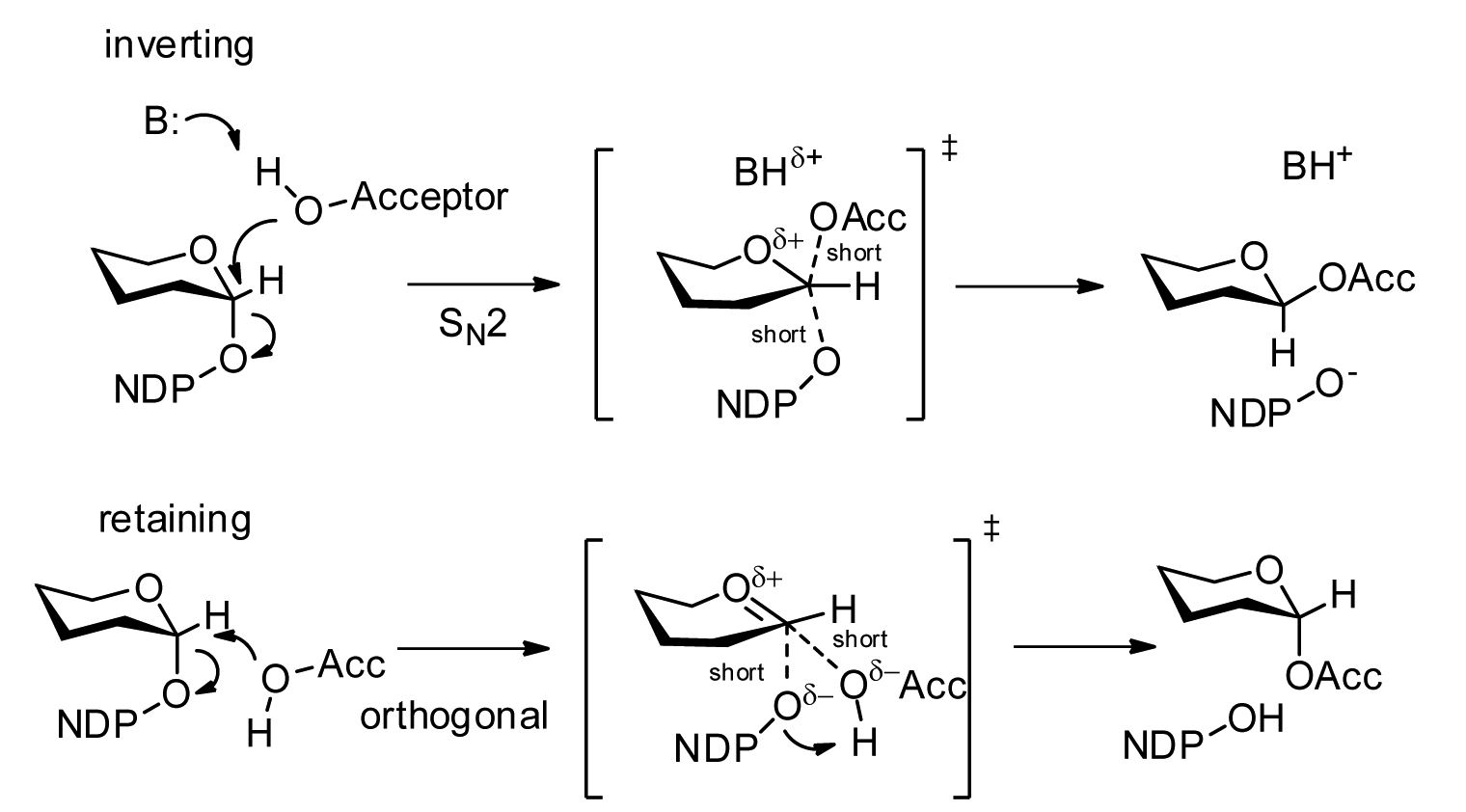
Glycosyltransferases (GTFs, Gtfs) are enzymes (EC 2.4) that establish natural glycosidic linkages. They catalyze the transfer of saccharide moieties from an activated nucleotide sugar (also known as the " glycosyl donor") to a nucleophilic glycosyl acceptor molecule, the nucleophile of which can be oxygen- carbon-, nitrogen-, or sulfur-based. The result of glycosyl transfer can be a carbohydrate, glycoside, oligosaccharide, or a polysaccharide. Some glycosyltransferases catalyse transfer to inorganic phosphate or water. Glycosyl transfer can also occur to protein residues, usually to tyrosine, serine, or threonine to give O-linked glycoproteins, or to asparagine to give N-linked glycoproteins. Mannosyl groups may be transferred to tryptophan to generate C-mannosyl tryptophan, which is relatively abundant in eukaryotes. Transferases may also use lipids as an acceptor, forming glycolipids, and even use lipid-linked sugar phosphate donors, such as dolichol phosphates in eukaryotic ... [...More Info...] [...Related Items...] OR: [Wikipedia] [Google] [Baidu] |
Golgi Apparatus
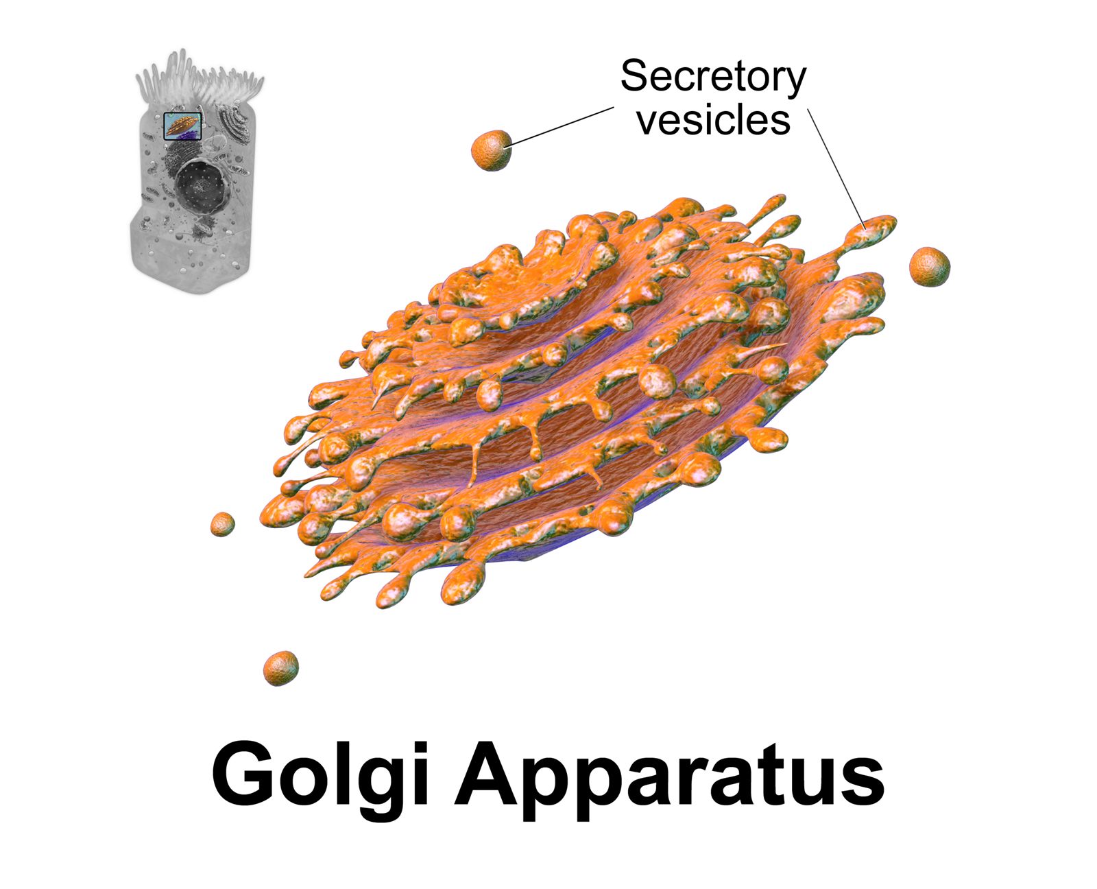
The Golgi apparatus (), also known as the Golgi complex, Golgi body, or simply the Golgi, is an organelle found in most eukaryotic cells. Part of the endomembrane system in the cytoplasm, it packages proteins into membrane-bound vesicles inside the cell before the vesicles are sent to their destination. It resides at the intersection of the secretory, lysosomal, and endocytic pathways. It is of particular importance in processing proteins for secretion, containing a set of glycosylation enzymes that attach various sugar monomers to proteins as the proteins move through the apparatus. It was identified in 1897 by the Italian scientist Camillo Golgi and was named after him in 1898. Discovery Owing to its large size and distinctive structure, the Golgi apparatus was one of the first organelles to be discovered and observed in detail. It was discovered in 1898 by Italian physician Camillo Golgi during an investigation of the nervous system. After first observing it under h ... [...More Info...] [...Related Items...] OR: [Wikipedia] [Google] [Baidu] |
Endoplasmic Reticulum
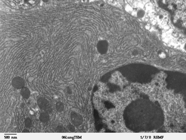
The endoplasmic reticulum (ER) is, in essence, the transportation system of the eukaryotic cell, and has many other important functions such as protein folding. It is a type of organelle made up of two subunits – rough endoplasmic reticulum (RER), and smooth endoplasmic reticulum (SER). The endoplasmic reticulum is found in most eukaryotic cells and forms an interconnected network of flattened, membrane-enclosed sacs known as cisternae (in the RER), and tubular structures in the SER. The membranes of the ER are continuous with the outer nuclear membrane. The endoplasmic reticulum is not found in red blood cells, or spermatozoa. The two types of ER share many of the same proteins and engage in certain common activities such as the synthesis of certain lipids and cholesterol. Different types of cells contain different ratios of the two types of ER depending on the activities of the cell. RER is found mainly toward the nucleus of cell and SER towards the cell membrane or pl ... [...More Info...] [...Related Items...] OR: [Wikipedia] [Google] [Baidu] |
Bursitis
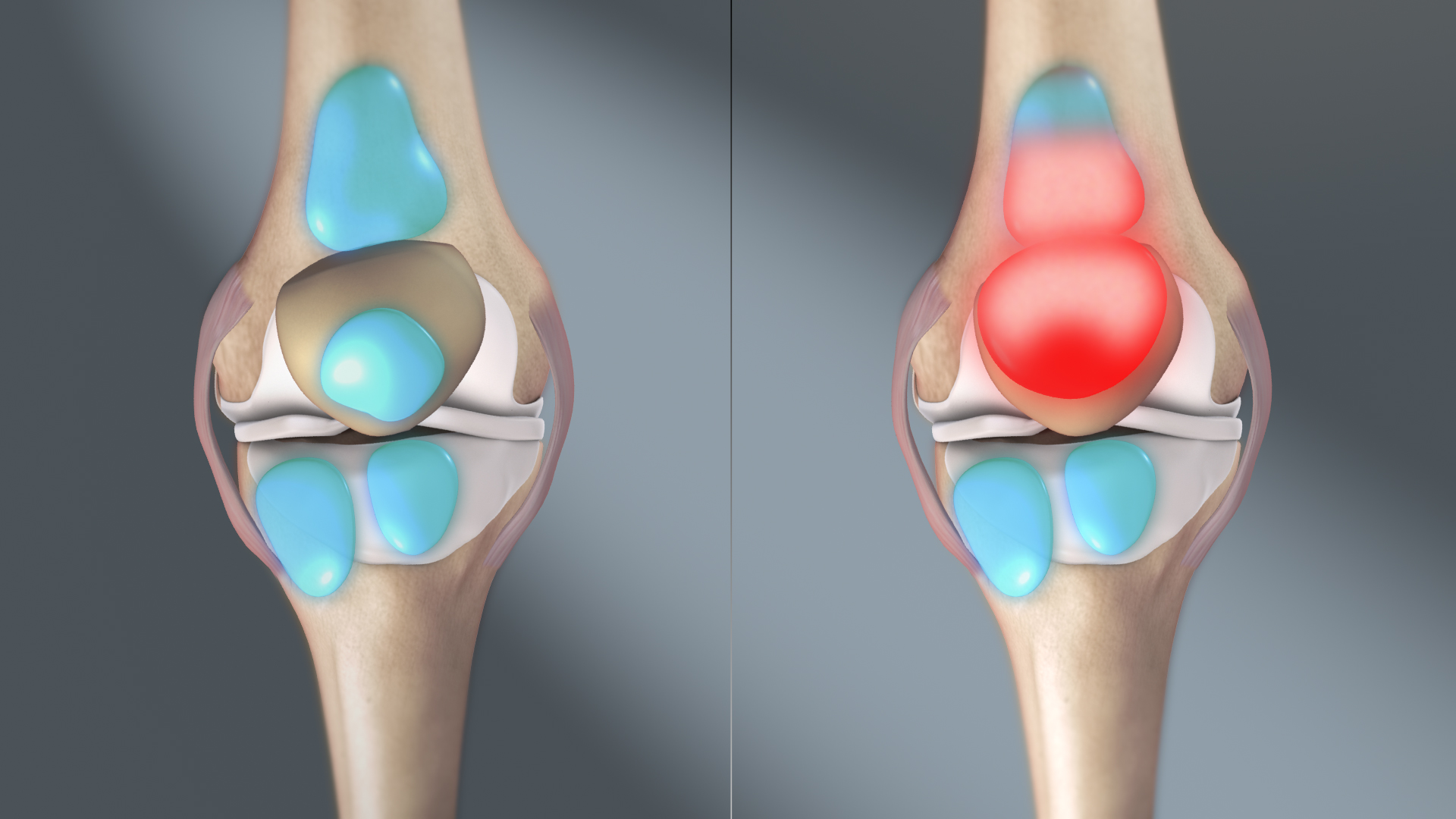
Bursitis is the inflammation of one or more bursae (fluid filled sacs) of synovial fluid in the body. They are lined with a synovial membrane that secretes a lubricating synovial fluid. There are more than 150 bursae in the human body. The bursae rest at the points where internal functionaries, such as muscles and tendons, slide across bone. Healthy bursae create a smooth, almost frictionless functional gliding surface making normal movement painless. When bursitis occurs, however, movement relying on the inflamed bursa becomes difficult and painful. Moreover, movement of tendons and muscles over the inflamed bursa aggravates its inflammation, perpetuating the problem. Muscle can also be stiffened. Signs and symptoms Bursitis commonly affects superficial bursae. These include the subacromial, prepatellar, retrocalcaneal, and ''pes anserinus'' bursae of the shoulder, knee, heel and shin, etc. (see below). Symptoms vary from localized warmth and erythema to joint pain and stiff ... [...More Info...] [...Related Items...] OR: [Wikipedia] [Google] [Baidu] |


