|
Oesophageal
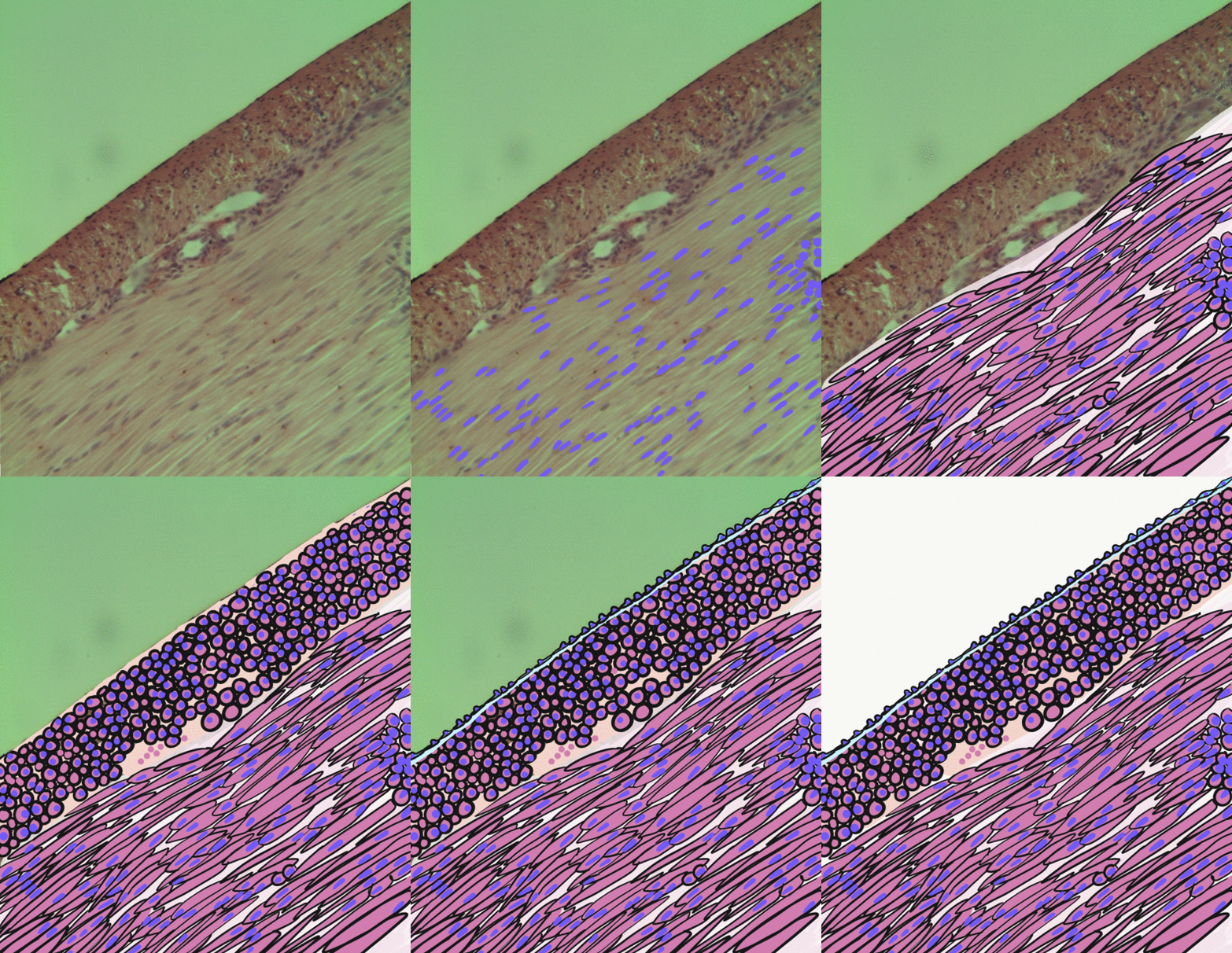
The esophagus (American English) or oesophagus (British English; both ), non-technically known also as the food pipe or gullet, is an organ in vertebrates through which food passes, aided by peristaltic contractions, from the pharynx to the stomach. The esophagus is a fibromuscular tube, about long in adults, that travels behind the trachea and heart, passes through the diaphragm, and empties into the uppermost region of the stomach. During swallowing, the epiglottis tilts backwards to prevent food from going down the larynx and lungs. The word ''oesophagus'' is from Ancient Greek οἰσοφάγος (oisophágos), from οἴσω (oísō), future form of φέρω (phérō, “I carry”) + ἔφαγον (éphagon, “I ate”). The wall of the esophagus from the lumen outwards consists of mucosa, submucosa (connective tissue), layers of muscle fibers between layers of fibrous tissue, and an outer layer of connective tissue. The mucosa is a stratified squamous epitheliu ... [...More Info...] [...Related Items...] OR: [Wikipedia] [Google] [Baidu] |
Stomach
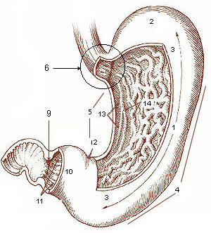
The stomach is a muscular, hollow organ in the gastrointestinal tract of humans and many other animals, including several invertebrates. The stomach has a dilated structure and functions as a vital organ in the digestive system. The stomach is involved in the gastric phase of digestion, following chewing. It performs a chemical breakdown by means of enzymes and hydrochloric acid. In humans and many other animals, the stomach is located between the oesophagus and the small intestine. The stomach secretes digestive enzymes and gastric acid to aid in food digestion. The pyloric sphincter controls the passage of partially digested food ( chyme) from the stomach into the duodenum, where peristalsis takes over to move this through the rest of intestines. Structure In the human digestive system, the stomach lies between the oesophagus and the duodenum (the first part of the small intestine). It is in the left upper quadrant of the abdominal cavity. The top of the stomach lies ag ... [...More Info...] [...Related Items...] OR: [Wikipedia] [Google] [Baidu] |
Esophageal Arteries
Esophageal (Oesophageal in British English) arteries are a group of arteries from disparate sources supplying the esophagus. The blood supply to the esophagus can roughly be divided into thirds, with anastamoses between each area of supply. More specifically, it can refer to: * Esophageal branches of inferior thyroid artery (top third) * Esophageal branches of thoracic part of aorta (middle third) * Esophageal branches of left gastric artery The esophageal branches of left gastric artery are branches which supply the esophagus The esophagus ( American English) or oesophagus (British English; both ), non-technically known also as the food pipe or gullet, is an organ in vertebra ... (bottom third) Arteries {{circulatory-stub ... [...More Info...] [...Related Items...] OR: [Wikipedia] [Google] [Baidu] |
Digestive Tract
The gastrointestinal tract (GI tract, digestive tract, alimentary canal) is the tract or passageway of the digestive system that leads from the mouth to the anus. The GI tract contains all the major organs of the digestive system, in humans and other animals, including the esophagus, stomach, and intestines. Food taken in through the mouth is digested to extract nutrients and absorb energy, and the waste expelled at the anus as feces. ''Gastrointestinal'' is an adjective meaning of or pertaining to the stomach and intestines. Most animals have a "through-gut" or complete digestive tract. Exceptions are more primitive ones: sponges have small pores ( ostia) throughout their body for digestion and a larger dorsal pore (osculum) for excretion, comb jellies have both a ventral mouth and dorsal anal pores, while cnidarians and acoels have a single pore for both digestion and excretion. The human gastrointestinal tract consists of the esophagus, stomach, and intestines, and is ... [...More Info...] [...Related Items...] OR: [Wikipedia] [Google] [Baidu] |
Thoracic Diaphragm
The thoracic diaphragm, or simply the diaphragm ( grc, διάφραγμα, diáphragma, partition), is a sheet of internal Skeletal striated muscle, skeletal muscle in humans and other mammals that extends across the bottom of the thoracic cavity. The diaphragm is the most important Muscles of respiration, muscle of respiration, and separates the thoracic cavity, containing the heart and lungs, from the abdominal cavity: as the diaphragm contracts, the volume of the thoracic cavity increases, creating a negative pressure there, which draws air into the lungs. Its high oxygen consumption is noted by the many mitochondria and capillaries present; more than in any other skeletal muscle. The term ''diaphragm'' in anatomy, created by Gerard of Cremona, can refer to other flat structures such as the urogenital diaphragm or Pelvic floor, pelvic diaphragm, but "the diaphragm" generally refers to the thoracic diaphragm. In humans, the diaphragm is slightly asymmetric—its right half is h ... [...More Info...] [...Related Items...] OR: [Wikipedia] [Google] [Baidu] |
Sphincter
A sphincter is a circular muscle that normally maintains constriction of a natural body passage or orifice and which relaxes as required by normal physiological functioning. Sphincters are found in many animals. There are over 60 types in the human body, some microscopically small, in particular the millions of precapillary sphincters. Sphincters relax at death, often releasing fluids and faeces. Functioning Each sphincter is associated with the lumen (opening) it surrounds. As long as the sphincter muscle is contracted, its length is shortened and the lumen is constricted (closed). Relaxation of the muscle causes it to lengthen, opening the lumen and allowing the passage of liquids, solids, or gases. This is evident, for example, in the blowholes of numerous marine mammals. Many sphincters are used every day in the normal course of digestion. For example, the lower oesophageal sphincter (or cardiac sphincter), which resides at the top of the stomach, is closed most of the time ... [...More Info...] [...Related Items...] OR: [Wikipedia] [Google] [Baidu] |
Striated Muscle
{{disambiguation ...
Striations means a series of ridges, furrows or linear marks, and is used in several ways: * Glacial striation * Striation (fatigue), in material * Striation (geology), a ''striation'' as a result of a geological fault * Striation Valley, in Antarctica * In hyperbolic geometry, a ''striation'' is a reflection across two parallel mirrors. * In anatomy, striated muscle * Striations can be found in certain glasses. These have been caused by turbulent flow during teeming (pouring) of the glass. * Striations can be observed in clouds. See Barber's pole. * Ballistic fingerprinting Forensic firearm examination is the forensic process of examining the characteristics of firearms or bullets left behind at a crime scene. Specialists in this field try to link bullets to weapons and weapons to individuals. They can raise and re ... [...More Info...] [...Related Items...] OR: [Wikipedia] [Google] [Baidu] |
Smooth Muscle
Smooth muscle is an involuntary non-striated muscle, so-called because it has no sarcomeres and therefore no striations (''bands'' or ''stripes''). It is divided into two subgroups, single-unit and multiunit smooth muscle. Within single-unit muscle, the whole bundle or sheet of smooth muscle cells contracts as a syncytium. Smooth muscle is found in the walls of hollow organs, including the stomach, intestines, bladder and uterus; in the walls of passageways, such as blood, and lymph vessels, and in the tracts of the respiratory, urinary, and reproductive systems. In the eyes, the ciliary muscles, a type of smooth muscle, dilate and contract the iris and alter the shape of the lens. In the skin, smooth muscle cells such as those of the arrector pili cause hair to stand erect in response to cold temperature or fear. Structure Gross anatomy Smooth muscle is grouped into two types: single-unit smooth muscle, also known as visceral smooth muscle, and multiunit smooth muscle. ... [...More Info...] [...Related Items...] OR: [Wikipedia] [Google] [Baidu] |
Simple Columnar Epithelia
Simple columnar epithelium is a single layer of columnar epithelial cells which are tall and slender with oval-shaped nuclei located in the basal region, attached to the basement membrane. In humans, simple columnar epithelium lines most organs of the digestive tract including the stomach, and intestines. Simple columnar epithelium also lines the uterus. Structure Simple columnar epithelium is further divided into two categories: ciliated and non-ciliated (glandular). The ciliated part of the simple columnar epithelium has tiny hairs which help move mucus and other substances up the respiratory tract. The shape of the simple columnar epithelium cells are tall and narrow giving a column like appearance. the apical surfaces of the tissue face the lumen of organs while the basal side faces the basement membrane. the nuclei are located closer along the basal side of the cell. Absorptive columnar epithelium is characterized as having a striated boarder on its apical side, this bor ... [...More Info...] [...Related Items...] OR: [Wikipedia] [Google] [Baidu] |
Stratified Squamous Epithelium
A stratified squamous epithelium consists of squamous (flattened) epithelial cells arranged in layers upon a basal membrane. Only one layer is in contact with the basement membrane; the other layers adhere to one another to maintain structural integrity. Although this epithelium is referred to as squamous, many cells within the layers may not be flattened; this is due to the convention of naming epithelia according to the cell type at the surface. In the deeper layers, the cells may be columnar or cuboidal. There are no intercellular spaces. This type of epithelium is well suited to areas in the body subject to constant abrasion, as the thickest layers can be sequentially sloughed off and replaced before the basement membrane is exposed. It forms the outermost layer of the skin and the inner lining of the mouth, esophagus and vagina. In the epidermis of skin in mammals, reptiles, and birds, the layer of keratin in the outer layer of the stratified squamous epithelial surface ... [...More Info...] [...Related Items...] OR: [Wikipedia] [Google] [Baidu] |
Fibrous Tissue
Fiber or fibre (from la, fibra, links=no) is a natural or artificial substance that is significantly longer than it is wide. Fibers are often used in the manufacture of other materials. The strongest engineering materials often incorporate fibers, for example carbon fiber and ultra-high-molecular-weight polyethylene. Synthetic fibers can often be produced very cheaply and in large amounts compared to natural fibers, but for clothing natural fibers can give some benefits, such as comfort, over their synthetic counterparts. Natural fibers Natural fibers develop or occur in the fiber shape, and include those produced by plants, animals, and geological processes. They can be classified according to their origin: *Vegetable fibers are generally based on arrangements of cellulose, often with lignin: examples include cotton, hemp, jute, flax, abaca, piña, ramie, sisal, bagasse, and banana. Plant fibers are employed in the manufacture of paper and textile (cloth), and diet ... [...More Info...] [...Related Items...] OR: [Wikipedia] [Google] [Baidu] |
Muscular Layer
The muscular layer (muscular coat, muscular fibers, muscularis propria, muscularis externa) is a region of muscle in many organs in the vertebrate body, adjacent to the submucosa. It is responsible for gut movement such as peristalsis. The Latin, tunica muscularis, may also be used. Structure It usually has two layers of smooth muscle: * inner and "circular" * outer and "longitudinal" However, there are some exceptions to this pattern. * In the stomach there are three layers to the muscular layer. Stomach contains an additional oblique muscle layer just interior to circular muscle layer. * In the upper esophagus, part of the externa is ''skeletal muscle'', rather than smooth muscle. * In the vas deferens of the spermatic cord, there are three layers: inner longitudinal, middle circular, and outer longitudinal. * In the ureter the smooth muscle orientation is opposite that of the GI tract. There is an inner longitudinal and an outer circular layer. The inner layer of the muscul ... [...More Info...] [...Related Items...] OR: [Wikipedia] [Google] [Baidu] |
Submucosa
The submucosa (or tela submucosa) is a thin layer of tissue (biology), tissue in various organ (anatomy), organs of the gastrointestinal tract, gastrointestinal, respiratory tract, respiratory, and genitourinary system, genitourinary tracts. It is the layer of dense irregular connective tissue that supports the mucosa (mucous membrane) and joins it to the muscular layer, the bulk of overlying smooth muscle (fibers running circularly within layer of longitudinal muscle). The submucosa (''wikt:sub-#Prefix, sub-'' + ''mucosa'') is to a mucous membrane what the subserosa (''wikt:sub-#Prefix, sub-'' + ''serosa'') is to a serous membrane. Structure Blood vessels, lymphatic vessels, and nerves (all supplying the mucosa) will run through here. In the intestinal wall, tiny parasympathetic ganglia are scattered around forming the submucous plexus (or "Meissner's plexus") where preganglionic parasympathetic neurons synapse with postganglionic nerve fibers that supply the muscularis mucosae. ... [...More Info...] [...Related Items...] OR: [Wikipedia] [Google] [Baidu] |


