|
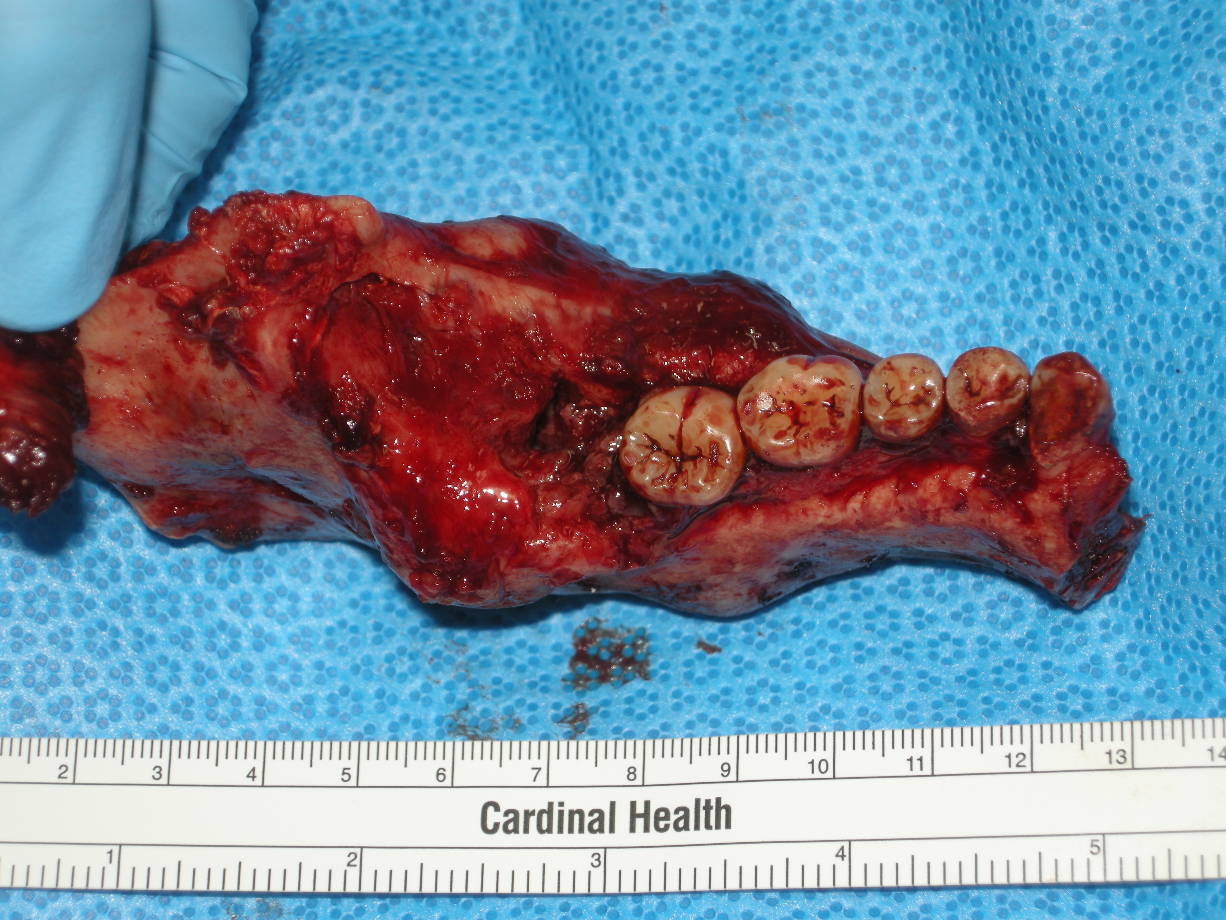
Odontogenic Keratocyst
An odontogenic keratocyst is a rare and benign but locally aggressive developmental cyst. It most often affects the posterior mandible and most commonly presents in the third decade of life. Odontogenic keratocysts make up around 19% of jaw cysts. In the WHO/IARC classification of head and neck pathology, this clinical entity had been known for years as the odontogenic keratocyst; it was reclassified as keratocystic odontogenic tumour (KCOT) from 2005 to 2017. In 2017 it reverted to the earlier name, as the new WHO/IARC classification reclassified OKC back into the cystic category. Under The WHO/IARC classification, Odontogenic Keratocyst underwent the reclassification as it is no longer considered a neoplasm due to a lack of quality evidence regarding this hypothesis, especially with respect to clonality. Within the Head and Neck pathology community there is still controversy surrounding the reclassification, with some pathologists still considering Odontogenic Keratocyst as a neo ... [...More Info...] [...Related Items...] OR: [Wikipedia] [Google] [Baidu] |
Micrograph
A micrograph or photomicrograph is a photograph or digital image taken through a microscope or similar device to show a magnified image of an object. This is opposed to a macrograph or photomacrograph, an image which is also taken on a microscope but is only slightly magnified, usually less than 10 times. Micrography is the practice or art of using microscopes to make photographs. A micrograph contains extensive details of microstructure. A wealth of information can be obtained from a simple micrograph like behavior of the material under different conditions, the phases found in the system, failure analysis, grain size estimation, elemental analysis and so on. Micrographs are widely used in all fields of microscopy. Types Photomicrograph A light micrograph or photomicrograph is a micrograph prepared using an optical microscope, a process referred to as ''photomicroscopy''. At a basic level, photomicroscopy may be performed simply by connecting a camera to a microscope, th ... [...More Info...] [...Related Items...] OR: [Wikipedia] [Google] [Baidu] |
Tumour Suppressor Gene
A tumor suppressor gene (TSG), or anti-oncogene, is a gene that regulates a cell during cell division and replication. If the cell grows uncontrollably, it will result in cancer. When a tumor suppressor gene is mutated, it results in a loss or reduction in its function. In combination with other genetic mutations, this could allow the cell to grow abnormally. The loss of function for these genes may be even more significant in the development of human cancers, compared to the activation of oncogenes. TSGs can be grouped into the following categories: caretaker genes, gatekeeper genes, and more recently landscaper genes. Caretaker genes ensure stability of the genome via DNA repair and subsequently when mutated allow mutations to accumulate. Meanwhile, gatekeeper genes directly regulate cell growth by either inhibiting cell cycle progression or inducing apoptosis. Lastly landscaper genes regulate growth by contributing to the surrounding environment, when mutated can cause an envir ... [...More Info...] [...Related Items...] OR: [Wikipedia] [Google] [Baidu] |
Dentigerous Cyst
Dentigerous cyst, also known as follicular cyst is an epithelial-lined developmental cyst formed by accumulation of fluid between the reduced enamel epithelium and crown of an unerupted tooth. It is formed when there is an alteration in the reduced enamel epithelium and encloses the crown of an unerupted tooth at the cemento-enamel junction. Fluid is accumulated between reduced enamel epithelium and the crown of an unerupted tooth. Dentigerous cyst is the second most common form of benign developmental odontogenic cysts. Dentigerous cyst is the second most prevalent type of odontogenic cysts after radicular cyst. 70 percent of the cases occurs in the mandible. Dentigerous cyst is usually painless. Patient usually comes with a concern of delayed tooth eruption or facial swelling. Dentigerous cyst can go unnoticed and may be discovered coincidentally on a regular radiographic examination. Pathogenesis Odontogenesis happens by means of a complex interaction between oral epitheliu ... [...More Info...] [...Related Items...] OR: [Wikipedia] [Google] [Baidu] |
Adenomatoid Odontogenic Tumor
The adenomatoid odontogenic tumor is an odontogenic tumor arising from the enamel organ or dental lamina. Signs and symptoms Two thirds of cases are located in the anterior maxilla, and one third are present in the anterior mandible. Two thirds of the cases are associated with an impacted tooth (usually being the canine). Diagnosis On radiographs, the adenomatoid odontogenic tumor presents as a radiolucency (dark area) around an unerupted tooth extending past the cementoenamel junction. It should be differentially diagnosed from a dentigerous cyst and the main difference is that the radiolucency in case of AOT extends apically beyond the cementoenamel junction. Radiographs will exhibit faint flecks of radiopacities surrounded by a radiolucent zone. It is sometimes misdiagnosed as a cyst A cyst is a closed sac, having a distinct envelope and cell division, division compared with the nearby Biological tissue, tissue. Hence, it is a cluster of Cell (biology), cells that have g ... [...More Info...] [...Related Items...] OR: [Wikipedia] [Google] [Baidu] |
Central Giant-cell Granuloma
Central giant-cell granuloma (CGCG) is a localised benign condition of the jaws. It is twice as common in females and is more likely to occur before age 30. Central giant-cell granulomas are more common in the anterior mandible, often crossing the midline and causing painless swellings. Signs and symptoms CGCG is the most common giant cell lesion of the jaws. These lesions are localised fibrous tissue tumours which contain osteoclasts and are usually several centimetres across. Frequently, a painless swelling that grows and expands rapidly is present. This growth can also erode through bone including the alveolar ridge, resulting in a soft tissue swelling that is purple in colour. Paresthesia of the lip has also been observed. Resorption of tooth roots is seen in 37% of cases compared to displacement of teeth in 50%. Two-thirds of lesions are found anterior to molars in the mandible, where teeth have deciduous predecessors. CGCGs are twice as likely to affect females an ... [...More Info...] [...Related Items...] OR: [Wikipedia] [Google] [Baidu] |
Ameloblastoma
Ameloblastoma is a rare, benign or cancerous tumor of odontogenic epithelium (ameloblasts, or outside portion, of the teeth during development) much more commonly appearing in the lower jaw than the upper jaw. It was recognized in 1827 by Cusack. This type of odontogenic neoplasm was designated as an ''adamantinoma'' in 1885 by the French physician Louis-Charles Malassez. It was finally renamed to the modern name ''ameloblastoma'' in 1930 by Ivey and Churchill. While these tumors are rarely malignant or metastatic (that is, they rarely spread to other parts of the body), and progress slowly, the resulting lesions can cause severe abnormalities of the face and jaw leading to severe disfiguration. Additionally, as abnormal cell growth easily infiltrates and destroys surrounding bony tissues, wide surgical excision is required to treat this disorder. If an aggressive tumor is left untreated, it can obstruct the nasal and oral airways making it impossible to breathe without oropharyng ... [...More Info...] [...Related Items...] OR: [Wikipedia] [Google] [Baidu] |
Odontogenic Myxoma
The odontogenic myxoma is an uncommon benign odontogenic tumor arising from embryonic connective tissue associated with tooth formation.Sapp, J. Philip., Lewis R. Eversole, and George P. Wysocki. Contemporary Oral and Maxillofacial Pathology. 2nd ed. St. Louis, MO: Mosby, 2002. 152-53. As a myxoma, this tumor consists mainly of spindle shaped cells and scattered collagen fibers distributed through a loose, mucoid material.Cawson, R. A., and E. W. Odell. Cawson's Essentials of Oral Pathology and Oral Medicine. 8th ed. Edinburgh: Churchill Livingstone, 2008. 145-46. Signs and symptoms Odontogenic myxomas have been found in patients ranging in age between 2 and 50 years, however, they are most commonly diagnosed in young adults (specifically between 25 and 35 years of age).Wood, Norman K., Paul W. Goaz, and Norman K. Wood. Differential Diagnosis of Oral and Maxillofacial Lesions. 5th ed. St. Louis: Mosby, 1997. 342-43.McDonald, Ralph E., David R. Avery, and Jeffrey A. Dean. Dentistry ... [...More Info...] [...Related Items...] OR: [Wikipedia] [Google] [Baidu] |
Ameloblastoma
Ameloblastoma is a rare, benign or cancerous tumor of odontogenic epithelium (ameloblasts, or outside portion, of the teeth during development) much more commonly appearing in the lower jaw than the upper jaw. It was recognized in 1827 by Cusack. This type of odontogenic neoplasm was designated as an ''adamantinoma'' in 1885 by the French physician Louis-Charles Malassez. It was finally renamed to the modern name ''ameloblastoma'' in 1930 by Ivey and Churchill. While these tumors are rarely malignant or metastatic (that is, they rarely spread to other parts of the body), and progress slowly, the resulting lesions can cause severe abnormalities of the face and jaw leading to severe disfiguration. Additionally, as abnormal cell growth easily infiltrates and destroys surrounding bony tissues, wide surgical excision is required to treat this disorder. If an aggressive tumor is left untreated, it can obstruct the nasal and oral airways making it impossible to breathe without oropharyng ... [...More Info...] [...Related Items...] OR: [Wikipedia] [Google] [Baidu] |
Hounsfield Unit
The Hounsfield scale , named after Sir Godfrey Hounsfield, is a quantitative scale for describing radiodensity. It is frequently used in CT scans, where its value is also termed CT number. Definition The Hounsfield unit (HU) scale is a linear transformation of the original linear attenuation coefficient measurement into one in which the radiodensity of distilled water at standard pressure and temperature (STP) is defined as 0 Hounsfield units (HU), while the radiodensity of air at STP is defined as −1000 HU. In a voxel with average linear attenuation coefficient \mu, the corresponding HU value is therefore given by: HU = 1000\times\frac where \mu_ and \mu_ are respectively the linear attenuation coefficients of water and air. Thus, a change of one Hounsfield unit (HU) represents a change of 0.1% of the attenuation coefficient of water since the attenuation coefficient of air is nearly zero. Calibration tests of HU with reference to water and other materials may be done to ensu ... [...More Info...] [...Related Items...] OR: [Wikipedia] [Google] [Baidu] |
Radiodensity
Radiodensity (or radiopacity) is opacity to the radio wave and X-ray portion of the electromagnetic spectrum: that is, the relative inability of those kinds of electromagnetic radiation to pass through a particular material. Radiolucency or hypodensity indicates greater passage (greater transradiancy) to X-ray photonsNovelline, Robert. ''Squire's Fundamentals of Radiology''. Harvard University Press. 5th edition. 1997. . and is the analogue of transparency and translucency with visible light. Materials that inhibit the passage of electromagnetic radiation are called radiodense or radiopaque, while those that allow radiation to pass more freely are referred to as radiolucent. Radiopaque volumes of material have white appearance on radiographs, compared with the relatively darker appearance of radiolucent volumes. For example, on typical radiographs, bones look white or light gray (radiopaque), whereas muscle and skin look black or dark gray, being mostly invisible (radiolucent). Th ... [...More Info...] [...Related Items...] OR: [Wikipedia] [Google] [Baidu] |
CT Scan
A computed tomography scan (CT scan; formerly called computed axial tomography scan or CAT scan) is a medical imaging technique used to obtain detailed internal images of the body. The personnel that perform CT scans are called radiographers or radiology technologists. CT scanners use a rotating X-ray tube and a row of detectors placed in a gantry (medical), gantry to measure X-ray Attenuation#Radiography, attenuations by different tissues inside the body. The multiple X-ray measurements taken from different angles are then processed on a computer using tomographic reconstruction algorithms to produce Tomography, tomographic (cross-sectional) images (virtual "slices") of a body. CT scans can be used in patients with metallic implants or pacemakers, for whom magnetic resonance imaging (MRI) is Contraindication, contraindicated. Since its development in the 1970s, CT scanning has proven to be a versatile imaging technique. While CT is most prominently used in medical diagnosis, ... [...More Info...] [...Related Items...] OR: [Wikipedia] [Google] [Baidu] |
Biopsy
A biopsy is a medical test commonly performed by a surgeon, interventional radiologist, or an interventional cardiologist. The process involves extraction of sample cells or tissues for examination to determine the presence or extent of a disease. The tissue is then fixed, dehydrated, embedded, sectioned, stained and mounted before it is generally examined under a microscope by a pathologist; it may also be analyzed chemically. When an entire lump or suspicious area is removed, the procedure is called an excisional biopsy. An incisional biopsy or core biopsy samples a portion of the abnormal tissue without attempting to remove the entire lesion or tumor. When a sample of tissue or fluid is removed with a needle in such a way that cells are removed without preserving the histological architecture of the tissue cells, the procedure is called a needle aspiration biopsy. Biopsies are most commonly performed for insight into possible cancerous or inflammatory conditions. History T ... [...More Info...] [...Related Items...] OR: [Wikipedia] [Google] [Baidu] |


