|
Nigrostriatal
The nigrostriatal pathway is a bilateral dopaminergic pathway in the brain that connects the substantia nigra pars compacta (SNc) in the midbrain with the dorsal striatum (i.e., the caudate nucleus and putamen) in the forebrain. It is one of the four major dopamine pathways in the brain, and is critical in the production of movement as part of a system called the basal ganglia motor loop. Dopaminergic neurons of this pathway release dopamine from axon terminals that synapse onto GABAergic medium spiny neurons (MSNs), also known as spiny projection neurons (SPNs), located in the striatum. Degeneration of dopaminergic neurons in the SNc is one of the main pathological features of Parkinson's disease, leading to a marked reduction in dopamine function and the symptomatic motor deficits of Parkinson's disease including hypokinesia, tremors, rigidity, and postural imbalance. Anatomy The connection between the substantia nigra pars compacta and the dorsal striatum is mediated via ... [...More Info...] [...Related Items...] OR: [Wikipedia] [Google] [Baidu] |
Dopaminergic Pathway
Dopaminergic pathways (dopamine pathways, dopaminergic projections) in the human brain are involved in both physiological and behavioral processes including movement, cognition, executive functions, reward, motivation, and neuroendocrine control. Each pathway is a set of projection neurons, consisting of individual dopaminergic neurons. The four major dopaminergic pathways are the mesolimbic pathway, the mesocortical pathway, the nigrostriatal pathway, and the tuberoinfundibular pathway.The mesolimbic pathway and the mesocortical pathway form the mesocorticolimbic system. Two other dopaminergic pathways to be considered are the hypothalamospinal tract and the incertohypothalamic pathway. Parkinson's disease, attention deficit hyperactivity disorder (ADHD), substance use disorders (addiction), and restless legs syndrome (RLS) can be attributed to dysfunction in specific dopaminergic pathways. The dopamine neurons of the dopaminergic pathways synthesize and release the neurotr ... [...More Info...] [...Related Items...] OR: [Wikipedia] [Google] [Baidu] |
Hypokinesia
Hypokinesia is one of the classifications of movement disorders, and refers to decreased bodily movement. Hypokinesia is characterized by a partial or complete loss of muscle movement due to a disruption in the basal ganglia. Hypokinesia is a symptom of Parkinson's disease shown as muscle rigidity and an inability to produce movement. It is also associated with mental health disorders and prolonged inactivity due to illness, amongst other diseases. The other category of movement disorder is hyperkinesia that features an exaggeration of unwanted movement, such as twitching or writhing in Huntington's disease or Tourette syndrome. Spectrum of disorders Hypokinesia describes a variety of more specific disorders: Causes The most common cause of Hypokinesia is Parkinson's disease, and conditions related to Parkinson's disease. Other conditions may also cause slowness of movements. These includhypothyroidism and severe depression.These conditions need to be carefully ruled out ... [...More Info...] [...Related Items...] OR: [Wikipedia] [Google] [Baidu] |
Pars Reticulata
The pars reticulata (SNpr) is a portion of the substantia nigra and is located lateral to the pars compacta. Most of the neurons that project out of the pars reticulata are inhibitory GABAergic neurons (i.e., these neurons release GABA, which is an inhibitory neurotransmitter). Anatomy Neurons in the pars reticulata are much less densely packed than those in the pars compacta (they were sometimes named pars diffusa). They are smaller and thinner than the dopaminergic neurons and conversely identical and morphologically similar to the pallidal neurons (see primate basal ganglia). Their dendrites as well as the pallidal are preferentially perpendicular to the striatal afferents. The massive striatal afferents correspond to the medial end of the nigrostriatal bundle. Nigral neurons have the same peculiar synaptology with the striatal axonal endings. They make connections with the dopamine neurons of the pars compacta whose long dendrites plunge deeply in the pars reticulata. The ... [...More Info...] [...Related Items...] OR: [Wikipedia] [Google] [Baidu] |
Dopamine Pathways
Dopamine (DA, a contraction of 3,4-dihydroxyphenethylamine) is a neuromodulatory molecule that plays several important roles in cells. It is an organic chemical of the catecholamine and phenethylamine families. Dopamine constitutes about 80% of the catecholamine content in the brain. It is an amine synthesized by removing a carboxyl group from a molecule of its precursor chemical, L-DOPA, which is synthesized in the brain and kidneys. Dopamine is also synthesized in plants and most animals. In the brain, dopamine functions as a neurotransmitter—a chemical released by neurons (nerve cells) to send signals to other nerve cells. Neurotransmitters are synthesized in specific regions of the brain, but affect many regions systemically. The brain includes several distinct dopamine pathways, one of which plays a major role in the motivational component of reward-motivated behavior. The anticipation of most types of rewards increases the level of dopamine in the brain, and many addi ... [...More Info...] [...Related Items...] OR: [Wikipedia] [Google] [Baidu] |
Substantia Nigra
The substantia nigra (SN) is a basal ganglia structure located in the midbrain that plays an important role in reward and movement. ''Substantia nigra'' is Latin for "black substance", reflecting the fact that parts of the substantia nigra appear darker than neighboring areas due to high levels of neuromelanin in dopaminergic neurons. Parkinson's disease is characterized by the loss of dopaminergic neurons in the substantia nigra pars compacta. Although the substantia nigra appears as a continuous band in brain sections, anatomical studies have found that it actually consists of two parts with very different connections and functions: the pars compacta (SNpc) and the pars reticulata (SNpr). The pars compacta serves mainly as a projection to the basal ganglia circuit, supplying the striatum with dopamine. The pars reticulata conveys signals from the basal ganglia to numerous other brain structures. Structure The substantia nigra, along with four other nuclei, is part ... [...More Info...] [...Related Items...] OR: [Wikipedia] [Google] [Baidu] |
Dorsal Striatum
The striatum, or corpus striatum (also called the striate nucleus), is a nucleus (a cluster of neurons) in the subcortical basal ganglia of the forebrain. The striatum is a critical component of the motor and reward systems; receives glutamatergic and dopaminergic inputs from different sources; and serves as the primary input to the rest of the basal ganglia. Functionally, the striatum coordinates multiple aspects of cognition, including both motor and action planning, decision-making, motivation, reinforcement, and reward perception. The striatum is made up of the caudate nucleus and the lentiform nucleus. The lentiform nucleus is made up of the larger putamen, and the smaller globus pallidus. Strictly speaking the globus pallidus is part of the striatum. It is common practice, however, to implicitly exclude the globus pallidus when referring to striatal structures. In primates, the striatum is divided into a ventral striatum, and a dorsal striatum, subdivisions that are base ... [...More Info...] [...Related Items...] OR: [Wikipedia] [Google] [Baidu] |
Pars Compacta
The pars compacta (SNpc) is a portion of the ''substantia nigra'', located in the midbrain. It is formed by dopaminergic neurons and located medial to the pars reticulata. Parkinson's disease is characterized by the death of dopaminergic neurons in this region. Anatomy In humans, the nerve cell bodies of the ''pars compacta'' are coloured black by the pigment neuromelanin. The degree of pigmentation increases with age. This pigmentation is visible as a distinctive black stripe in brain sections and is the origin of the name given to this volume of the brain. The neurons have particularly long and thick dendrites (François et al.). The ventral dendrites, particularly, go down deeply in the pars reticulata. Other similar neurons are more sparsely distributed in the midbrain and constitute "groups" with no well-defined borders, although continuous to the pars compacta, in a prerubral position. These have been given, in early works in rats (with not much respect for the anatomical ... [...More Info...] [...Related Items...] OR: [Wikipedia] [Google] [Baidu] |
Medial Forebrain Bundle
The medial forebrain bundle (MFB), is a neural pathway containing axon, fibers from the basal olfactory regions, the periamygdaloid region and the septal nuclei, as well as fibers from brainstem regions, including the ventral tegmental area and nigrostriatal pathway. Anatomy The MFB passes through the lateral hypothalamus and the basal forebrain in a rostral-caudal direction. The MFB has its main projections to these regions of Brodmann areas (BA) 8, 9, 10, 11, 11m. The superior frontal region of MFB projects to BA 8, 9, 10; the rostral middle frontal projects to dorsolateral prefrontal cortex (BA 9, 10); lateral orbitofrontal of MFB shows its projections to nucleus accumbens septi (NAC) and ventral striatum as subcortical reward associated structures. It contains both ascending and descending fibers. The mesolimbic pathway, which is a collection of dopaminergic neurons that projects from the ventral tegmental area to the nucleus accumbens, is a component pathway of the MFB. The M ... [...More Info...] [...Related Items...] OR: [Wikipedia] [Google] [Baidu] |
Mesolimbic Pathway
The mesolimbic pathway, sometimes referred to as the reward pathway, is a dopaminergic pathway in the brain. The pathway connects the ventral tegmental area in the midbrain to the ventral striatum of the basal ganglia in the forebrain. The ventral striatum includes the nucleus accumbens and the olfactory tubercle. The release of dopamine from the mesolimbic pathway into the nucleus accumbens regulates incentive salience (e.g. motivation and desire for rewarding stimuli) and facilitates reinforcement and reward-related motor function learning; it may also play a role in the subjective perception of pleasure. The dysregulation of the mesolimbic pathway and its output neurons in the nucleus accumbens plays a significant role in the development and maintenance of an addiction. Anatomy The mesolimbic pathway is a collection of dopaminergic (i.e., dopamine-releasing) neurons that project from the ventral tegmental area (VTA) to the ventral striatum, which includes the nucleus accumben ... [...More Info...] [...Related Items...] OR: [Wikipedia] [Google] [Baidu] |
Ventral Tegmental Area
The ventral tegmental area (VTA) (tegmentum is Latin for ''covering''), also known as the ventral tegmental area of Tsai, or simply ventral tegmentum, is a group of neurons located close to the midline on the floor of the midbrain. The VTA is the origin of the dopaminergic cell bodies of the mesocorticolimbic dopamine system and other dopamine pathways; it is widely implicated in the drug and natural reward circuitry of the brain. The VTA plays an important role in a number of processes, including reward cognition ( motivational salience, associative learning, and positively-valenced emotions) and orgasm, among others, as well as several psychiatric disorders. Neurons in the VTA project to numerous areas of the brain, ranging from the prefrontal cortex to the caudal brainstem and several regions in between. Structure Neurobiologists have often had great difficulty distinguishing the VTA in humans and other primate brains from the substantia nigra (SN) and surrounding nucl ... [...More Info...] [...Related Items...] OR: [Wikipedia] [Google] [Baidu] |
Caudate Nucleus
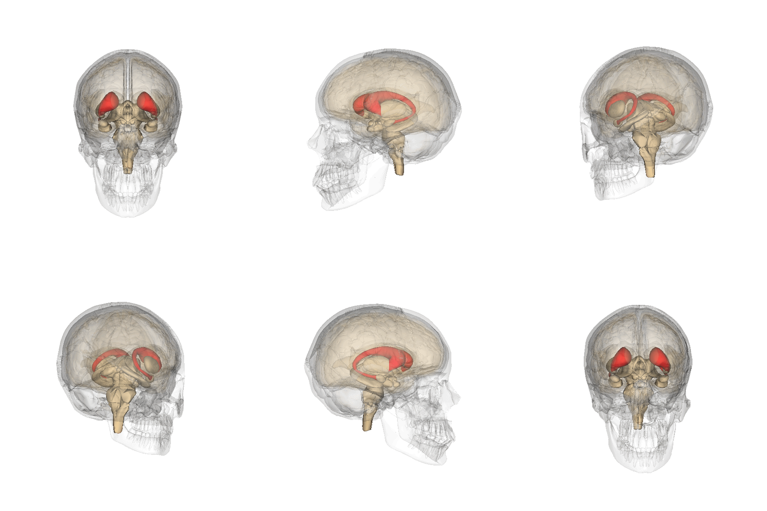
The caudate nucleus is one of the structures that make up the corpus striatum, which is a component of the basal ganglia in the human brain. While the caudate nucleus has long been associated with motor processes due to its role in Parkinson's disease, it plays important roles in various other nonmotor functions as well, including procedural learning, associative learning and inhibitory control of action, among other functions. The caudate is also one of the brain structures which compose the reward system and functions as part of the cortico–basal ganglia– thalamic loop. Structure Together with the putamen, the caudate forms the dorsal striatum, which is considered a single functional structure; anatomically, it is separated by a large white matter tract, the internal capsule, so it is sometimes also referred to as two structures: the medial dorsal striatum (the caudate) and the lateral dorsal striatum (the putamen). In this vein, the two are functionally distinct not a ... [...More Info...] [...Related Items...] OR: [Wikipedia] [Google] [Baidu] |
Striatum
The striatum, or corpus striatum (also called the striate nucleus), is a nucleus (a cluster of neurons) in the subcortical basal ganglia of the forebrain. The striatum is a critical component of the motor and reward systems; receives glutamatergic and dopaminergic inputs from different sources; and serves as the primary input to the rest of the basal ganglia. Functionally, the striatum coordinates multiple aspects of cognition, including both motor and action planning, decision-making, motivation, reinforcement, and reward perception. The striatum is made up of the caudate nucleus and the lentiform nucleus. The lentiform nucleus is made up of the larger putamen, and the smaller globus pallidus. Strictly speaking the globus pallidus is part of the striatum. It is common practice, however, to implicitly exclude the globus pallidus when referring to striatal structures. In primates, the striatum is divided into a ventral striatum, and a dorsal striatum, subdivisions that are ... [...More Info...] [...Related Items...] OR: [Wikipedia] [Google] [Baidu] |


