|
Substantia Nigra
The substantia nigra (SN) is a basal ganglia structure located in the midbrain that plays an important role in reward and movement. ''Substantia nigra'' is Latin for "black substance", reflecting the fact that parts of the substantia nigra appear darker than neighboring areas due to high levels of neuromelanin in dopaminergic neurons. Parkinson's disease is characterized by the loss of dopaminergic neurons in the substantia nigra pars compacta. Although the substantia nigra appears as a continuous band in brain sections, anatomical studies have found that it actually consists of two parts with very different connections and functions: the pars compacta (SNpc) and the pars reticulata (SNpr). The pars compacta serves mainly as a projection to the basal ganglia circuit, supplying the striatum with dopamine. The pars reticulata conveys signals from the basal ganglia to numerous other brain structures. Structure The substantia nigra, along with four other nuclei, is part ... [...More Info...] [...Related Items...] OR: [Wikipedia] [Google] [Baidu] |
Superior Colliculus
In neuroanatomy, the superior colliculus () is a structure lying on the roof of the mammalian midbrain. In non-mammalian vertebrates, the homologous structure is known as the optic tectum, or optic lobe. The adjective form ''tectal'' is commonly used for both structures. In mammals, the superior colliculus forms a major component of the midbrain. It is a paired structure and together with the paired inferior colliculi forms the corpora quadrigemina. The superior colliculus is a layered structure, with a pattern that is similar to all mammals. The layers can be grouped into the superficial layers ( stratum opticum and above) and the deeper remaining layers. Neurons in the superficial layers receive direct input from the retina and respond almost exclusively to visual stimuli. Many neurons in the deeper layers also respond to other modalities, and some respond to stimuli in multiple modalities. The deeper layers also contain a population of motor-related neurons, capable of activat ... [...More Info...] [...Related Items...] OR: [Wikipedia] [Google] [Baidu] |
Basal Ganglia Circuits
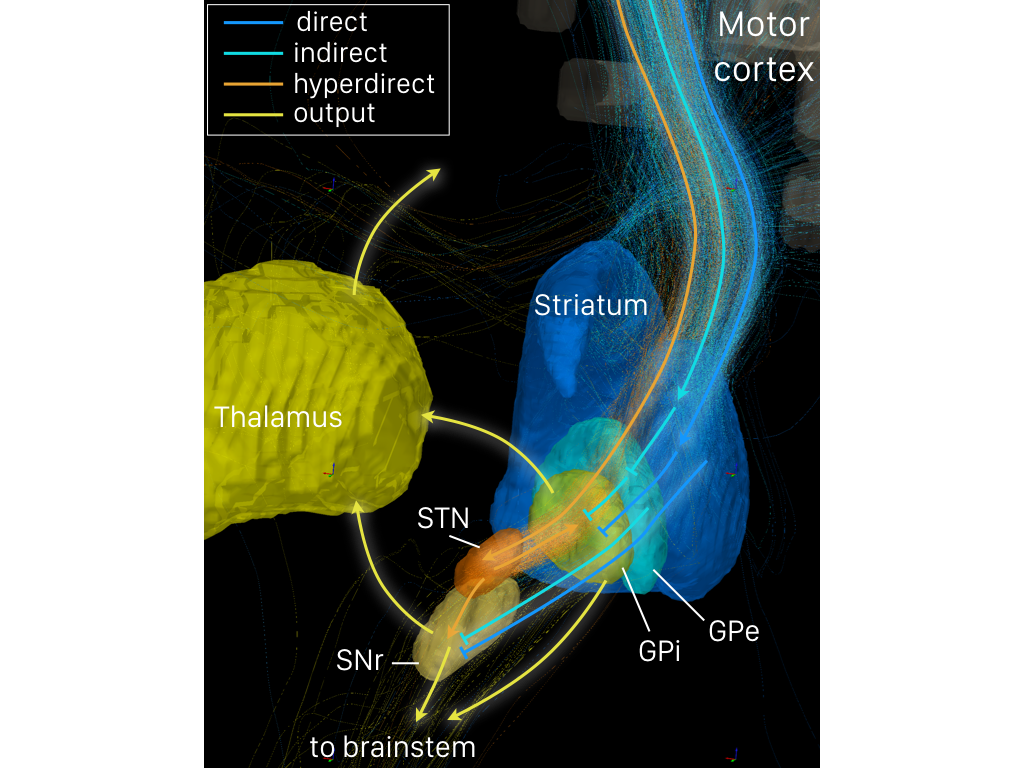
The basal ganglia (BG), or basal nuclei, are a group of subcortical nuclei, of varied origin, in the brains of vertebrates. In humans, and some primates, there are some differences, mainly in the division of the globus pallidus into an external and internal region, and in the division of the striatum. The basal ganglia are situated at the base of the forebrain and top of the midbrain. Basal ganglia are strongly interconnected with the cerebral cortex, thalamus, and brainstem, as well as several other brain areas. The basal ganglia are associated with a variety of functions, including control of voluntary motor movements, procedural learning, habit learning, conditional learning, eye movements, cognition, and emotion. The main components of the basal ganglia – as defined functionally – are the striatum, consisting of both the dorsal striatum (caudate nucleus and putamen) and the ventral striatum (nucleus accumbens and olfactory tubercle), the globus pallidus, th ... [...More Info...] [...Related Items...] OR: [Wikipedia] [Google] [Baidu] |
Learning
Learning is the process of acquiring new understanding, knowledge, behaviors, skills, value (personal and cultural), values, attitudes, and preferences. The ability to learn is possessed by humans, animals, and some machine learning, machines; there is also evidence for some kind of learning in certain plants. Some learning is immediate, induced by a single event (e.g. being burned by a Heat, hot stove), but much skill and knowledge accumulate from repeated experiences. The changes induced by learning often last a lifetime, and it is hard to distinguish learned material that seems to be "lost" from that which cannot be retrieved. Human learning starts at birth (it might even start before in terms of an embryo's need for both interaction with, and freedom within its environment within the womb.) and continues until death as a consequence of ongoing interactions between people and their environment. The nature and processes involved in learning are studied in many established fi ... [...More Info...] [...Related Items...] OR: [Wikipedia] [Google] [Baidu] |
Motor System
The motor system is the set of central and peripheral structures in the nervous system that support motor functions, i.e. movement. Peripheral structures may include skeletal muscles and neural connections with muscle tissues. Central structures include cerebral cortex, brainstem, spinal cord, pyramidal system including the upper motor neurons, extrapyramidal system, cerebellum, and the lower motor neurons in the brainstem and the spinal cord. Pyramidal motor system The pyramidal motor system, also called the pyramidal tract or the corticospinal tract, start in the motor center of the cerebral cortex.Rizzolatti G, Luppino G (2001) The Cortical Motor System. ''Neuron'' 31: 889-90SD/ref> There are upper and lower motor neurons in the corticospinal tract. The motor impulses originate in the giant pyramidal cells or Betz cells of the motor area; i.e., precentral gyrus of cerebral cortex. These are the upper motor neurons (UMN) of the corticospinal tract. The axons of these cel ... [...More Info...] [...Related Items...] OR: [Wikipedia] [Google] [Baidu] |
Eye Movement (sensory)
Eye movement includes the voluntary or involuntary movement of the eyes. Eye movements are used by a number of organisms (e.g. primates, rodents, flies, birds, fish, cats, crabs, octopus) to fixate, inspect and track visual objects of interests. A special type of eye movement, rapid eye movement, occurs during REM sleep. The eyes are the visual organs of the human body, and move using a system of six muscles. The retina, a specialised type of tissue containing photoreceptors, senses light. These specialised cells convert light into electrochemical signals. These signals travel along the optic nerve fibers to the brain, where they are interpreted as vision in the visual cortex. Primates and many other vertebrates use three types of voluntary eye movement to track objects of interest: smooth pursuit, vergence shifts and saccades. These types of movements appear to be initiated by a small cortical region in the brain's frontal lobe. This is corroborated by removal of the fr ... [...More Info...] [...Related Items...] OR: [Wikipedia] [Google] [Baidu] |
Glutamatergic
Glutamatergic means "related to glutamate". A glutamatergic agent (or drug) is a chemical that directly modulates the excitatory amino acid (glutamate/ aspartate) system in the body or brain. Examples include excitatory amino acid receptor agonists, excitatory amino acid receptor antagonists, and excitatory amino acid reuptake inhibitors. See also * Adenosinergic * Adrenergic * Cannabinoidergic * Cholinergic * Dopaminergic * GABAergic * GHBergic * Glycinergic * Histaminergic * Melatonergic * Monoaminergic * Opioidergic * Serotonergic * Sigmaergic Sigma receptors (σ-receptors) are protein cell surface receptors that bind ligands such as 4-PPBP (4-phenyl-1-(4-phenylbutyl) piperidine), SA 4503 (cutamesine), ditolylguanidine, dimethyltryptamine, and siramesine. There are two subtypes, ... References Neurochemistry Neurotransmitters {{nervous-system-drug-stub ... [...More Info...] [...Related Items...] OR: [Wikipedia] [Google] [Baidu] |
Subthalamic Nucleus
The subthalamic nucleus (STN) is a small lens-shaped nucleus in the brain where it is, from a functional point of view, part of the basal ganglia system. In terms of anatomy, it is the major part of the subthalamus. As suggested by its name, the subthalamic nucleus is located ventral to the thalamus. It is also dorsal to the substantia nigra and medial to the internal capsule. It was first described by Jules Bernard Luys in 1865, and the term ''corpus Luysi'' or ''Luys' body'' is still sometimes used. Anatomy Structure The principal type of neuron found in the subthalamic nucleus has rather long, sparsely spiny dendrites. In the more centrally located neurons, the dendritic arbors have a more ellipsoidal shape. The dimensions of these arbors (1200 μm, 600 μm, and 300 μm) are similar across many species—including rat, cat, monkey and human—which is unusual. However, the number of neurons increases with brain size as well as the external dimensions of th ... [...More Info...] [...Related Items...] OR: [Wikipedia] [Google] [Baidu] |
GABAergic
In molecular biology and physiology, something is GABAergic or GABAnergic if it pertains to or affects the neurotransmitter GABA. For example, a synapse is GABAergic if it uses GABA as its neurotransmitter, and a GABAergic neuron produces GABA. A substance is GABAergic if it produces its effects via interactions with the GABA system, such as by stimulating or blocking neurotransmission. A GABAergic or GABAnergic agent is any chemical that modifies the effects of GABA in the body or brain. Some different classes of GABAergic drugs include agonists, antagonists, modulators, reuptake inhibitors and enzymes. See also * GABA reuptake inhibitor * Adenosinergic * Adrenergic * Cannabinoidergic * Cholinergic * Dopaminergic * Glycinergic * Histaminergic * Melatonergic * Monoaminergic * Opioidergic * Serotonergic Serotonergic () or serotoninergic () means "pertaining to or affecting serotonin". Serotonin is a neurotransmitter. A synapse is serotonergic if it uses serotonin as its neurotr ... [...More Info...] [...Related Items...] OR: [Wikipedia] [Google] [Baidu] |
Indirect Pathway
The indirect pathway, sometimes known as the indirect pathway of movement, is a neuronal circuit through the basal ganglia and several associated nuclei within the central nervous system (CNS) which helps to prevent unwanted muscle contractions from competing with voluntary movements. It operates in conjunction with the direct pathway. Overview of Neuronal Connections and Normal Function The indirect pathway passes through the caudate, putamen, and globus pallidus, which are parts of the basal ganglia. It traverses the subthalamic nucleus, a part of the diencephalon, and enters the substantia nigra, a part of the midbrain. In a resting individual, a specific region of the globus pallidus, known as the internus, and a portion of the substantia nigra, known as the pars reticulata, send spontaneous inhibitory signals to the ventrolateral nucleus (VL) of the thalamus, through the release of GABA, an inhibitory neurotransmitter. Inhibition of the excitatory neurons within VL, whi ... [...More Info...] [...Related Items...] OR: [Wikipedia] [Google] [Baidu] |
Direct Pathway
The direct pathway, sometimes known as the direct pathway of movement, is a neural pathway within the central nervous system (CNS) through the basal ganglia which facilitates the initiation and execution of voluntary movement. It works in conjunction with the indirect pathway. Both of these pathways are part of the cortico-basal ganglia-thalamo-cortical loop. Overview of neuronal connections and normal function The direct pathway passes through the caudate nucleus, putamen, and globus pallidus, which are parts of the basal ganglia. It also involves another basal ganglia component the substantia nigra, a part of the midbrain. In a resting individual, a specific region of the globus pallidus, the internal globus pallidus (GPi), and a part of the substantia nigra, the pars reticulata (SNpr), send spontaneous inhibitory signals to the ventral lateral nucleus (VL) of the thalamus, through the release of GABA, an inhibitory neurotransmitter. Inhibition of the inhibitory neurons that p ... [...More Info...] [...Related Items...] OR: [Wikipedia] [Google] [Baidu] |
Internal Capsule
The internal capsule is a white matter structure situated in the inferomedial part of each cerebral hemisphere of the brain. It carries information past the basal ganglia, separating the caudate nucleus and the thalamus from the putamen and the globus pallidus. The internal capsule contains both ascending and descending axons, going to and coming from the cerebral cortex. It also separates the caudate nucleus and the putamen in the dorsal striatum, a brain region involved in motor and reward pathways. The corticospinal tract constitutes a large part of the internal capsule, carrying motor information from the primary motor cortex to the lower motor neurons in the spinal cord. Above the basal ganglia the corticospinal tract is a part of the corona radiata. Below the basal ganglia the tract is called cerebral crus (a part of the cerebral peduncle) and below the pons it is referred to as the corticospinal tract. Structure The internal capsule consists of three parts and is V-shap ... [...More Info...] [...Related Items...] OR: [Wikipedia] [Google] [Baidu] |


