|
Niche Cell
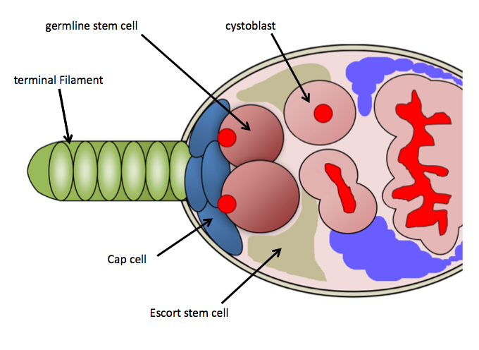
Stem-cell niche refers to a microenvironment, within the specific anatomic location where stem cells are found, which interacts with stem cells to regulate cell fate. The word 'niche' can be in reference to the ''in vivo'' or ''in vitro'' stem-cell microenvironment. During embryonic development, various niche factors act on embryonic stem cells to alter gene expression, and induce their proliferation or differentiation for the development of the fetus. Within the human body, stem-cell niches maintain adult stem cells in a quiescent state, but after tissue injury, the surrounding micro-environment actively signals to stem cells to promote either self-renewal or differentiation to form new tissues. Several factors are important to regulate stem-cell characteristics within the niche: cell–cell interactions between stem cells, as well as interactions between stem cells and neighbouring differentiated cells, interactions between stem cells and adhesion molecules, extracellular matrix co ... [...More Info...] [...Related Items...] OR: [Wikipedia] [Google] [Baidu] |
Stem Cell
In multicellular organisms, stem cells are undifferentiated or partially differentiated cells that can differentiate into various types of cells and proliferate indefinitely to produce more of the same stem cell. They are the earliest type of cell in a cell lineage. They are found in both embryonic and adult organisms, but they have slightly different properties in each. They are usually distinguished from progenitor cells, which cannot divide indefinitely, and precursor or blast cells, which are usually committed to differentiating into one cell type. In mammals, roughly 50–150 cells make up the inner cell mass during the blastocyst stage of embryonic development, around days 5–14. These have stem-cell capability. '' In vivo'', they eventually differentiate into all of the body's cell types (making them pluripotent). This process starts with the differentiation into the three germ layers – the ectoderm, mesoderm and endoderm – at the gastrulation stage. Howev ... [...More Info...] [...Related Items...] OR: [Wikipedia] [Google] [Baidu] |
Histology
Histology, also known as microscopic anatomy or microanatomy, is the branch of biology which studies the microscopic anatomy of biological tissues. Histology is the microscopic counterpart to gross anatomy, which looks at larger structures visible without a microscope. Although one may divide microscopic anatomy into ''organology'', the study of organs, ''histology'', the study of tissues, and '' cytology'', the study of cells, modern usage places all of these topics under the field of histology. In medicine, histopathology is the branch of histology that includes the microscopic identification and study of diseased tissue. In the field of paleontology, the term paleohistology refers to the histology of fossil organisms. Biological tissues Animal tissue classification There are four basic types of animal tissues: muscle tissue, nervous tissue, connective tissue, and epithelial tissue. All animal tissues are considered to be subtypes of these four principal tissue type ... [...More Info...] [...Related Items...] OR: [Wikipedia] [Google] [Baidu] |
Melanoma
Melanoma, also redundantly known as malignant melanoma, is a type of skin cancer that develops from the pigment-producing cells known as melanocytes. Melanomas typically occur in the skin, but may rarely occur in the mouth, intestines, or eye ( uveal melanoma). In women, they most commonly occur on the legs, while in men, they most commonly occur on the back. About 25% of melanomas develop from moles. Changes in a mole that can indicate melanoma include an increase in size, irregular edges, change in color, itchiness, or skin breakdown. The primary cause of melanoma is ultraviolet light (UV) exposure in those with low levels of the skin pigment melanin. The UV light may be from the sun or other sources, such as tanning devices. Those with many moles, a history of affected family members, and poor immune function are at greater risk. A number of rare genetic conditions, such as xeroderma pigmentosum, also increase the risk. Diagnosis is by biopsy and analysis of any sk ... [...More Info...] [...Related Items...] OR: [Wikipedia] [Google] [Baidu] |
Marrow Adipose Tissue
Bone marrow adipose tissue (BMAT), sometimes referred to as marrow adipose tissue (MAT), is a type of fat deposit in bone marrow. It increases in states of low bone density - osteoporosis, anorexia nervosa/ caloric restriction, skeletal unweighting such as that which occurs in space travel, and anti-diabetes therapies. BMAT decreases in anaemia, leukaemia, and hypertensive heart failure; in response to hormones such as oestrogen, leptin, and growth hormone; with exercise-induced weight loss or bariatric surgery; in response to chronic cold exposure; and in response to pharmacological agents such as bisphosphonates, teriparatide, and metformin. Anatomy Bone marrow adipocytes (BMAds) originate from mesenchymal stem cell (MSC) progenitors that also give rise to osteoblasts, among other cell types. Thus, it is thought that BMAT results from preferential MSC differentiation into the adipocyte, rather than osteoblast, lineage in the setting of osteoporosis. Since BMAT is increas ... [...More Info...] [...Related Items...] OR: [Wikipedia] [Google] [Baidu] |
Adipocyte
Adipocytes, also known as lipocytes and fat cells, are the cells that primarily compose adipose tissue, specialized in storing energy as fat. Adipocytes are derived from mesenchymal stem cells which give rise to adipocytes through adipogenesis. In cell culture, adipocyte progenitors can also form osteoblasts, myocytes and other cell types. There are two types of adipose tissue, white adipose tissue (WAT) and brown adipose tissue (BAT), which are also known as white and brown fat, respectively, and comprise two types of fat cells. Structure White fat cells White fat cells contain a single large lipid droplet surrounded by a layer of cytoplasm, and are known as unilocular. The nucleus is flattened and pushed to the periphery. A typical fat cell is 0.1 mm in diameter with some being twice that size, and others half that size. However, these numerical estimates of fat cell size depend largely on the measurement method and the location of the adipose tissue. The f ... [...More Info...] [...Related Items...] OR: [Wikipedia] [Google] [Baidu] |
Monocyte
Monocytes are a type of leukocyte or white blood cell. They are the largest type of leukocyte in blood and can differentiate into macrophages and conventional dendritic cells. As a part of the vertebrate innate immune system monocytes also influence adaptive immune responses and exert tissue repair functions. There are at least three subclasses of monocytes in human blood based on their phenotypic receptors. Structure Monocytes are amoeboid in appearance, and have nongranulated cytoplasm. Thus they are classified as agranulocytes, although they might occasionally display some azurophil granules and/or vacuoles. With a diameter of 15–22 μm, monocytes are the largest cell type in peripheral blood. Monocytes are mononuclear cells and the ellipsoidal nucleus is often lobulated/indented, causing a bean-shaped or kidney-shaped appearance. Monocytes compose 2% to 10% of all leukocytes in the human body. Development Monocytes are produced by the bone marrow from precursors ... [...More Info...] [...Related Items...] OR: [Wikipedia] [Google] [Baidu] |
Fibroblast
A fibroblast is a type of biological cell that synthesizes the extracellular matrix and collagen, produces the structural framework ( stroma) for animal tissues, and plays a critical role in wound healing. Fibroblasts are the most common cells of connective tissue in animals. Structure Fibroblasts have a branched cytoplasm surrounding an elliptical, speckled nucleus having two or more nucleoli. Active fibroblasts can be recognized by their abundant rough endoplasmic reticulum. Inactive fibroblasts (called fibrocytes) are smaller, spindle-shaped, and have a reduced amount of rough endoplasmic reticulum. Although disjointed and scattered when they have to cover a large space, fibroblasts, when crowded, often locally align in parallel clusters. Unlike the epithelial cells lining the body structures, fibroblasts do not form flat monolayers and are not restricted by a polarizing attachment to a basal lamina on one side, although they may contribute to basal lamina components in so ... [...More Info...] [...Related Items...] OR: [Wikipedia] [Google] [Baidu] |
Sinusoidal Endothelial Cells
A liver sinusoid is a type of capillary known as a sinusoidal capillary, discontinuous capillary or sinusoid, that is similar to a fenestrated capillary, having discontinuous endothelium that serves as a location for mixing of the oxygen-rich blood from the hepatic artery and the nutrient-rich blood from the portal vein. The liver sinusoid has a larger caliber than other types of capillaries and has a lining of specialised endothelial cells known as the liver sinusoidal endothelial cells (LSECs), and Kupffer cells. The cells are porous and have a scavenging function. The LSECs make up around half of the non-parenchymal cells in the liver and are flattened and fenestrated. LSECs have many fenestrae that gives easy communication between the sinusoidal lumen and the space of Disse. They play a part in filtration, endocytosis, and in the regulation of blood flow in the sinusoids. The Kupffer cells can take up and destroy foreign material such as bacteria. Hepatocytes are separated f ... [...More Info...] [...Related Items...] OR: [Wikipedia] [Google] [Baidu] |
Bone Marrow
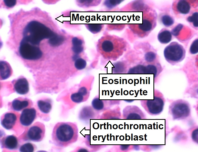
Bone marrow is a semi-solid tissue found within the spongy (also known as cancellous) portions of bones. In birds and mammals, bone marrow is the primary site of new blood cell production (or haematopoiesis). It is composed of hematopoietic cells, marrow adipose tissue, and supportive stromal cells. In adult humans, bone marrow is primarily located in the ribs, vertebrae, sternum, and bones of the pelvis. Bone marrow comprises approximately 5% of total body mass in healthy adult humans, such that a man weighing 73 kg (161 lbs) will have around 3.7 kg (8 lbs) of bone marrow. Human marrow produces approximately 500 billion blood cells per day, which join the systemic circulation via permeable vasculature sinusoids within the medullary cavity. All types of hematopoietic cells, including both myeloid and lymphoid lineages, are created in bone marrow; however, lymphoid cells must migrate to other lymphoid organs (e.g. thymus) in order to complete ... [...More Info...] [...Related Items...] OR: [Wikipedia] [Google] [Baidu] |
Hematopoietic Stem Cell
Hematopoietic stem cells (HSCs) are the stem cells that give rise to other blood cells. This process is called haematopoiesis. In vertebrates, the very first definitive HSCs arise from the ventral endothelial wall of the embryonic aorta within the (midgestational) aorta-gonad-mesonephros region, through a process known as endothelial-to-hematopoietic transition. In adults, haematopoiesis occurs in the red bone marrow, in the core of most bones. The red bone marrow is derived from the layer of the embryo called the mesoderm. Haematopoiesis is the process by which all mature blood cells are produced. It must balance enormous production needs (the average person produces more than 500 billion blood cells every day) with the need to regulate the number of each blood cell type in the circulation. In vertebrates, the vast majority of hematopoiesis occurs in the bone marrow and is derived from a limited number of hematopoietic stem cells that are multipotent and capable of extens ... [...More Info...] [...Related Items...] OR: [Wikipedia] [Google] [Baidu] |
Gap Junction
Gap junctions are specialized intercellular connections between a multitude of animal cell-types. They directly connect the cytoplasm of two cells, which allows various molecules, ions and electrical impulses to directly pass through a regulated gate between cells. One gap junction channel is composed of two protein hexamers (or hemichannels) called connexons in vertebrates and innexons in invertebrates. The hemichannel pair connect across the intercellular space bridging the gap between two cells. Gap junctions are analogous to the plasmodesmata that join plant cells. Gap junctions occur in virtually all tissues of the body, with the exception of adult fully developed skeletal muscle and mobile cell types such as sperm or erythrocytes. Gap junctions are not found in simpler organisms such as sponges and slime molds. A gap junction may also be called a ''nexus'' or ''macula communicans''. While an ephapse has some similarities to a gap junction, by modern definition t ... [...More Info...] [...Related Items...] OR: [Wikipedia] [Google] [Baidu] |
CDH1 (gene)
Cadherin-1 or Epithelial cadherin (E-cadherin), (not to be confused with the APC/C activator protein CDH1) is a protein that in humans is encoded by the ''CDH1'' gene. Mutations are correlated with gastric, breast, colorectal, thyroid, and ovarian cancers. CDH1 has also been designated as CD324 ( cluster of differentiation 324). It is a tumor suppressor gene. History The discovery of cadherin cell-cell adhesion proteins is attributed to Masatoshi Takeichi, whose experience with adhering epithelial cells began in 1966. His work originally began by studying lens differentiation in chicken embryos at Nagoya University, where he explored how retinal cells regulate lens fiber differentiation. To do this, Takeichi initially collected media that had previously cultured neural retina cells (CM) and suspended lens epithelial cells in it. He observed that cells suspended in the CM media had delayed attachment compared to cells in his regular medium. His interest in cell adherence was ... [...More Info...] [...Related Items...] OR: [Wikipedia] [Google] [Baidu] |


.png)
.jpg)


