|
Nephrosclerosis
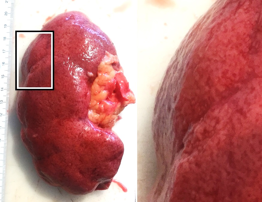
Hypertensive kidney disease is a medical condition referring to damage to the kidney due to chronic high blood pressure. It manifests as hypertensive nephrosclerosis (sclerosis referring to the stiffening of renal components). It should be distinguished from renovascular hypertension, which is a form of secondary hypertension, and thus has opposite direction of causation. Signs and symptoms Signs and symptoms of chronic kidney disease, including loss of appetite, nausea, vomiting, itching, sleepiness or confusion, weight loss, and an unpleasant taste in the mouth, may develop. Causes "Hypertensive" refers to high blood pressure and "nephropathy" means damage to the kidney; hence this condition is where chronic high blood pressure causes damages to kidney tissue; this includes the small blood vessels, glomeruli, kidney tubules and interstitial tissues. The tissue hardens and thickens which is known as nephrosclerosis. The narrowing of the blood vessels means less blood is goin ... [...More Info...] [...Related Items...] OR: [Wikipedia] [Google] [Baidu] |
Arteriolosclerosis
Arteriolosclerosis is a form of cardiovascular disease involving hardening and loss of elasticity of arterioles or small arteries and is most often associated with hypertension and diabetes mellitus. Types include hyaline arteriolosclerosis and hyperplastic arteriolosclerosis, both involved with vessel wall thickening and luminal narrowing that may cause downstream ischemic injury. The following two terms whilst similar, are distinct in both spelling and meaning and may easily be confused with arteriolosclerosis. * Arteriosclerosis is any hardening (and loss of elasticity) of medium or large arteries (from the Greek '' arteria'', meaning ''artery'', and '' sclerosis'', meaning ''hardening'') * Atherosclerosis is a hardening of an artery specifically due to an atheromatous plaque. The term ''atherogenic'' is used for substances or processes that cause atherosclerosis. Hyaline arteriolosclerosis Also arterial hyalinosis and arteriolar hyalinosis refers to thickening of the walls ... [...More Info...] [...Related Items...] OR: [Wikipedia] [Google] [Baidu] |
Proteinuria
Proteinuria is the presence of excess proteins in the urine. In healthy persons, urine contains very little protein; an excess is suggestive of illness. Excess protein in the urine often causes the urine to become foamy (although this symptom may also be caused by other conditions). Severe proteinuria can cause nephrotic syndrome in which there is worsening swelling of the body. Signs and symptoms Proteinuria often causes no symptoms and it may only be discovered incidentally. Foamy urine is considered a cardinal sign of proteinuria, but only a third of people with foamy urine have proteinuria as the underlying cause. It may also be caused by bilirubin in the urine ( bilirubinuria), retrograde ejaculation, pneumaturia (air bubbles in the urine) due to a fistula, or drugs such as pyridium. Causes There are three main mechanisms to cause proteinuria: * Due to disease in the glomerulus * Because of increased quantity of proteins in serum (overflow proteinuria) * Due to low r ... [...More Info...] [...Related Items...] OR: [Wikipedia] [Google] [Baidu] |
Micrograph
A micrograph or photomicrograph is a photograph or digital image taken through a microscope or similar device to show a magnified image of an object. This is opposed to a macrograph or photomacrograph, an image which is also taken on a microscope but is only slightly magnified, usually less than 10 times. Micrography is the practice or art of using microscopes to make photographs. A micrograph contains extensive details of microstructure. A wealth of information can be obtained from a simple micrograph like behavior of the material under different conditions, the phases found in the system, failure analysis, grain size estimation, elemental analysis and so on. Micrographs are widely used in all fields of microscopy. Types Photomicrograph A light micrograph or photomicrograph is a micrograph prepared using an optical microscope, a process referred to as ''photomicroscopy''. At a basic level, photomicroscopy may be performed simply by connecting a camera to a microscope, th ... [...More Info...] [...Related Items...] OR: [Wikipedia] [Google] [Baidu] |
Interstitial Fibrosis
An interstitial space or interstice is a space between structures or objects. In particular, interstitial may refer to: Biology * Interstitial cell tumor * Interstitial cell, any cell that lies between other cells * Interstitial collagenase, enzyme that breaks the peptide bonds in collagen * Interstitial cystitis * Interstitium, the contiguous fluid-filled space existing between the skin and body organs * Interstitial fluid, a solution that bathes and surrounds the cells of multicellular animals * Interstitial granulomatous dermatitis * Interstitial infusion * Interstitial keratitis * Interstitial lung disease * Interstitial nephritis * Interstitial pregnancy Other uses To describe the spaces within particulate matter such sands, gravels, cobbles, grain, etc. that lie between the discrete particles. * Interstitial art * Interstitial condensation, in construction * Interstitial site, in chemistry * Interstitial defect, in chemistry * Interstitial television show, in televi ... [...More Info...] [...Related Items...] OR: [Wikipedia] [Google] [Baidu] |
Mesangium
The glomerulus (plural glomeruli) is a network of small blood vessels (capillaries) known as a ''tuft'', located at the beginning of a nephron in the kidney. Each of the two kidneys contains about one million nephrons. The tuft is structurally supported by the mesangium (the space between the blood vessels), composed of intraglomerular mesangial cells. The blood is filtered across the capillary walls of this tuft through the glomerular filtration barrier, which yields its filtrate of water and soluble substances to a cup-like sac known as Bowman's capsule. The filtrate then enters the renal tubule of the nephron. The glomerulus receives its blood supply from an afferent arteriole of the renal arterial circulation. Unlike most capillary beds, the glomerular capillaries exit into efferent arterioles rather than venules. The resistance of the efferent arterioles causes sufficient hydrostatic pressure within the glomerulus to provide the force for ultrafiltration. The glomerulus an ... [...More Info...] [...Related Items...] OR: [Wikipedia] [Google] [Baidu] |
Sclerosis (medicine)
Sclerosis (from Greek σκληρός ''sklērós'', "hard") is the stiffening of a tissue or anatomical feature, usually caused by a replacement of the normal organ-specific tissue with connective tissue. The structure may be said to have undergone sclerotic changes or display sclerotic lesions, which refers to the process of sclerosis. Common medical conditions whose pathology involves sclerosis include: * Amyotrophic lateral sclerosis—also known as Lou Gehrig's disease or motor neurone disease—a progressive, incurable, usually fatal disease of motor neurons. * Atherosclerosis, a deposit of fatty materials, such as cholesterol, in the arteries which causes hardening. * Focal segmental glomerulosclerosis is a disease that attacks the kidney's filtering system (glomeruli) causing serious scarring and thus a cause of nephrotic syndrome in children and adolescents, as well as an important cause of kidney failure in adults. * Hippocampal sclerosis, a brain damage often seen in ... [...More Info...] [...Related Items...] OR: [Wikipedia] [Google] [Baidu] |
Basement Membrane
The basement membrane is a thin, pliable sheet-like type of extracellular matrix that provides cell and tissue support and acts as a platform for complex signalling. The basement membrane sits between Epithelium, epithelial tissues including mesothelium and endothelium, and the underlying connective tissue. Structure As seen with the electron microscope, the basement membrane is composed of two layers, the basal lamina and the reticular lamina. The underlying connective tissue attaches to the basal lamina with collagen VII anchoring fibrils and fibrillin microfibrils. The basal lamina layer can further be subdivided into two layers based on their visual appearance in electron microscopy. The lighter-colored layer closer to the epithelium is called the lamina lucida, while the denser-colored layer closer to the connective tissue is called the lamina densa. The Electron microscope, electron-dense lamina densa layer is about 30–70 nanometers thick and consists of an underlying ... [...More Info...] [...Related Items...] OR: [Wikipedia] [Google] [Baidu] |
Tunica Media
The tunica media (New Latin "middle coat"), or media for short, is the middle tunica (layer) of an artery or vein. It lies between the tunica intima on the inside and the tunica externa on the outside. Artery Tunica media is made up of smooth muscle cells, elastic tissue and collagen. It lies between the tunica intima on the inside and the tunica externa on the outside. The middle coat (tunica media) is distinguished from the inner (tunica intima) by its color and by the transverse arrangement of its fibers. * In the ''smaller arteries'' it consists principally of smooth muscle fibers in fine bundles, arranged in lamellæ and disposed circularly around the vessel. These lamellæ vary in number according to the size of the vessel; the smallest arteries having only a single layer, and those slightly larger three or four layers - up to a maximum of six layers. It is to this coat that the thickness of the wall of the artery is mainly due. * In the ''larger arteries'', as the ilia ... [...More Info...] [...Related Items...] OR: [Wikipedia] [Google] [Baidu] |
Tunica Intima
The tunica intima (New Latin "inner coat"), or intima for short, is the innermost tunica (layer) of an artery or vein. It is made up of one layer of endothelial cells and is supported by an internal elastic lamina. The endothelial cells are in direct contact with the blood flow. The three layers of a blood vessel are an inner layer (the tunica intima), a middle layer (the tunica media), and an outer layer (the tunica externa). In dissection, the inner coat (tunica intima) can be separated from the middle (tunica media) by a little maceration, or it may be stripped off in small pieces; but, because of its friability, it cannot be separated as a complete membrane. It is a fine, transparent, colorless structure which is highly elastic, and, after death, is commonly corrugated into longitudinal wrinkles. Structure The structure of the tunica intima depends on the blood vessel type. Elastic arteries – A single layer of Endothelial and a supporting layer of elastin-rich collagen. ... [...More Info...] [...Related Items...] OR: [Wikipedia] [Google] [Baidu] |
Albuminuria
Albuminuria is a pathological condition wherein the protein albumin is abnormally present in the urine. It is a type of proteinuria. Albumin is a major plasma protein (normally circulating in the blood); in healthy people, only trace amounts of it are present in urine, whereas larger amounts occur in the urine of patients with kidney disease. For a number of reasons, clinical terminology is changing to focus on albuminuria more than proteinuria. Signs and symptoms It is usually asymptomatic but whitish foam may appear in urine. Swelling of the ankles, hands, belly or face may occur if losses of albumin are significant and produce low serum protein levels ( nephrotic syndrome). Causes The kidneys normally do not filter large molecules into the urine, so albuminuria can be an indicator of damage to the kidneys or excessive salt intake. It can also occur in patients with long-standing diabetes, especially type 1 diabetes. Recent international guidelinesKDIGO 2012 reclassified chron ... [...More Info...] [...Related Items...] OR: [Wikipedia] [Google] [Baidu] |
Nephron
The nephron is the minute or microscopic structural and functional unit of the kidney. It is composed of a renal corpuscle and a renal tubule. The renal corpuscle consists of a tuft of capillaries called a glomerulus and a cup-shaped structure called Bowman's capsule. The renal tubule extends from the capsule. The capsule and tubule are connected and are composed of epithelial cells with a lumen. A healthy adult has 1 to 1.5 million nephrons in each kidney. Blood is filtered as it passes through three layers: the endothelial cells of the capillary wall, its basement membrane, and between the foot processes of the podocytes of the lining of the capsule. The tubule has adjacent peritubular capillaries that run between the descending and ascending portions of the tubule. As the fluid from the capsule flows down into the tubule, it is processed by the epithelial cells lining the tubule: water is reabsorbed and substances are exchanged (some are added, others are removed); first with t ... [...More Info...] [...Related Items...] OR: [Wikipedia] [Google] [Baidu] |
Endothelium
The endothelium is a single layer of squamous endothelial cells that line the interior surface of blood vessels and lymphatic vessels. The endothelium forms an interface between circulating blood or lymph in the lumen and the rest of the vessel wall. Endothelial cells form the barrier between vessels and tissue and control the flow of substances and fluid into and out of a tissue. Endothelial cells in direct contact with blood are called vascular endothelial cells whereas those in direct contact with lymph are known as lymphatic endothelial cells. Vascular endothelial cells line the entire circulatory system, from the heart to the smallest capillaries. These cells have unique functions that include fluid filtration, such as in the glomerulus of the kidney, blood vessel tone, hemostasis, neutrophil recruitment, and hormone trafficking. Endothelium of the interior surfaces of the heart chambers is called endocardium. An impaired function can lead to serious health issues throug ... [...More Info...] [...Related Items...] OR: [Wikipedia] [Google] [Baidu] |


